Brain (1999), 122, 2337–2344
The neural correlates of verb and noun processing A PET study Daniela Perani,1 Stefano F. Cappa,2 Tatiana Schnur,1 Marco Tettamanti,1 Simona Collina,1 Ma`rio Miguel Rosa3 and Ferruccio Fazio1 1Istituto
di Neuroscienze e Bioimmagini CNR, Scientific Institute, H San Raffaele, University of Milan-Bicocca, Milan, 2University of Brescia Medical School, Italy and 3Centro de Estudios Egas Moniz, Hospital de Santa Maria, Lisbon, Portugal
Correspondence to: Daniela Perani MD, INB-CNR, H San Raffaele, Via Olgettina 60, 20132, Milano, Italy E-mail:
[email protected]
Summary The hypothesis that categorical information, distinguishing among word classes, such as nouns, verbs, etc., is an organizational principle of lexical knowledge in the brain, is supported by the observation of aphasic subjects who are selectively impaired in the processing of nouns and verbs. The study of lesion location in these patients has suggested that the left temporal lobe plays a crucial role in processing nouns, while the left frontal lobe is necessary for verbs. To delineate the brain areas involved in the processing of different word classes, we used PET to measure regional cerebral activity during tasks requiring reading of concrete and abstract nouns and verbs for lexical decision. These tasks activated an extensive network of brain areas, mostly in the left frontal and temporal cortex, which represents the neural correlate of single word processing. Some left hemispheric areas,
including the dorsolateral frontal and lateral temporal cortex, were activated only by verbs, while there were no brain areas more active in response to nouns. Furthermore, the comparison of abstract and concrete words indicated that abstract word processing was associated with selective activations (right temporal pole and amygdala, bilateral inferior frontal cortex), while no brain areas were more active in response to concrete words. There were no significant interaction effects between word class and concreteness. Taken together, these findings are compatible with the view that lexical–semantic processing of words is mediated by an extensive, predominantly left hemispheric network of brain structures. Additional brain activations appear to be related to specific semantic content, or, in the case of verbs, may be associated with the automatic access of syntactic information.
Keywords: grammatical categories; verbs; nouns; reading; lexical decision; PET Abbreviations: BA ⫽ Brodmann area; SPM ⫽ statistical parametric mapping
Introduction Several clinical observations have suggested that different cerebral areas are involved in the processing of nouns and verbs. There is ample evidence that aphasic patients may be selectively impaired in the naming of objects or of actions. A double dissociation between object and action naming performance in individual cases has been reported (Miceli et al., 1988; Miozzo et al., 1994). A difference in the cerebral localization of lesions has been suggested to underlie this behavioural dissociation: patients with a selective disorder for object naming have usually had lesions centred on the left temporal lobe; conversely, a selective impairment in action naming has been associated with large lesions, usually extending to the left frontal cortex (for a review, see Daniele et al., 1994). These observations have led to the hypothesis (Damasio and Tranel, 1993) that the neural networks subser© Oxford University Press 1999
ving noun and verb retrieval are distinct, with the left frontal convexity playing a crucial role in the case of verbs. Converging evidence has been provided by the study of degenerative conditions: in particular, a severe impairment in action naming has been found in patients with frontal dementia (Cappa et al., 1998). Functional imaging studies have provided only a limited evidence for selective activations associated with noun and verb processing. There are several PET and functional MRI studies of verb generation, i.e. a task in which, in response to the presentation of an object name, the subjects must produce an appropriate verb (i.e. cake–eat). These studies have consistently shown the activation of the left prefrontal cortex, as well as of other left hemispheric areas (Petersen et al., 1988). However, the same pattern of activation is also
2338
D. Perani et al.
present with name generation (Warburton et al., 1996). Martin and colleagues asked subjects to generate either the appropriate colour or an appropriate action, while viewing an achromatic object drawing (Martin et al., 1995). A network of left hemispheric areas was activated by both of these generation tasks, in addition to those observed with visual naming; however, the area of activation specifically associated with verb generation was not in the left prefrontal cortex, but in the left middle temporal lobe. The lack of consistency between clinical and PET findings might, at least in part, be due to task differences. Most functional imaging studies have been based on spontaneous generation tasks, which are associated with a prominent activation of the dorsolateral frontal cortex, and this is also the case for non-verbal conditions (Frith et al., 1991). The utilization of an effortful lexical–semantic retrieval task (Fiez, 1997) may then obscure any relatively subtle difference in activation related to grammatical category effect, in particular in the frontal lobe area. On the other hand, most patient studies have used naming tasks, and in particular the naming of actions. Verbs are an extremely complex and heterogeneous category (Jackendoff, 1983; Pinker, 1989); while it may be speculated that action verbs are processed in brain regions involved in motor function, such as the frontal lobe, other verb categories, such as abstract verbs related to psychological functions, may involve different neurological substrates (Bushell and Martin, 1997). In order to overcome these limitations, we have used noun and verb stimuli from matched semantic categories in a task which did not require effortful lexical retrieval. The semantic categories were, for nouns: tool names and abstract words; for verbs: manipulation actions and verbs related to psychological states. The decision to include abstract nouns and verbs was motivated by the claim that grammatical category effects may be due to the inadequate matching of noun and verbs for concreteness, the latter being, in general, more abstract (Allport and Funnell, 1981). The task was visual lexical decision. Regional cerebral perfusion was measured with PET and the results analysed with a factorial design in order to assess the cerebral activations selectively associated with word class, semantic category and their interaction.
Methods Subjects The experimental subjects were 14 right-handed male volunteers (age range 22–26 years) who gave written informed consent prior to the experiment. All subjects had no history of neurological or psychiatric disorders. Right-handedness was verified using the Edinburgh Inventory (Oldfield, 1971). The experimental protocol was approved by the local hospital ethics committee and subjects.
Tasks Baseline task In the baseline task, 40 letter strings without an ‘X’ and five letter strings containing an ‘X’, randomly located, were
presented for 1200 ms in isolation on a computer screen, with an inter-stimulus interval of 300 ms. Letter string length was matched with experimental word length. Subjects were asked to read the consonant letter strings and to respond by pressing the button on a response box with the right hand when they saw a letter string containing an ‘X’.
Experimental tasks The subjects were presented with four categories of words: (i) concrete verbs, related to object manipulation (to cut, to comb, to brush, to rake, to write . . .); (ii) abstract verbs, related to psychological states (to think, to hope, to believe, to desire . . .); (iii) concrete nouns (manipulable tools: comb, hammer, screwdriver, scissors . . .); (iv) abstract nouns (future, justice, hope, meaning . . .). It must be pointed out that, because of the properties of the Italian language, verbs and nouns referring to related concepts are never completely homonymous (e.g. ‘hope’, ‘speranza’; ‘to hope’, ‘sperare’; ‘sleep’, ‘sonno’; ‘to sleep’, ‘dormire’). For each task, the words of one category consisting of 40 words and five pseudowords, randomly located, were presented for 1200 ms each, with an inter-stimulus interval of 300 ms. The tasks were designed to elicit a lexical decision: the subjects were asked to read each word silently and when a legal pseudoword appeared on the screen to respond by pressing the button on a response box with the right hand. The stimuli for both verbs and nouns were selected from different sources: all stimuli were high-frequency (De Mauro et al., 1993) and balanced across scans for length and number of syllables. They were presented to nine normal subjects outside the PET experiment. Reaction times were collected and analysed with a two-factor ANOVA (analysis of variance). No significant differences were present for this very easy task (mean reaction times: abstract verbs 488.7 ms; abstract nouns 464.1 ms; concrete verbs 502.9 ms; concrete nouns 469.9 ms). All five experimental conditions were repeated twice for each subject, for a total of 10 scans.
PET data acquisition Regional cerebral blood flow was measured by recording the distribution of radioactivity following an intravenous injection of 15O-labelled water (H215O) with a GE-Advance scanner (General Electric Medical Systems, Milwaukee, Wis., USA) which has a field of view of 15.2 cm. Data were acquired by scanning in 3D mode. A 5 mCi slow bolus of H215O, 4 ml in 20 s plus 4 ml of saline solution in 20 s, was injected (Silbersweig et al., 1993). After attenuation correction (measured by a transmission scan using a pair of rotating pin sources filled with 68Ge), the data were reconstructed as 35 transaxial planes by 3D filtered back projection with a Hanning filter (cut-off 4 mm filter width) in the transaxial plane, and a Ramp filter (cut-off 8.5 mm) in the axial direction. The integrated counts collected for 90 s, starting
A PET study of verb and noun processing 30 s after injection time, were used as an index of regional cerebral blood flow.
Data analysis Image manipulations and statistical analysis were performed in MATLAB 4.2 (Math Works, Natick, Mass., USA) using statistical parametric mapping (SPM 96, Wellcome Department of Cognitive Neurology, London, UK). The original brain images were first realigned and then transformed into a standard stereotactic space; global differences in cerebral blood flow were covaried out for all voxels and comparisons across conditions were made using t-statistics with appropriate linear contrasts (Friston et al., 1995a, b). In order to increase signal to noise ratio and accommodate normal variability in functional gyral anatomy each image was smoothed in three dimensions with a Gaussian filter (16 ⫻ 16 ⫻ 16 mm). A repeated-measures ANCOVA (analysis of covariance) was used for the comparison of different tasks, in which every subject was studied under all conditions. The SPM package allows the comparison of one experimental condition relative to a pair or a group of control conditions, specifying a set of weights that are used to add the means together with a sum set to zero. The contrast is divided by the adjusted root mean square error and the resulting value has the t-distribution under the null hypothesis of no differences. This set of t-values comprises the statistical parametric map (SPM{t}). Only regional activations significant at P ⬍ 0.001 (uncorrected for multiple non-independent comparisons) were considered. The following contrasts were used. (i) Contrast 1. Main effect of all lexical stimuli in comparison with the letter string baseline: (VC ⫹ VA ⫹ NC ⫹ NA) – LS, where VC ⫽ concrete verbs; VA ⫽ abstract verbs; NC ⫽ concrete nouns; NA ⫽ abstract nouns; LS ⫽ letter strings. (ii) Contrast 2. Regions common to all four activation conditions were then identified using the masking procedure in SPM. This eliminates voxels that are not significant at P ⬍ 0.05 in each contrast specified. Here the overall main effect was masked with each of the four individual contrasts: (VC ⫹ VA ⫹ NC ⫹ NA) – LS masked with (VC – LS), (VA – LS), (NC – LS), (NA – LS). (iii) Contrast 3. Main effect of grammatical category: (VC ⫹ VA) – (NC ⫹ NA); (NC ⫹ NA) – (VC ⫹ VA). (iv) Contrast 4. Main effect of concreteness: (VC ⫹ NC) – (VA ⫹ NA); (VA ⫹ NA) – (VC ⫹ NC) (v). Contrast 5. The interaction effects between word class and concreteness were also sought: (VC - NC) (VA – NA); (NC – VC) – (NA – VA) masked on the main effect of concreteness; (VA – NA) – (VC – NC); (NA – VA) –(NC – VC) masked on the main effect of abstractness. These sets of comparisons were designed to reveal differences and commonalities in the anatomical foci associated with reading of verbs and nouns, as well as the effects of word class and concreteness.
2339
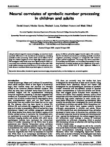
Results Areas specific for lexical stimuli Main effect of words relative to baseline The lexical decision tasks yielded an extensive pattern of brain activations, which involved the left frontal [Brodmann area (BA) 44, 45, 47, 6], temporal (BA 22/38, 22/21, 20, 37), temporoparietal (BA 22/39) and occipital cortex (BA 18), as well as the right cerebellum and some right frontal areas (BA 47, 32) (Table 1A and Fig. 1A).
Areas common to all lexical conditions The main effect masked with each of the individual contrasts shows the commonalities in the patterns of brain activation of the four lexical conditions. In particular, there was a selective left hemispheric involvement with activations in the inferior frontal gyrus (BA 44, 45, 47), the superior temporal sulcus (BA 22/21), the posterior-superior temporal gyrus (BA 22/21) and inferior temporal gyrus (BA 20) (Table 1B and Fig. 1B).
Areas specific for verbs The direct comparison between the condition in which the subjects were performing lexical decision for verbs and that where the stimuli were nouns (both abstract and concrete), showed that several areas were significantly more active when processing verbs (Table 2A and Fig. 1C). These included the left dorsolateral frontal cortex (BA 45, 46, 46/9), as well as superior parietal (BA 7), anterior temporal (BA 22/38), middle temporal (BA 21/37) and occipital (BA 18) areas. The reverse comparison (nouns versus verbs) failed to reveal areas which were significantly more active during noun processing.
Areas specific for abstract words A similar direct comparison was performed between the conditions in which the lexical decision was made on abstract words and those on concrete words (both nouns and verbs) (Table 2C and Fig. 1D). Bilateral activations were found in the lateral ventral frontal cortex (BA 47). In the right hemisphere, there were activation foci in the temporal pole (BA 38), the parieto-occipital junction (BA 39/19), the anterior cingulate gyrus (BA 32) and the amygdala. On the left there was a focus in the upper and anterior part of the superior temporal gyrus (BA 22). The reverse comparison (concrete versus abstract) failed to show any significant activation related to concrete word processing. The interaction effects between grammatical category and concreteness were not significant.
2340
D. Perani et al.
Fig. 1 The areas of activation have been rendered on to the lateral surfaces of the cortex of the brain used by SPM 96 to illustrate the distribution of the major sites of activation (P ⬍ 0.001). (A) The areas activated when all lexical conditions pooled together are compared with the letter string baseline. Within these areas, the commonalities (i.e. regions equally activated by all stimuli, irrespective of word class or concreteness) (B), the regions specifically associated with the processing of verbs (C) or abstract words (D) are shown (see text for details).
A PET study of verb and noun processing Table 2 Main effects of categories
Table 1 Main effects of lexical conditions Area
x
2341
y
(A) Combined lexical conditions versus letter string Left inferior front gyrus (44/45) –50 20 Left inferior frontal gyrus (45) –38 24 Left inferior frontal gyrus (45) –48 18 Left inferior frontal gyrus (47) –52 20 Left inferior frontal gyrus (47/11) –32 26 Left precentral gyrus (6) –50 –10 Left superior temporal gyrus (22/38) –50 10 Left superior temporal sulcus (22/21) –54 –2 Left posterior superior temporal gyrus –56 –38 (22/21) Left insula –34 24 Left temporoparietal junction (22/39) –60 –50 Left fusiform gyrus (37) –32 –38 Left inferior temporal gyrus (20) –42 –18 Left lingual gyrus (18) –10 –70 Right inferior frontal gyrus (47) 42 18 Right anterior cingulate gyrus (32) 6 20 Right lingual gyrus (18) 10 –74 Right cerebellum 10 –58
z Z-score 16 8 12 –8 –20 48 –12 –12 4
4.6 4.4 5.1 4.1 3.7 3.6 4.2 5.0 4.5
8 12 –24 –32 4 0 44 4 –36
3.9 3.9 3.9 3.7 3.9 4.9 3.3 3.6 4.0
(B) Main effects of combined lexical conditions versus letter string (masked with the individual contrasts) Left inferior frontal gyrus (44/45) –50 20 16 4.6 Left inferior frontal gyrus (44/45) –56 22 4 4.0 Left inferior frontal gyrus (47) –56 20 –4 4.0 Left inferior frontal gyrus (47/11) –34 26 –16 3.6 Left superior temporal sulcus (22/21) –54 –2 –12 5.0 Left superior temporal sulcus (22/21) –58 –8 –8 3.7 Left posterior superior temporal gyrus –56 –38 4 4.5 (22/21) Left inferior temporal gyrus (20) –42 –12 –36 3.6 Left inferior temporal gyrus (20) –42 –4 –32 3.3 Numbers in parentheses refer to BAs.
Discussion The presence of extensive activation of the language areas of the left hemisphere during the silent reading of words in order to perform a lexical decision task can be considered to represent the functional brain correlate of automatic lexical– semantic processing. This activation encompassed all the areas which have been shown to be involved in lexical– semantic processing by functional imaging studies (Price, 1998). Moreover, the results of the factorial analysis support the notion that, beyond a common pattern of activation centred on the left perisylvian language cortex, some areas appear to be specifically engaged by verb and abstract word processing. These results must be interpreted with reference to neuropsychological evidence. In aphasic patients, verb processing impairments are associated with lesions which involve the left dorsolateral frontal cortex, but usually extend to other parts of the left perisylvian language cortex (Daniele et al., 1994). On the other hand, patients with selective sparing of verb processing have lesions which are usually limited to the temporal lobe (Silveri and Di Betta, 1997). Our PET results indicate that, in the left hemisphere, some
Area (A) Verbs versus nouns Left middle frontal gyrus (46/9) Left inferior frontal gyrus (45/46) Left superior parietal lobule (7) Left superior temporal gyrus (22/38) Left middle temporal gyrus (21/37) Left inferior occipital gyrus (18) Left lingual gyrus (18) Right lenticular nucleus
x
y
–28 –36 –8 –48 –48 –42 –22 26
28 30 –42 6 –48 –90 –70 –8
z Z-score 28 20 80 –8 4 –20 4 8
3.4 3.1 3.8 3.3 3.2 3.6 4.0 3.4
(B) Nouns versus verbs No voxels above threshold (C) Abstractness versus concreteness Left inferior frontal gyrus (47) –44 14 –4 4.5 Left superior temporal gyrus (22/38) –58 8 –16 3.2 Right inferior frontal gyrus (47) 52 20 –12 4.1 Right temporal pole (38) 42 16 –36 3.9 Right parieto-occipital junction (39/19) 40 –70 36 3.4 Right anterior cingulate gyrus (32) 6 16 40 3.3 Right amygdala 30 –4 –8 3.9 (D) Concreteness versus abstractness No voxels above threshold Numbers in parentheses refer to BAs.
areas, not only in the dorsolateral frontal cortex (BA 45, 46), but also in the temporal, parietal and occipital lobe are activated only when verbs are processed. Given that verbs and nouns were matched for length and lexical frequency, word class differences may be considered to arise at the syntactic or at the semantic level. Verbs are in general richer in structural information than nouns, including the nature and number of arguments, such as agent, theme and goal, which must be directly mapped in the surface syntactic structure of the sentence (Grimshaw, 1990). The differential activations associated with verb processing might thus be, at least in part, related to the automatic access of structural (syntactic) information associated with active verbs. The frontal activation seems to be a likely candidate in this respect. BA 45 (in conjunction with area 44) was observed to be involved in many single-word processing tasks, such as verb retrieval (Warburton et al., 1996), silent word generation (Wise et al., 1991; McGuire et al., 1996) and semantic judgement (Demonet et al., 1992). There is now also evidence that the anterior part of Broca’s area (BA 45, 46) may be related to syntactic processing, as suggested by PET experiments involving sentential contexts (Caplan et al., 1998). The posterior (opercular) part of Broca’s area (BA 44) appears to play a crucial role in phonological processing (Paulesu et al., 1997). From a different standpoint, the activation of this region has been suggested to be related to action recognition rather than (solely) speech processing. Rizzolatti and colleagues reported activation of the left inferior frontal gyrus during the observation of grasping
2342
D. Perani et al.
movements and they conjectured that this region corresponds to a system for their representation, functionally similar to the monkey ventral area 6 where ‘mirror’ neurons were recorded (Rizzolatti et al., 1996). Indeed, a similar region is activated during mental simulation of hand actions (Decety et al., 1994). This area was also activated in other studies related to language for action, such as generation of action verbs (Martin et al., 1995). We found an additional focus in the left middle temporal gyrus (BA 21/37) which was also activated in the PET study by Martin and colleagues in which subjects generated action words (Martin et al., 1995). Thus, the activation of these areas, as well as of the superior parietal lobule, may be explained within the framework of recognition and memory storage of the semantic aspects of action (Jeannerod, 1997). However, the fact that in the present study the activations were observed for both manipulation and psychological verbs, as indicated by the lack of a significant interaction with concreteness, seems to rule out any strictly ‘semantic’ interpretation in terms of a relationship to tools and their utilization. Another area, in the anterior part of the superior temporal gyrus, was also involved during the tasks related to verb processing (Table 2A and Fig. 1C). It is noteworthy that an area with similar coordinates, extending into the left temporal pole, was also found to be activated by tasks requiring language comprehension of extended narratives, but not by word lists (Mazoyer et al., 1993; Perani et al., 1998); this may suggest its involvement in syntactic processing. It is more difficult to comment on the occipital activations related to verb processing. Further evidence is probably necessary before an interpretation can be attempted. No areas were more active for nouns than for verbs. In other words, PET imaging does not show a neurological correlate for the double dissociation of verb and noun processing observed clinically. A possible explanation for the failure to find areas selectively activated by noun processing might again be related to the semantic categories used in the present experiment. There are now several imaging studies showing that stimuli belonging to different semantic categories are associated with different patterns of brain activation. In particular, there is evidence for a crucial role of the occipital and occipitotemporal cortex in the identification and naming of living entities (Perani et al., 1995; Damasio et al., 1996; Martin et al., 1996); on the other hand, the processing of non-living items, such as tools, is associated with left perisylvian cortex activation (Perani et al., 1995). While it has been suggested that the semantic organization of verbs is different from nouns, in that generally it affords a relational rather then a decompositional analysis (Miller and Fellbaum, 1991), it is likely that the choice of stimuli largely overlapping in semantic content (nouns related to tools and to abstract entities, verbs related to manipulative actions and psychological states) may have minimized differences related to semantic factors in the noun–verb comparison. Another result of the present experiment was the finding
of activations specific to abstract word processing. The neurological correlates of the concreteness effect are largely unknown. A left-hemispheric superiority for abstract word processing has been hypothesized, largely on the basis of split-field studies in normal subjects. However, the presence of a right hemifield advantage has not always been confirmed (Saffran et al., 1980). Studies in brain-damaged patients have also failed to provide univocal evidence. Most aphasic patients after left hemispheric lesions are more impaired with abstract word processing; this ‘standard’ effect is particularly evident in the case of deep dyslexia, which has been suggested to reflect the reading abilities of the intact right hemisphere (Coltheart et al., 1980). However, unusual cases showing a reversal of this effect, i.e. better performance with abstract words, have been reported in association with left hemispheric perisylvian damage (Warrington, 1981) or bilateral temporal involvement due to progressive dementia (Breedin et al., 1994). The evidence from functional imaging is limited. During episodic memory tasks, the processing of high imagery (largely concrete) words is associated with activation of the precuneus region (Fletcher et al., 1996). Mellet and colleagues (Mellet et al., 1998) have reported that listening to vocabulary definitions of abstract words, compared with concrete words, results in bilateral temporal activations, with a right-sided extension towards the temporal pole. The temporal pole was also found to be activated in the present experiment. This region is anatomically and functionally linked to the limbic system, in particular to the amygdala, which has a crucial role in emotional processing (Ledoux, 1995). It might be suggested that in the present case the activation of these areas, with a clear right hemispheric prevalence, may be related to the emotional value of the stimuli, which can be considered in general greater for abstract words than for words related to tools and their manipulation.
Acknowledgements We wish to thank Professors Andrea Moro and Alfonso Caramazza for helpful comments. This work was supported by CNR grants.
References Allport DA, Funnell E. Components of the mental lexicon. Philos Trans R Soc Lond B Biol Sci 1981; 295: 397–410. Breedin SD, Saffran EM, Coslett HB. Reversal of the concreteness effect in a patient with semantic dementia. Cogn Neuropsychol 1994; 11: 617–60. Bushell CM, Martin A. Automatic semantic priming of nouns and verbs in patients with Alzheimer’s disease. Neuropsychologia 1997; 35: 1059–67. Caplan D, Alpert N, Waters G. Effects of syntactic structure and propositional number on patterns of regional cerebral blood flow. J Cogn Neurosci 1998; 10: 541–52. Cappa SF, Binetti G, Pezzini A, Padovani A, Rozzini L, Trabucchi
A PET study of verb and noun processing M. Object and action naming in Alzheimer’s disease and frontotemporal dementia [see comments]. Neurology 1998; 50: 351–5. Comment in: Neurology 1998; 50: 324–5. Coltheart M, Patterson K, Marshall JC. Deep dyslexia. London: Routledge and Kegan Paul; 1980. Damasio AR, Tranel D. Nouns and verbs are retrieved with differently distributed neural systems. Proc Natl Acad Sci USA 1993; 90: 4957–60. Damasio H, Grabowski TJ, Tranel D, Hichwa RD, Damasio AR. A neural basis for lexical retrieval [see comments] [published erratum appears in Nature 1996; 381: 810]. Nature 1996; 380: 499–505. Comment in: Nature 1996; 380: 485–6. Daniele A, Giustolisi L, Silveri MC, Colosimo C, Gainotti G. Evidence for a possible neuroanatomical basis for lexical processing of nouns and verbs. Neuropsychologia 1994; 32: 1325–41.
2343
correlates of category-specific knowledge. Nature 1996; 379: 649–52. Mazoyer BM, Tzourio N, Frak V, Syrota A, Murayama N, Levrier O, et al. The cortical representation of speech. J Cogn Neurosci 1993; 5: 467–79. McGuire PK, Silbersweig DA, Frith CD. Functional neuroanatomy of verbal self-monitoring. Brain 1996; 119: 907–17. Mellet E, Tzourio N, Denis M, Mazoyer B. Cortical anatomy of mental imagery of concrete nouns based on their dictionary definition. Neuroreport 1998; 9: 803–8. Miceli G, Silveri MC, Nocentini U, Caramazza A. Patterns of dissociation in comprehension and production of nouns and verbs. Aphasiology 1988; 2: 251–8. Miller GA, Fellbaum C. Semantic networks of English. Cognition 1991; 41: 197–229.
De Mauro T, Mancini F, Vedovelli M, Voghera M. Lessico di frequenza dell’ italiano parlato. Rome: Etaslibri; 1993.
Miozzo A, Soardi M, Cappa SF. Pure anomia with spared action naming due to a left temporal lesion. Neuropsychologia 1994; 32: 1101–9.
Decety J, Perani D, Jeannerod M, Bettinardi V, Tadary B, Woods R, et al. Mapping motor representations with positron emission tomography. Nature 1994; 371: 600–2.
Oldfield RC. The assessment and analysis of handedness: the Edinburgh inventory. Neuropsychologia 1971; 9: 97–113.
Demonet JF, Chollet F, Ramsay S, Cardebat D, Nespoulous JL, Wise R, et al. The anatomy of phonological and semantic processing in normal subjects. Brain 1992; 115: 1753–68.
Paulesu E, Goldacre B, Scifo P, Cappa SF, Gilardi M, Castiglioni I, et al. Differential activation of left frontal cortex during phonemic and semantic word fluency. An EPI-fMRI activation study. Neuroreport 1997; 8: 2011–16.
Fiez JA. Phonology, semantics and the role of the left inferior prefrontal cortex. Hum Brain Mapp 1997; 5: 79–83. Fletcher PC, Shallice T, Frith CD, Frackowiak RS, Dolan RJ. Brain activity during memory retrieval. The influence of imagery and semantic cueing. Brain 1996; 119: 1587–96. Friston KJ, Ashburner J, Frith CD, Poline JB, Heather JD, Frackowiak RSJ. Spatial registration and normalization of images. Hum Brain Mapp 1995a; 3: 165–89. Friston KJ, Holmes AP, Worsley KJ, Poline JB, Frith CD, Frackowiak RSJ. Statistical parametric maps in functional imaging: a general linear approach. Hum Brain Mapp 1995b; 2: 189–210. Frith CD, Friston K, Liddle PF, Frackowiak RSJ. Willed action and the prefrontal cortex in man: a study with PET. Proc Roy Soc Lond B Biol Sci 1991; 244: 241–6. Grimshaw JB. Argument structure. Cambridge (MA): MIT Press; 1980. Jackendoff RS. Semantics and cognition. Cambridge (MA): MIT Press; 1983. Jeannerod M. The cognitive neuroscience of action. Oxford: Blackwell; 1997. Ledoux JE. In search of an emotional system in the brain: leaping from fear to emotion and consciousness. In: Gazzaniga MS, editor. The cognitive neurosciences. Cambridge (MA): MIT Press; 1995: 1049–61. Martin A, Haxby JV, Lalonde FM, Wiggs CL, Ungerleider LG. Discrete cortical regions associated with knowledge of color and knowledge of action. Science 1995; 270: 102–5. Martin A, Wiggs CL, Ungerleider LG, Haxby JV. Neural
Perani D, Cappa SF, Bettinardi V, Bressi S, Gorno-Tempini M, Matarrese M, et al. Different neural systems for the recognition of animals and man-made tools. Neuroreport 1995; 6: 1637–41. Perani D, Paulesu E, Galles NS, Dupoux E, Dehaene S, Bettinardi V, et al. The bilingual brain. Proficiency and age of acquisition of the second language. Brain 1998; 121: 1841–52. Petersen SE, Fox PT, Posner MI, Mintun M, Raichle ME. Positron emission tomographic studies of the cortical anatomy of single-word processing. Nature 1988; 331: 585–9. Pinker S. Learnability and cognition. The acquisition of argument structure. Cambridge (MA): MIT Press; 1989. Price CJ. The functional anatomy of word comprehension and production. Trends Cogn Sci 1998; 2: 281–8. Rizzolatti G, Fadiga L, Matelli M, Bettinardi V, Paulesu E, Perani D, et al. Localization of grasp representations in humans by PET: 1. Observation versus execution. Exp Brain Res 1996; 111: 246–52. Saffran EM, Bogyo LC, Schwartz MF, Marin OSM. Does deep dyslexia reflect right-hemisphere reading? In: Coltheart M, Patterson K, Marshall JC, editors. Deep dyslexia. London: Routledge & Kegan Paul; 1980. p. 381–406. Silbersweig DA, Stern E, Frith CD, Cahill C, Schnorr L, Grootoonk S, et al. Detection of thirty-second cognitive activations in single subjects with positron emission tomography: a new low-dose H2(15)O regional cerebral blood flow three-dimensional imaging technique. J Cereb Blood Flow Metab 1993; 13: 617–29. Silveri MC, Di Betta AM. Noun-verb dissociations in braindamaged patients: further evidence. Neurocase 1997; 3: 477–88.
2344
D. Perani et al.
Warburton E, Wise RJ, Price CJ, Weiller C, Hadar U, Ramsay S, et al. Noun and verb retrieval by normal subjects. Studies with PET. [Review] Brain 1996; 119: 159–79. Warrington EK. Concrete word dyslexia. Br J Psychol 1981; 72: 175–96. Wise R, Chollet F, Hadar U, Friston K, Hoffner E, Frackowiak
RSJ. Distribution of cortical neural networks involved in word comprehension and word retrieval. Brain 1991; 114: 1803–17.
Received April 16, 1999. Revised May 26, 1999. Accepted June 14, 1999