Taxonomic status of the reptile genus Procolophon from the Gondwanan Triassic Juan Carlos Cisneros* Bernard Price Institute for Palaeontological Research, University of the Witwatersrand, Private Bag 3, WITS, Johannesburg, 2050 South Africa Recieved 29 January 2007. Accepted 29 May 2007
The specific composition of the genus Procolophon in Brazil, South Africa and Antarctica is discussed in the light of new data. It is found that P. pricei and P. brasiliensis, two species described from Brazil, fit within the pattern of ontogenetic variation of the type species P. trigoniceps, and they are here considered junior synonyms. The South African species P. laticeps, characterized by the presence of a temporal fenestra, is no longer considered valid. The peculiar temporal openings of this species are regarded here as an anomalous condition without taxonomic significance. The only complete skull known from Antarctica shows a unique feature consisting of an elliptical depression in the palate. The interpretation of this structure is ambiguous because it may also be attributable to individual variation, and this specimen is provisionally kept within P. trigoniceps. Therefore, only the type species, P. trigoniceps, is recognized in Gondwana. This species occupies a wide geographic range, from the Paraná Basin to the Transantarctic Mountains. Keywords: Procolophonidae, Procolophon, temporal fenestrae, Triassic, Gondwana.
INTRODUCTION Procolophon is a member of the Procolophonidae, a clade of small parareptiles that flourished across all Pangaea during the Triassic, becoming extinct during the time of the T/J boundary. Procolophon is known from hundreds of specimens, most of them found in the Lower Triassic of the South African Karoo, were it is outnumbered only by the ubiquitous dicynodont Lystrosaurus (Kitching 1977; Groenewald & Kitching 1995). The genus possesses a robust, wide torso and enlarged chisel-like teeth, both adaptations suggesting a high-fibre diet (Hotton et al. 1997); prominent quadratojugal processes confer a bizarre, triangular shape to the head (Fig. 1). Limb adaptations suggest that Procolophon was capable of burrowing (deBraga 2003) and burrow casts from the Lystrosaurus Assemblage Zone (AZ) of South Africa have been ascribed to this genus (Groenewald 1991). The genus is noteworthy because of the presence of some individuals with temporal openings, sometimes regarded as representing a different species (Hamley & Thulborn 1993). Procolophon has also been recovered from the Fremouw Formation of the Transantarctic Basin (Kitching et al. 1972; Colbert & Kitching 1975) and the Sanga do Cabral Formation of the Paraná Basin in Brazil (Barberena et al. 1981; Lavina 1983). Two Procolophon species have been described from Brazil (Lavina 1983; Cisneros & Schultz 2002). In the light of new data, this study reviews the taxonomic status of the Antarctic material, the Brazilian species, and the specimens with temporal openings from South Africa. TAXONOMIC HISTORY Together with the description of the type species Procolophon trigoniceps (by page priority, Fig. 2), Owen (1876) proposed P. minor based on a juvenile skull. This was followed by five more species proposed by Seeley (1878, 1905): P. griersoni, P. cuneiceps, P. laticeps, *Present address: Departamento de Paleontologia e Estratigrafia, Universidade Federal do Rio Grande do Sul, and CNPq, Porto Alegre, CP 15001, 91501-970, Brazil. E-mail:
[email protected]
ISSN 0078-8554 Palaeont. afr. (April 2008) 43: 7–17
P. platyrhinus and P. sphenorhinus. Some of the features used to distinguish these species are better explained by taphonomic, ontogenetic or individual variation. Besides, all seven of Owen’s and Seeley’s holotypes were collected by D. White from Donnybrook Farm, in the Eastern Cape Province of South Africa, leading Broom (1936) to recognize only the type species, P. trigoniceps. The non-validity of the additional species has been virtually unquestioned by later authors. Broom (1905) in turn, proposed another species, P. baini, based on a well-preserved, almost complete skeleton collected by J.M. Bain or T. Bain at an unknown locality. This species was said to differ from P. trigoniceps mainly in the number of marginal teeth, but this character is known to be ontogenetically variable in the genus (Gow 1977). P. baini was also sunk into junior synonymy with P. trigoniceps by Colbert & Kitching (1975). The discovery of a Lystrosaurus fauna in the Fremouw Formation of Antarctica yielded the first Procolophon remains from that continent (Kitching et al. 1972). These remains were represented by one well-preserved, articulated skeleton plus a number of fragmentary specimens. Colbert & Kitching (1975) noted a number of peculiarities in the best Antarctic specimen, including very small quadratojugal horns and relatively robust limbs, but decided that it fitted within the range of variation of the South African P. trigoniceps and referred all Antarctic material to this species. In Brazil, Procolophon was reported by Barberena et al. (1981) in the Sanga do Cabral Formation, in the state of Rio Grande do Sul, southern Brazil. This find consisted of one partial cranium and mandible, a second mandible and non-associated vertebral elements. These specimens were later described by Lavina (1983) as a new species, P. pricei. A Procolophon cranium collected subsequently from a different locality was described by Cisneros & Schultz (2002) as P. brasiliensis. Both Brazilian species were founded mainly on characters relating to the arrangement of the palatal dentition. Hamley & Thulborn (1993) reviewed the status of the species P. laticeps. This species was said to differ from 7
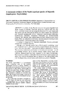
Figure 1. Life reconstruction of Procolophon trigoniceps. Note the presence of quadratojugal processes covered by long keratinous spines and large digging-claws. The skeleton of Procolophon supports these features (Carroll & Lindsay 1985; deBraga 2003). Numerous minor keratinous spines are common over large or stocky-bodied modern lizards; these structures do not leave traces in the skeleton (e.g. Iguana, Phrynosoma, Uromastix; pers. obs.). The long quadratojugal and supratemporal spines of Procolophon may have acted as an anti-predatory mechanism, as in phrynosomatid lizards (Young et al. 2004).
P. trigoniceps by the presence of a small temporal fenestra (Seeley 1878, 1905). However, the authenticity of these small openings, which are present only in the holotype and a referred specimen of a genus that in South Africa is known from several specimens, was questioned almost from the beginning. They were regarded by some authors as an artefact of preparation or preservation (Broom 1936; von Huene 1912) and the species was neglected, a view endorsed by most later authors (e.g. Colbert 1946; Kuhn 1969; Colbert & Kitching 1975; Carroll & Lindsay 1985). Nevertheless, Hamley & Thulborn (1993) supported the view that the temporal openings were a natural feature in the hypodigm. The temporal fenestra was shown to be particularly well preserved in the holotype, and these authors resurrected the species P. laticeps. In addition, Hamley & Thulborn (1993) found four additional apomorphies to support the diagnosis of P. laticeps. Recent fieldwork by Johann Neveling in the Lystrosaurus AZ of South Africa has provided a new Procolophon specimen in which the presence of small temporal fenestrae is unequivocal; this specimen is discussed here. MATERIALS AND METHODS Preparation by conventional methods (i.e. mechanical preparation using air-scribes and fine needles) was carried out on several specimens, including recently collected material and the holotypes of P. trigoniceps, P. laticeps and P. pricei. A large number of Procolophon specimens were examined, most of them in South African collections, although only some are cited in the text. Specimens from other taxa studied for comparative purposes include the following: BP/1/4299, holotype of Teratophon spinigenis, BP/1/4587 paratype of Teratophon spinigenis, BP/1/4586 paratype of Thelerpeton oppressus, BP/1/4538 holotype of Thelerpeton oppressus, IVPP V6064 holotype of Eumetabolodon bathycephalus, IVPP V6175 Eumetabolodon bathycephalus. 8
Figure 2. BMNH R1726, holotype of Procolophon trigoniceps, cranium in dorsal and left lateral views. Scale bar = 10 mm. ISSN 0078-8554 Palaeont. afr. (April 2008) 43: 7–17
COMPARISONS AND DISCUSSION The genus Procolophon in the Paraná Basin The genus Procolophon is commonly found in the conglomeratic layers of the Sanga do Cabral Formation of Rio Grande do Sul State, southern Brazil (Langer & Lavina 2000; Dias-da-Silva et al. 2006). However, these remains consist of disarticulated and reworked bones. The finding of two partial skulls from different localities, that differed from each other mainly in the arrangement of the palatal dentition, was the basis for the proposal of the new species P. pricei (Lavina 1983) and P. brasiliensis (Cisneros & Schultz 2002). These holotypes are discussed below. Lavina (1983) distinguished P. pricei (Fig. 3A) from P. trigoniceps by the following characters: (i) snout shorter and more rounded; (ii) vomerine dentition arranged as an inverted ‘V’; (iii) posteromedial palatine tooth row not extending to the anterolateral region of the pterygoid; and (iv) interpterygoid vacuity distinctly larger. The first character is an artefact of taphonomy because the premaxillae of UFRGS PV0231T have been weathered, giving the snout a more rounded aspect. Character (ii) is consistent with the morphology of several P. trigoniceps specimens (Fig. 4), being distinctive only when compared with the stylized reconstruction of the palate of P. trigoniceps provided by Broili & Schröder (1936, fig. 6). Character (iii) is variable, and is found in juvenile P. trigoniceps specimens (e.g. AMNH 5693, NM QR1447, Fig. 4D,E). An enlarged interpterygoid vacuity (character (iv)) is also seen in some P. trigoniceps specimens, especially in juveniles and subadults (e.g. specimen CGP 1-89, Fig. 4A). Thus, the supposedly diagnostic characters of P. pricei can be rejected as of a taphonomic nature, or due to individual variation or ontogeny. Cisneros & Schultz (2002) distinguished P. brasiliensis (Fig. 3B) from other species of Procolophon by the following characters: (i) vomer possessing a single tooth row extending over the entire bone, and reaching the pterygoid– vomer contact; (ii) a small diastema (or hiatus) in the posterior third of the row; and (iii) three vomerine fangs in the anterior part of the bone, arranged in a ‘V’ shape, pointing forwards. Characters (i) and (ii) result from a misinterpretation of the position of the vomer–pterygoid contact in MCN PV1904. This suture was erroneously traced on what is in fact a transverse fracture of the anterior part of the pterygoid. The actual suture occurs anteriorly, in the place interpreted by these authors as a tooth hiatus of the vomerine row. Thus, the vomerine tooth row does not reach the vomer–pterygoid suture. Character (iii) is somewhat subjective. The three anterior vomerine teeth are large elements in the vomerine tooth row, but the anteriormost tooth is the largest element, its diameter at the base exceeding by c. 50% that of the two following enlarged teeth. The number of enlarged teeth in the vomer is actually variable in P. trigoniceps, and some old individuals have a number of them (e.g. BP/1/966, BP/1/4248, Fig. 4C,F). Thus, character (iii) is not a valid autapomorphy. Re-examination of both diagnoses shows that there are no current grounds to retain P. pricei or P. brasiliensis as ISSN 0078-8554 Palaeont. afr. (April 2008) 43: 7–17
Figure 3. A, UFRGS PV231T, holotype of Procolophon pricei, cranium in palatal view. B, MCN PV1904, holotype of Procolophon brasiliensis, cranium in palatal view. Scale bar = 10 mm.
valid species. Both Brazilian holotypes fit within the range of individual and ontogenetic variation of P. trigoniceps. The arrangement of the palatal dentition is particularly variable in P. trigoniceps and juveniles differ substantially from adults (pers. obs.). A comprehensive description of the pattern of tooth succession in the palate of P. trigoniceps is in preparation. Specimens with temporal fenestrae from the Karoo Basin The species P. laticeps was resurrected by Hamley & Thulborn (1993) on the basis that the temporal fenestrae noted in the two known specimens were real features. In addition, these authors pointed out four further autapomorphies for this taxon. The temporal region of these two individuals, and of a new specimen showing temporal fenestrae recently collected, all from South Africa, is discussed below. BMNH R3583. The holotype of P. laticeps is a weathered but otherwise unaltered cranium and mandible (Fig. 5C). The temporal region is better preserved on the left side of the skull, where the fenestra can be seen. This region is not visible on the right side of the cranium, where it is covered with resin. The temporal fenestra (Fig. 6E,F) is subcircular, located between the postorbital, jugal, quadratojugal and squamosal bones. These bones contribute almost equally to the margins of the temporal opening. The postorbital 9
Figure 4. Palate of Procolophon trigoniceps specimens from the South African Karoo. A, CGP 1-89; B, BP/1/4014; C, BP/1/4248; D, NM QR1447; E, AMNH 5693; F, BP/1/966. Arrows in C and F indicate posterior enlarged vomerine teeth. Arrows in D and E indicate the last tooth in the pterygoid-palatine tooth row. The mandible is in occlusion in BP/1/4248, NM QR1447 and AMNH 5963. Scale bars = 10 mm, smaller scale bar is for F.
bone forms the anterodorsal margin of the fenestra. Ventrally, this bone has a thin contact with the jugal, which forms the anteroventral edge of the opening. The quadratojugal forms the posteroventral border, and the squamosal the posterodorsal border. As noted by Hamley & Thulborn (1993), part of the dorsal rim of the opening, between the postorbital and squamosal bones, was slightly damaged in preparation. The rest of the structure is, however, well preserved. BMNH R1949. This specimen was referred to P. laticeps by Seeley (1905) and Hamley & Thulborn (1993), and it has been repeatedly illustrated and discussed (see Seeley 1889, 1905; von Huene 1912; Carroll & Lindsay 1985). The specimen consists of an articulated partial skeleton of a large-sized individual (Fig. 4B). The cranium lacks the tip of the snout and the left quadratojugal process, and some areas are weathered. The temporal fenestra (Fig. 6F–I) is preserved on both sides of the cranium. This structure is elliptical, and elongated in a posterodorsal to anteroventral direction. It is located between the postorbital, jugal and squamosal bones. The postorbital delimits the anterior and anterodorsal borders of the fenestra. On both sides of the skull the margins of this bone have been slightly damaged by over-preparation. The openings should therefore be moderately more constricted than 10
they appear now. Anteroventrally, the fenestra is bordered by a basin of the posterodorsal projection of the jugal. The jugal contribution to the margin of the fenestra is the shortest of the three bones that form this structure. The squamosal contribution is the longest, delimiting the posterodorsal to posteroventral margins. Posterodorsally, the squamosal contribution to the margin is concave, and ventrally it forms a straight, thin ventral projection that contacts the jugal. This process excludes the quadratojugal from the margin of the opening. CGP 1-127. A well-preserved medium-sized specimen (Fig. 5A), consisting of cranium and anterior part of postcranium, collected by Johann Neveling (for stratigraphic context, see Neveling 2004). The temporal region is well preserved on both sides of the cranium. The opening (Fig. 6A–D) is subcircular and smaller than in other specimens, composed by the margins of the postorbital, jugal and squamosal bones. The postorbital forms the anterodorsal rim of the opening. Dorsally, this bone has a short contact with the squamosal that prevents the supratemporal from contributing to the dorsal margin of the fenestra. The squamosal forms the posterior border of the opening, and is less convex than the other two bones that form this structure. The squamosal sutures ventrally to the jugal, this short contact excludes the quadratojugal ISSN 0078-8554 Palaeont. afr. (April 2008) 43: 7–17
Figure 5. Procolophon specimens with temporal fenestrae. A, CGP 1-127; B, BMNH R1949; C, BMNH R 3583 holotype of P. laticeps. Scale bar = 10 mm, arrows indicate temporal fenestrae.
from the rim. The jugal forms the markedly convex ventral border of the orbit. In the right fenestra, this bone is positioned slightly more anteriorly than on the left side. On the validity of Procolophon laticeps The temporal fenestrae show clear discrepancies in the specimens discussed here. In the holotype, the temporal fenestra is considerably wide, being some 50% larger than in CGP 1-127. This difference does not seem to be ontogenetic, because the skulls of both individuals are of comparable length (maximum cranial length: BMNH R3583 = 41.5 mm; CGP 1-127 = 42.4 mm), suggesting comparable ontogenetic stages. In the much larger cranium BMNH R1949, the temporal openings are elongated, being longer dorsoventrally than in the holotype, but shorter anteroposteriorly. The quadratojugal contribution to the rim of the orbit is present only in the holotype. Furthermore, in this specimen the quadratojugal contributes to the rim to a larger extent than the jugal or the squamosal. In the remaining specimens, the temporal openings are formed exclusively by the jugal, the squamosal, and the postorbital. It is difficult to infer an ontogenetic mechanism that can account for this variation in size, shape and composition of the temporal openings in all specimens, and it seems unlikely that they can be regarded as merely due to individual variation. Besides the presence of temporal fenestrae, Hamley & Thulborn (1993) proposed four additional autapomorphies for P. laticeps. Because the bone surface of the skull of the holotype of P. laticeps is heavily weathered, these characters were mostly obtained from BMNH R1949, and presumably, by contrast with the holotype of P. trigoniceps and other specimens held at the Natural History Museum in London. Taking into account the new specimen with ISSN 0078-8554 Palaeont. afr. (April 2008) 43: 7–17
temporal openings, CGP 1-127, as well as a broader sample of P. trigoniceps, however, none of these characters seems diagnostic. These characters are: (i) contact between parietal and squamosal bones; (ii) ventrolateral extension of parietal on occipital surface; (iii) arched tip of paroccipital process; and (iv) contact between prefrontal and postfrontal. A contact between parietal and squamosal bones (character (i)) is not obvious in BMNH R1949. On both sides of this specimen, the anterodorsal margins of the squamosals are not well preserved, nor are the lateral margins of the parietals. No contact can be traced between these bones. The bone labelled as “parietal” in Hamley and Thulborn (1993, “pa” in fig. 3) is in fact the supratemporal. Character (ii), ventrolateral extension of parietal on occipital surface, is in fact present in BMNH R1949, but is also present in specimens with no temporal fenestrae, e.g. BMNH R4087 (see Carroll & Lindsay 1985, fig. 5). The character is, therefore, not confined to specimens that bear temporal openings. With regard to character (iii), BMNH R1949 does posses an arched tip of the paroccipital process of the opisthotic (Fig. 7A), but this character is not present in the new specimen CGP 1-127 (Fig. 7B), where the paroccipital process is well preserved and has been carefully prepared. Hence, this character cannot be considered diagnostic for P. laticeps. The peculiar morphology of the opisthotic in BMNH R1949 may be due to individual variation. In ventrolateral view, a very short contact is present between the tips of the prefrontal and postfrontal bones (Character (iv)) in BMNH R1949. This prefrontal–postfrontal contact is a highly variable character in Procolophon. It is not present in all specimens with temporal openings, and it is present in some specimens that do not possess temporal openings (e.g. CGP 1-108, pers. obs.). 11
Figure 6. Temporal fenestrae in Procolophon. A–D, CGP 1-127. E, F, BMNH R3583 holotype of P. laticeps. G–J, BMNH R1949. A, C, E, G, I, left lateral views. B, D, F, H, J, right lateral views.
Specimen CGP 1-127 was collected from the same locality and horizon as CGP 1-256 (J. Neveling, pers. comm. 2005), the latter being a Procolophon specimen with no temporal fenestrae. Further more, the holotypes of both P. trigoniceps and P. laticeps were collected by D. White at Donnybrook Farm, Queenstown District, presumably from the same localized band of dark red mudstone, approximately one metre thick, within the Katberg Formation. This is the only stratum where Procolophon is known to occur at Donnybrook (Kitching 1977). Unfortunately, no precise locality information is available for BMNH R1949. Thus, at least two of the three specimens referable to P. laticeps come from sites and horizons that have also produced P. trigoniceps. This implies sympatry for the two species, at least in two localities in the Eastern Cape Province of South Africa. A number of factors, when combined, strongly suggest that P. laticeps is not a valid species: (1) the rarity of speci-
Figure 7. Paroccipital process of the right opisthotic in Procolophon, in lateral view. A, BMNH R1949. B, CGP 1-127. C, BMNH R4087. A and B are individuals with temporal openings. Scale bar = 5 mm. 12
mens with temporal openings, within a genus that is known from several well preserved crania; (2) the high variation in the morphology of the temporal openings among the specimens that have them; (3) the absence of additional autapomorphies for P. laticeps; and (4) the co-occurrence with P. trigoniceps of at least two of the three individuals known. The phenomenon of sympatry is known to occur when there is a non-geographical mechanism of reproductive isolation among the species populations (Mayr 1970; Dobzhansky 1970). Sympatry, however, is an exceptional event in nature. Brooks & McLennan (2002) report a frequency of only 9.1% of sympatric speciation, and sibling species are more likely to occupy different geographic areas (Mayr 1970; Dobzhansky 1970). Thus, P. laticeps is here regarded as a junior synonym of P. trigoniceps. The temporal fenestration of Procolophon is considered herein to represent an anomalous or pathological feature of the three individuals in which it is known. When considering a large sample of individuals – as is the case in Procolophon (some hundred well preserved crania recovered at the Karoo Basin) – anomalous specimens should be expected. The temporal openings of Procolophon are smaller and much simpler than those of the procolophonoid Candelaria barbouri (see Cisneros et al. 2004), and no more than a small genetic change would be necessary to activate the development of these openings in Procolophon. The mere embryological failure to close sutures of the relevant bones in the temporal region ISSN 0078-8554 Palaeont. afr. (April 2008) 43: 7–17
Figure 8. AMNH 9506, Procolophon trigoniceps from Shackleton Glacier, Transantarctic Mountains. A, skeleton in dorsal view. B, detail of palate in ventral view, arrow points to the depression between the vomers. Scale bars = 5 mm (A) and 2 mm (B).
would result in this feature (Tarsitano et al. 2001). This unusual condition was not necessarily disadvantageous, because judging by its size, BMNH R1949 represents an old individual. The genus Procolophon in the Transantarctic Mountains Among the Procolophon remains from the Transantarctic Mountains described by Colbert & Kitching (1975), one specimen is notable for its completeness and fine preservation, and offers the best comparisons with non-Antarctic Procolophon material. AMNH 9506 is a subadult individual, consisting of a cranium and articulated postcranium, including the 21st presacral vertebrae, ribs, pectoral girdle and forelimbs (Fig. 8A). Colbert & Kitching (1975) noted some peculiarities of AMNH 9506 when compared to known specimens of P. trigoniceps; including the fact that it has rather small quadratojugal processes and relatively robust forelimbs. They considered, however, that these traits fitted within the range of ontogenetic, individual or sexual variation of the type species, a view also endorsed here. Recent preparation of the palate in AMNH 9506 has revealed an unusual feature of this Antarctic specimen, the only one in the collection of AMNH in which the palate is preserved. It consists of a distinctive depression between the vomers (Fig. 8B). This structure is located in the posteromedial region of these bones, is elliptical and symmetrical, and the medial vomerine suture divides it through the midline. It is shallow (less than one millimetre in depth), but taking into account the postmortem dorsoventral compression of the cranium, the depression ISSN 0078-8554 Palaeont. afr. (April 2008) 43: 7–17
must have been deeper, although narrower, in life. The surface and margins of this inter-vomerine depression, except for small areas of subperiosteum damaged during preparation, are well preserved. It possesses regular borders and is roughly symmetrical, suggesting that it was a natural feature of the Antarctic procolophonid, not an artefact of taphonomy or preparation. No other Procolophon specimen from South Africa or southern Brazil shows this depression. AMNH 9506 is also peculiar because of the large number of foramina in the vomers. Although the number of these foramina is variable in Procolophon, no specimen examined in this study possesses a number similar to AMNH 9506. It is possible, however, that the remarkable preservation of the palate in this specimen allows one to see more foramina than in other Procolophon specimens where this area has been prepared. Although it is possible that this depression in the vomers may be of taxonomic significance, especially taking into account the provenance of AMNH 9506, far from both the Karoo and Paraná basins, it is considered unjustified to propose a new species based on this single small feature that could also be a result of mere individual variation. A similar depression was noted by Dias-da-Silva et al. (2006) in a large Brazilian specimen, however, its palate is very damaged and the character may not be a natural feature (pers. obs.). As mentioned above, other particular features seen in the Antarctic specimen are considered to fit within the known range of variation of Procolophon trigoniceps (Colbert & Kitching 1975). If future work reveals that the palatal depression is characteristic of Procolophon specimens from Antarctica, then it may be necessary to propose a new taxon to accommodate them. 13
SYSTEMATIC PALAEONTOLOGY Parareptilia Olson, 1947 Procolophonoidea Lydekker, 1890 Procolophonidae Lydekker, 1890 Procolophoninae Lydekker, 1890 Procolophon Owen, 1876 Type species. Procolophon trigoniceps Owen, 1876 Revised diagnosis. Robust procolophonid with adults possessing six to eight large bicuspidate molariform teeth with mesiodistally-compressed, chisel-like crowns. In Eumetabolodon from China, and Thelerpeton and Teratophon from South Africa, adults may posses a similar number of molariform teeth, roughly comparable in morphology to those of Procolophon. In these taxa, however, molariform teeth are less compressed and more bulbous than in Procolophon (Fig. 9). In addition, the maxillary tooth row of Procolophon does not increase distally in labio–lingual breadth as in Teratophon and Thelerpeton. Procolophon can also be distinguished from all procolophonids except Thelerpeton by the presence of a single, prominent, posterolaterally directed quadratojugal spine, that does not exceed the orbitotemporal maximum width. Other procolophonids (e.g. Eumetabolodon, Timanophon) possess a single but much smaller, posterolaterally directed quadratojugal spine. Conversely, Teratophon possesses a single spine that greatly exceeds the maximum orbitotemporal width. Remarks. DeBraga (2003) listed other distinctive, probably diagnostic features for the postcranium of Procolophon. Unfortunately this author based his observations largely on SAM PK-7711, a specimen which is unlikely to belong to Procolophon. SAM PK-7711 is a huge postcranium collected on the farm Erf 1, near Aliwal North, Free State Province. Erf 1 is the type locality of the archosauromorph Euparkeria. The fossiliferous exposures on this farm belong to the Burgersdorp Formation, and are referred to the Cynognathus subzone B (Hancox et al. 1995). Hence, the age of the specimen is Middle Triassic, exceeding by a considerable margin the LAD of Procolophon based on diagnostic material (see below). Four procolophonids, formerly grouped within the genus ‘Thelegnathus’, are known from this horizon, all established on cranial characters: Thelephon contritus, Theledectes perforatus, Thelerpeton oppressus, and Teratophon spinigenis (Modesto & Damiani 2003). The unusually large size of SAM PK-7711 (see Table 1) suggests that is referable to Teratophon spinigenis, the largest procolophonid known from South Africa. A specimen of Teratophon spinigenis recently collected by Roger Smith (Fig. 10, Table 1) is closely comparable in size with SAM PK-7711. Referred material. The Procolophon material comprises hundreds of specimens, being too numerous to be listed here. Major collections are held at the following South African institutions: AM, BPI, CGP, NM and SAM. Localities. Sanga do Cabral Formation (Paraná Basin), Rio Grande do Sul State, Brazil; Katberg and lowermost Burgersdorp formations (Karoo Basin), Free State, Eastern Cape and KwaZulu Natal provinces, South Africa; and 14
Figure 9. Comparison between A, Teratophon spinigenis (BP/1/4587), B, Eumetabolodon bathycephalus (IVPP V6064) and C, Procolophon trigoniceps (BP/1/5927b); showing differences in dentition. Scale bar = 5 mm for A, B and 3.5 mm for C.
Lower Fremouw Formation (Transantarctic Basin), Shackleton Glacier, Transantarctic Mountains. (for a list of localities of specimens cited in the text, see Appendix). Horizon. The temporal range of Procolophon is best documented for the Karoo Basin. The FAD of Procolophon is represented by a maxilla (RS 265) recorded at 116 m above the P/T boundary, in the Lower Katberg Formation (Botha
Figure 10. Teratophon spinigenis SAM PK-K10174, collected by Roger Smith at the farm Lemoenfontein 44 (Free State Province, South Africa) in the Cynognathus subzone B (Middle Triassic). Scale bar = 10 mm. ISSN 0078-8554 Palaeont. afr. (April 2008) 43: 7–17
Table 1. Selected postcranial maximum measurements (in millimetres) from Procolophon trigoniceps (AMNH 9506, CGP 1-127, BMNH R1949, BP/1/962, CGP 1-1), SAM PK-7711 and Teratophon spinigenis (SAM PK-K10174). Only adult or subadult material has been included. Maximum transversal length of postzygapophyses was measured on a mid-trunk vertebrae. R, right, L, left. Specimens
Postzygapophyses transversal length
AMNH 9506 CGP 1-127 BMNH R1949 BP/1/962 CGP 1-1 SAM PK-7711 SAM PK-K10174
Interclavicle transversal length
11.7 9.3
17 19.5 20
26.7 28.8 29 49.5
and Smith 2006), being located much higher than the FAD of the dicynodont Lystrosaurus (c. 41 m below the P/T boundary, Smith and Ward 2001). The LAD of Procolophon is represented by a partial maxilla (CGP 1-9) and a partial left mandible (CGP 1-7) found in mudstones at the base of the Burgersdorp horizon 1, a unit that represents the lower part of the Burgersdorp Formation in the proximal sector (Neveling 2004). The genus surpasses the LAD of Lystrosaurus and co-existed at least briefly with the reptile Palacrodon and the amphibian Trematosuchus (Neveling 1999, 2004; Damiani et al. 2000), components of the Cynognathus Subzone A (Hancox et al. 1995). The temporal range of Procolophon, therefore, spans the upper part of the Lystrosaurus AZ (including the informal ‘Procolophon Abundance Zone’ sensu Neveling 2004) to the lowermost Langbergia Subzone (Early Triassic). Procolophon trigoniceps Owen, 1876, Figs 2–8, 9C, 10 v* 1876 1876 1878 1878 v* 1878 v 1903 1905 1905 v* 1905 1914 1936 1974 1975 vp 1977 p 1974 v* 1979 v* 1983 1985 v* v v* vp
1987 1993 2002 2003
Procolophon trigoniceps Owen p. 25, pl. 20, figs 4–7 Procolophon minor Owen p. 26, pl. 20, figs 8–12 Procolophon griersoni Seeley p. 797, pl. 22, figs 1–3 Procolophon cuneiceps Seeley p. 799, pl. 22, figs 7, 8 Procolophon laticeps Seeley p. 801, pl 22, figs 4–6 Procolophon trigoniceps Broom figs 4–6 Procolophon platyrhinus Seeley p. 226, text-fig. 35 Procolophon sphenorhinus Seeley p. 226, text-fig. 36 Procolophon baini Broom p. 332 Procolophon trigoniceps Watson pls 1–3, text-figs 1–5 Procolophon trigoniceps Broili & Schröeder figs 1–10, pls; pl. 3, figs 2–3; pl. 4–6 Procolophon trigoniceps Kemp pl. 1, figs 1–4 Procolophon trigoniceps Colbert & Kitching figs 1–24 Procolophon trigoniceps Gow text-figs 1–3, 5, 7 [non fig. 6 = new undescribed procolophonid] Procolophon van Heerden pls 1,2, 5–8, figs 1, 3–5 [non pls 3, 4, fig. 2 = temnospondyl] Procolophonoides baini Ivachnenko p. 13 Procolophon pricei Lavina p. 54, figs 1–9 Procolophon trigoniceps Carroll & Lindsay figs 1, 3–14 Procolophonoides baini Ivachnenko p. 52 Procolophon laticeps Hamley & Thulborn figs 2–4 Procolophon brasiliensis Cisneros & Schultz figs 1, 2 Procolophon trigoniceps deBraga figs 1–3, 6–9, 18 [non figs 4, 5, 10–17 = cf. Teratophon spinigenis]
Holotype. BMNH R1726, a small, almost complete cranium and mandible in occlusion. Collected by D. White at ISSN 0078-8554 Palaeont. afr. (April 2008) 43: 7–17
Interclavicle anteroposterior length
Humerus length
31.7 31.9 36
~24(R) 25.7(R)/27.2(L)
60
25.5(R) 39(R) 48(R)/50(L)
Femur length
26(L) ~42(L) 50(R)/~49(L) ~53(R)
Donnybrook Farm, Eastern Cape Province, South Africa. Diagnosis. See generic diagnosis above. BIOGEOGRAPHICAL CONSIDERATIONS It is a valid question to ask whether a species of small terrestrial reptile could have such a wide geographic range in Gondwana, through the Paraná, Karoo and Transantarctic basins. Some modern lizards provide examples of wide distributions at species level for small reptiles that are comparable with, or even wider than, the geographic range that is proposed for Procolophon trigoniceps in this study. A number of Australian species, including the skinks Tiliqua scincoides, T. rugosa, T. occipitalis, Menetia grey, and the gecko Heteronotia binoei, have ranges that cover most of the continent, being excluded from the most arid regions of the Australian Desert (Cogger 1979). The fact that these species are spread right across Australia suggests that they could have broader ranges if a larger area was available. An even larger distribution is shown by the agamid Agama agama; this species covers all Equatorial Africa (Enge et al. 2004). The lacertid Zootoca vivipara (formerly Lacerta vivipara) has probably the widest range of any modern lizard. It is found all across North Eurasia, from the British Isles east to the Japanese Archipelago (Surget-Groba et al. 2002). The range of this species, that is well known for having both viviparous and oviparous populations, includes high altitudes in the Alps, the Pyrénées and the Urals. The modern distribution of Zootoca vivipara is far wider than the distribution proposed for Procolophon trigoniceps in this study. It would not be unexpected though, that Procolophon trigoniceps may yet be found in a yet wider area than that currently known. CONCLUSIONS The holotypes of P. pricei and P. brasiliensis fit within the pattern of individual, and specially, ontogenetic variation of P. trigoniceps, and are here considered junior synonyms. The pattern of palatal dentition of P. trigoniceps, thus, is more complex than previously suspected, and juveniles may differ substantially from adults. The only complete Antarctic skull known differs from South African and Brazilian specimens in having an elliptical depression in the vomers. The interpretation of this structure is ambiguous as it may also be regarded as due to individual variation, and this specimen is provisionally kept within P. trigoniceps. Further evidence from Antarctica would be 15
necessary to confirm if this structure is present in other specimens, and if it should be regarded as taxonomically important. The peculiar temporal openings of the South African species P. laticeps are here regarded as an authentic feature of these specimens, but their presence is interpreted as anomalous or pathological, and hence without taxonomic significance. The occurrence of temporal openings in Procolophon is intriguing, due to their presence in a number of other parareptile lineages (Cisneros et al. 2004). Only the type species P. trigoniceps is recognized in Gondwana, on the basis of available evidence. This species occupied a large geographical range, from the Paraná Basin to the Transantarctic Mountains. Acknowledgements to Bruce Rubidge and Ross Damiani for advice and support. To Sheena Kaal (SAM), Elize Butler (NM) and Billy de Klerk (AM) for allowing the study of fossils under their care. To Doctor M. Mbense (BP), John Nyaphuli (NM), Scott Moore-Fay (BMNH) and Amy Davidson (AMNH) for their careful preparation. Johann Neveling, Roger Smith and Jennifer Botha are recognized for vital discussion on stratigraphy. Adam Yates is acknowledged for discussion on lizards. Two anonymous referees and Mike Raath provided valuable comments on the manuscript. The author is recipient of a grant from the Palaeontology Scientific Trust, in South Africa. A visit to AMNH was made possible by a Collection Study Grant from that institution.
ABBREVIATIONS Institutional AM Albany Museum, Grahamstown, South Africa. AMNH American Museum of Natural History, New York, USA. BMNH Natural History Museum, London, United Kingdom. BP Bernard Price Institute for Palaeontological Research. Johannesburg, South Africa. CGP Council for Geosciences, Pretoria, South Africa. IVPP Institute of Vertebrate Palaeontology and Palaeoanthropology, Beijing, China. MCN Museu de Ciências Naturáis, Porto Alegre, Brazil. NM National Museum, Bloemfontein, South Africa. SAM South African Museum, Cape Town, South Africa. RS South African Museum (field number), Cape Town, South Africa. UFRGS Universidade Federal do Rio Grande do Sul, Porto Alegre, Brazil. Anatomical bo basioccipital ec ectopterygoid eo exoccipital f vomerine ‘fang’ iv interperygoid vacuity j jugal m maxilla op opisthotic pl palatine pm premaxilla po postorbital pp paroccipital process of the opisthotic ps parasphenoid pt pterygoid q quadrate qj quadratojugal sf suborbital foramen so supraoccipital sq squamosal st supratemporal v vomer
REFERENCES BARBERENA, M.C., LAVINA, E.L. & BECKER, M.R. 1981. Sobre a presença de tetrápodos na Formação Sanga do Cabral (Grupo Rosário do Sul), Triássico do Rio Grande do Sul, Brasil. Anais do 2o Congresso Latino-Americano de Paleontologia, 1981, Porto Alegre 1, 295–306. BOTHA, J. & SMITH, R.M.H. 2006. Rapid vertebrate recuperation in the Karoo Basin of South Africa following the End-Permian extinction. Journal of African Earth Sciences 45, 502–514. 16
BROILI, F. & SCHRÖDER, J. 1936. Beobachtungen an Wirbeltieren der Karrooformation. XXI: Über Procolophon Owen. Sitzungsberichte der Bayerischen Akademie der Wissenschaften, mathematisch-naturwissenschaftliche Abteilung 1936, 239–256, pls 3–6. BROOKS, D.R. & McLENNAN, D.A. 2002. The Nature of Diversity: An Evolutionary Voyage of Discovery. Chicago, University of Chicago Press. BROOM, R. 1903. On the remains of Procolophon in the Albany Museum. Albany Museum Records 1, 8–24, pl. 1. BROOM, R. 1905. Notice on some new fossil reptiles from the Karoo Beds of South Africa. Albany Museum Records 1, 331–339. BROOM, R. 1936. The South African Procolophonia. Annals of the Transvaal Museum 18, 387–391. CARROLL, R.L. & LINDSAY, W. 1985. Cranial anatomy of the primitive reptile Procolophon. Canadian Journal of Earth Sciences 22, 1571–1587. CISNEROS, J.C. & SCHULTZ, C.L. 2002. Procolophon brasiliensis n. sp., a new procolophonid reptile from the Lower Triassic of southern Brazil. Neues Jahrbuch für Geologie und Paläontologie, Monatshefte 2002(11), 641–648. CISNEROS, J.C., DAMIANI, R., SCHULTZ, C., DA ROSA, Á., SCHWANKE, C., NETO, L.W. & AURÉLIO, P.L. 2004. A procolophonoid reptile with temporal fenestration from the Middle Triassic of Brazil. Proceedings of the Royal Society of London, Series B, Biological Sciences 271, 1541–1546. COGGER, H.G. 1979. Reptiles and Amphibians of Australia (2nd edn). Reed, Sydney. COLBERT, E.H. 1946. Hypsognathus, a Triassic reptile from New Jersey. Bulletin of the American Museum of Natural History 86, 227–274. COLBERT, E.H. & KITCHING, J.W. 1975. The Triassic reptile Procolophon in Antarctica. American Museum Novitates 2566, 1–23. DAMIANI, R., NEVELING, J., HANCOX, J. & RUBIDGE, B. 2000. First trematosaurid temnospondyl from the Lystrosaurus Assemblage Zone of South Africa and its biostratigraphic implications. Geological Magazine 137, 659–665. DEBRAGA, M. 2003. The postcranial skeleton, phylogenetic position and probable lifestyle of the Early Triassic reptile Procolophon trigoniceps. Canadian Journal of Earth Sciences 40, 527–556. DOBZHANSKY, T.D. 1970. Genetics of the Evolutionary Process. New York, Columbia University Press. DIAS-DA-SILVA, S., MODESTO, S.P. & SCHULTZ, C.L. 2006. New material of Procolophon (Parareptilia: Procolophonoidea) from the Lower Triassic of Brazil, with remarks on the ages of the Sanga do Cabral and Buena Vista formations of South America. Canadian Journal of Earth Sciences 43, 1685–1693. ENGE, K.M., KRYSKO, K.L. & TALLEY, B.L. 2004. Distribution and ecology of the introduced African rainbow lizard, Agama agama africana (Sauria: Agamidae), in Florida. Florida Scientist 67, 303–310. GOW, C.E. 1977. Tooth function and succession in the Triassic reptile Procolophon trigoniceps. Palaeontology 20, 695–704. GROENEWALD, G.H. 1991. Burrow casts from the Lystrosaurus-Procolophon Assemblage Zone. Koedoe 34, 13–22. GROENEWALD, G.H. & KITCHING, J.W. 1995. Biostratigraphy of the Lystrosaurus Assemblage Zone. In: Rubidge, B. (ed.), Biostratigraphy of the Beaufort Group (Karoo Supergroup). South African Committee for Stratigraphy, Biostratigraphic Series 1, 35–39. HAMLEY, T. & THULBORN, T. 1993. Temporal fenestration in the primitive Triassic reptile Procolophon. In: Lucas, S.G & Morales, M. (eds), The Nonmarine Triassic, Bulletin of the New Mexico Museum of Natural History and Science 3, 171–174. HANCOX, P.J., SHISHKIN, M.A., RUBIDGE, B.S. & KITCHING, J.W. 1995. A threefold subdivision of the Cynognathus Assemblage Zone (Beaufort Group, South Africa) and its palaeogeographic implications. South African Journal of Science 91, 143–144. HOTTON III, N., OLSON, E.C. & BEERBOWER, R. 1997. The amniote transition and the discovery of herbivory. In: Sumida, S.S. & Martin, K.L.M. (eds), Amniote Origins; Completing the Transition to Land, 207–264. San Diego, Academic Press. HUENE, F. VON. 1912. Die Cotylosaurier der Trias. Palaeontographica 59, 69–102, 6 pls. IVACHNENKO, M.F. 1979. Permian and Triassic procolophonians of the Russian Platform. Trudy Paleontologicheskogo Instituta, Academiia Nauka SSSR 164, 1–80. [in Russian] IVACHNENKO, M.F. 1987. Permian parareptiles of the USSR. Trudy Paleontologicheskogo Instituta, Academiia Nauka SSSR 233, 1–159. [in Russian] KITCHING, J.W., COLLINSON, J.W., ELLIOT, D.W. & COLBERT. E.H. 1972. Lystrosaurus Zone (Triassic) fauna from Antarctica. Science 175, 524–527. KITCHING, J.W. 1977. The distribution of the Karroo vertebrate fauna. Memoir of the Bernard Price Institute for Palaeontological Research, University of the Witwatersrand 1, 1–131, 1 map. ISSN 0078-8554 Palaeont. afr. (April 2008) 43: 7–17
KUHN, O. 1969. Cotylosauria. Handbuch der Paläoherpetologie Teil 6. Stuttgart, Gustav Fischer Verlag. LANGER, M.C. & LAVINA, E.L. 2000. Os amniotas do Neopermiano e Eotriássico da Bacia do Paraná – répteis e ‘répteis mamaliformes’. In: Holz, M. & de Ros, L.F. (eds), Paleontologia do Rio Grande do Sul, 210–235. Centro de Investigações do Gondwana, Universidade Federal do Rio Grande do Sul, Porto Alegre. LAVINA, E.L. 1983. Procolophon pricei sp. n., um novo réptil procolophonídeo do Triássico do Rio Grande do Sul. Iheringia, série Geologia 9, 51–78. LYDEKKER, R. 1890. Catalogue of the Fossil Reptilia and Amphibia in the British Museum (Natural History). Part IV. Containing the Orders Anomodontia, Eucaudata, Caudata, and Labyrinthodontia; and supplement. London, British Museum (Natural History). MAYR, E. 1970. Populations, Species and Evolution. Cambridge, Harvard University Press. MODESTO, S.P. & DAMIANI, R.J. 2003. Taxonomic status of Thelegnathus browni Broom, a procolophonid reptile from the South African Triassic. Annals of the Carnegie Museum 72, 53–64. NEVELING, J. 2004. Stratigraphic and sedimentological investigation of the contact between the Lystrosaurus and the Cynognathus assemblage zones (Beaufort Group: Karoo Supergroup). Bulletin of the Council for Geoscience, Pretoria 137, 1–165. NEVELING, J., RUBIDGE, B.S. & HANCOX, P.J. 1999. A lower Cynognathus Assemblage Zone fossil from the Katberg Formation (Beaufort Group, South Africa). South African Journal of Science 95, 555– 556.mm OLSON, E.C. 1947. The family Diadectidae and its bearing on the classification of reptiles. Fieldiana: Geology 11, 1–53. OWEN, R. 1876. Descriptive and Illustrated Catalogue of the Fossil Reptilia of
South Africa in the Collection of the British Museum. London, British Museum (Natural History). ROMER, A.S. 1956. Osteology of the Reptiles. Chicago, University of Chicago Press. SEELEY, H.G. 1878. On new species of Procolophon from the Cape Colony preserved in Dr. Grierson’s Museum, Thornhill, Dumfriesshire; with some remarks on the affinities of the genus. Geological Society of London Quarterly Journal 34, 797–807. SEELEY, H.G. 1889. Researches on the structure, organization and classification of the fossil Reptilia. VI. On the Anomodont Reptilia and their allies. Philosophical transactions of the Royal Society of London, B 180, 215–296. SEELEY, H.G. 1905. On the primitive reptile Procolophon. Proceedings of the Zoological Society of London (unnumbered volume), 218–230. SMITH, R.M.H & WARD, P.D. 2001. Pattern of vertebrate extinctions across an event bed at the Permian–Triassic boundary in the Karoo Basin of South Africa. Geology 29, 1147–1150. SURGET-GROBA, Y., HEULIN, B., GHIELMI, S., GUILLAUME, C-P. & VOGRIN, N. 2002. Phylogeography and conservation of the populations of Zootoca vivipara carniolica. Biological Conservation 106, 365–372. TARSITANO, S.F., OELOFSEN, B., FREY, E. & RIESS, J. 2001. The origin of temporal fenestrae. South African Journal of Science 97, 334–336. VAN HEERDEN, J. 1974. A short note on some natural casts of the cotylosaurian reptile Procolophon. Navorsinge van die Nasionale Museum 2, 417–428. WATSON, D.M.S. 1914. Procolophon trigoniceps, a cotylosaurian reptile from South Africa. Proceedings of the Zoological Society of London 1914, 735–747. YOUNG, K.V., BRODIE Jr., E.D. & BRODIE III, E.D. 2004. How the horned lizard got its horns. Science 304, 65.
Appendix Provenance of Procolophon specimens cited in the text Specimen
Locality
AMNH 5693 AMNH 9506 BMNH R1726 BMNH R3583 BMNH R1949 BMNH R4087 BP/1/962 BP/1/966 BP/1/4014 BP/1/4248 BP/1/5927b CGP 1-1 CGP 1-7 CGP 1-9 CGP 1-89 CGP 1-108 CGP 1-127 CGP 1-256 MCN PV1905 NM QR1447 RS 265 UFRGS PV231T
Unknown locality, South Africa Kitching Ridge, E of Shackleton Glacier, Transantarctic Mountains, 85°13’S/177°E Donnybrook, Queenstown, Eastern Cape Province, South Africa Donnybrook, Queenstown, Eastern Cape Province, South Africa Unknown locality, Free State Province, South Africa Haslope Hill, Tarkastad, Eastern Cape Province, South Africa Prinsfontein, Tarkastad, Eastern Cape Province, South Africa Middelkraal, Tarkastad, Eastern Cape Province, South Africa Hobbs Hill (Windvogelsberg), Cathcart, Eastern Cape Province, South Africa Klipfontein 340 (‘Procolophon Hill’), Bethulie, Free State Province, South Africa, 30°28’S/26°08’E Klipfontein 340 (‘Procolophon Hill’), Bethulie, Free State Province, South Africa, 30°28’S/26°08’E Goedemed prison grounds, Rouxville, Free State Province, South Africa Odendaalstroom, Burgersdorp, Free State Province, South Africa Odendaalstroom, Burgersdorp, Free State Province, South Africa Elandskop 116, Tarkastad, Eastern Cape Province, South Africa Palmietfontein 94, Tarkastad, Eastern Cape Province, South Africa Hill & Dale 156, Tarkastad, Eastern Cape Province, South Africa Hill & Dale 156, Tarkastad, Eastern Cape Province, South Africa Rincão dos Weiss, Mata, Rio Grande do Sul State, Brazil, 29°33’27.35”S/53°26’56.43”W Klipfontein 340 (‘Procolophon Hill’), Bethulie, Free State Province, South Africa, 30°28’S/26°08’E Farm Donald 207, Bethulie, Free State Province, South Africa, 30°24’52”S, 26°15’00”E Dilermando de Aguiar, Santa Maria, Rio Grande do Sul State, Brazil, 29°49’37”S/54°13’55”W
ISSN 0078-8554 Palaeont. afr. (April 2008) 43: 7–17
17