Ann Surg Oncol (2009) 16:1526–1536 DOI 10.1245/s10434-008-0239-x
ORIGINAL ARTICLE – MELANOMAS
Surgical Management of Melanoma-In-Situ Using a Staged Marginal and Central Excision Technique Mecker G. Mo¨ller, MD1 , Effie Pappas-Politis, MD2, Jonathan S. Zager, MD1,3, Luis A. Santiago, MD2, Daohai Yu, PhD3,4,5, Amy Prakash, MD6, Adam Kinal, MD7, Graham S. Clark, MD7, Weiwei Zhu, MS4, Christopher A. Puleo, PA-C1, L. Frank Glass, MD1,7, Jane L. Messina, MD1,6,7, Vernon K. Sondak, MD1,2,3, and C. Wayne Cruse, MD1,2 1 Department of Cutaneous Oncology, H. Lee Moffitt Cancer Center and Research Institute, 12902 Magnolia Drive, SRB 24016, Tampa, FL 33612, USA; 2Department of Surgery, University of South Florida College of Medicine, Tampa, FL, USA; 3Department of Oncologic Sciences, University of South Florida College of Medicine, Tampa, FL, USA; 4 Department of Biostatistics, H. Lee Moffitt Cancer Center and Research Institute, Tampa, FL, USA; 5Department of Epidemiology and Biostatisctics, University of South Florida College of Medicine, Tampa, FL, USA; 6Department of Pathology and Cell Biology, University of South Florida College of Medicine, Tampa, FL, USA; 7Department of Dermatology and Cutaneous Surgery, University of South Florida College of Medicine, Tampa, FL, USA
ABSTRACT Melanoma-in-situ (MIS) represents 45% of all melanomas. The margins of MIS are often poorly defined with extensive subclinical disease. Standard fusiform excision with 5-mm margins results in positive margins in up to a third of cases. To decrease the incidence of involved margins, we use a staged excision approach for MIS. First, patients undergo excision under local anesthesia of a 2- to 3-mm ‘‘contoured’’ rim of tissue optimally 5 mm beyond the visible extent of the lesion. Formalin-fixed paraffin-embedded en face sections from this excision are then evaluated, if necessary with the aid of immunohistochemical stains. Any positive margins are further excised. When all margins are negative, the central area is then excised and reconstructed. A total of 61 patients with MIS or lentigo maligna melanoma underwent staged contoured excisions from 2004 to 2007 at Moffitt Cancer Center. We analyzed data only from patients with MIS of the head and neck. Patients with known invasive melanoma or non–head and neck primary disease were excluded. Demographics, tumor characteristics, margin status, number of stages, and type of reconstruction and recurrences were evaluated. Forty-nine patients with MIS of the head and neck, 28 (57%) male and 21 (43%) female, 42 to 88-years-old (median 72; mean 70), underwent staged contoured margin excision before definitive central tumor excision and reconstruction. The final surgical defect size Ó Society of Surgical Oncology 2008
ranged from 2 to 130 cm2 (median 16 cm2). Twelve patients (24%) required reexcision of at least one margin; the median number of reexcisions was 1 (range 1–2). There seemed to be a positive association between lesion size and margin status (as well as number of excisions needed to clear the margin). Unsuspected invasive melanoma was found in the central specimen in six patients (12%). Even small tumors could have unsuspected invasive melanoma: invasive cancer was seen in 4 (21%) of 19 tumors B2 cm in greatest dimension and 2 (7%) of 30 [ 2 cm, respectively. Surgical defects were reconstructed with flaps in 18 (37%), full-thickness grafts in 20 (41%), and split-thickness grafts in 10 patients (20%). Median time from first margin excision to completion/final reconstruction was 7 days (range 7–63 days). No local recurrences have been reported at a median follow-up of 14 months (range 1–36 months). This technique allows for careful margin analysis and subsequent central tumor excision with simultaneous reconstruction. This approach minimizes the need for a second major operation, which would have been necessary in 24% of our patients if treated by a one-stage excisional approach. It is noteworthy that 12% of MIS patients had invasive melanoma in the final excision specimen. This reinforces the importance of adequate fullthickness biopsies of suspicious pigmented lesions before any type of surgical management. With short follow-up, local control has been achieved by this technique in 100% of cases.
First Received: 7 May 2008; Published Online: 3 December 2008 M. G. Mo¨ller, MD e-mail:
[email protected]
Lentigo maligna (LM) is a subtype of melanoma-in-situ (MIS) that arises in the setting of chronic solar exposure,
Surgical Management of Melanoma-In-Situ
with a predilection for skin of the head, neck, and distal extremities.1,2 LM is histologically defined as a proliferation of atypical melanocytes confined to the epidermis that fulfills the criteria for MIS, including confluent intraepidermal and often appendageal growth of atypical, hyperchromatic, angulated melanocytes, and occasional pagetoid extension.1,2 It is associated with a variety of genetic and environmental factors; the most important risk factor is exposure to ultraviolet (UV) radiation from sunlight. The true risk of progression from LM to its invasive counterpart, lentigo maligna melanoma (LMM), is unknown, but it has been estimated to be as low as 5% and as high as 30–50% based on epidemiologic data.2–7 This risk is thought to increase with increasing lesional diameter.3,8 An epidemiologic analysis by Weinstock and Sober that used incidence and prevalence data estimated an approximate 5% lifetime risk of LMM in patients diagnosed with LM at the age of 45 years.9 Prior studies have demonstrated an increased age-specific incidence of both LM and LMM.10,11 National epidemiological data collected between 1990 and 2000 were compared with regional trends from Northern California between 1995 and 2000, and revealed that LM and LMM increase at a higher rate compared with other subtypes of melanoma for patients aged 45–64 and [65 years.12 Together, LM and LMM represent approximately 10% of all melanomas and 10–26% of head and neck melanomas.8 They have the same prognosis as other forms of melanoma after accounting for Breslow depth.3 Surgical excision with 5-mm margins remains the standard of care for all forms of MIS, including LM, as established by the National Institutes of Health Consensus Conference in 1992 and subsequently incorporated into the cutaneous melanoma treatment guidelines published by the National Comprehensive Cancer Network.13,14 These recommendations are based on two prospective multicenter trials, which specifically addressed invasive melanomas of the trunk and extremities.15,16 No prospective trials have been conducted to evaluate resection margins in any form of MIS. A mounting body of evidence suggests that margins of 5 mm are frequently inadequate for the treatment of LM of the face; clearance rates ranging from 24% to 70% have been reported.17–19 In fact, for clearance rates of [94%, margins as large as 9, 10, or 15 mm have been required.20–22 Recent studies have demonstrated that the standard 5-mm recommendation is adequate in \50% of cases.23 Not surprisingly, recurrence rates for standard conventional surgical treatment are high, ranging from 7% to 20%.19,24,25 LM and LMM of the face represent a unique and important surgical and histological challenge. The clinical margins are poorly defined because they are often masked by ephelides (freckles), pigmented actinic keratoses, lentigines, nevi, or seborrheic keratoses. This creates difficulty in assessing the
1527
clinical margins of the lesion. In addition, difficulty exists because of the propensity for subclinical peripheral and periadnexal horizontal extension of the atypical junctional melanocytic hyperplasia beyond the visible edge of the lesion.7,16,26,27 Wood’s lamp (UV or black light) illumination may help better delineate the edges of the MIS. These lesions have a predilection for functionally and aesthetically important areas such as skin around the eyes, nose, and mouth, where tissue preservation is imperative, making peripheral margin control and tumor clearance an intricate endeavor. Many lesions diagnosed as MIS at initial biopsy are found to have an invasive component on reexcision.5,23,27–29 Dawn et al. examined 10 studies with a total of 570 lesions between 1968 and 2005, and they determined that almost 25% have an invasive melanoma component at reexcision (range 5–67%).30 Pathologic evaluation of LM is challenging because of extensive histologic overlap with the melanocytic hyperplasia that occurs in the setting of chronic solar damage. Immunohistochemical stains such as S-100, MITF, MelanA, and Pan-melanoma cocktail (MART1, HMB-45, tyrosinase) have been developed to assist the pathologists not only in adequate delineation of histological margins when atypical junctional melanocytic hyperplasia is present, but also to detect the presence of an invasive component.5,18,20,22,31–33 Megahed et al. reported on 104 cases of MIS, with a 29% rate of invasive melanoma detected by immunohistochemical staining.34 Several nonsurgical and surgical techniques have emerged to deal with LM. Destructive therapies such as cryosurgery, lasers (argon, Q-switched Nd:YAG, ruby, alexandrite, short pulsed dye), radiation, dermabrasion, electrodesiccation, and curettage, and intralesional or topical treatments such as imiquimod, fluorouracil, and isotretinoin are associated with local recurrence rates ranging from 10% to 100% at 5 years.5,8,35,36 The high recurrence rates associated with destructive/ablative therapies are most likely due to several factors, including inadequate surface area treated leading to perimeter recurrences, insufficient penetration of treatment modality to the depth of the frequently involved appendageal epithelium, and resistance of the atypical melanocytes to treatment.36 Complete excision with histologically negative margins is the treatment of choice compared with other destructive modalities, resulting in a much lower recurrence rate.5,21,35,37–41 In a study of 1351 patients by Zalaudek et al., the 5-year recurrence rate was 6.8 ± 1.3% for surgically treated patients and 31.3 ± 8.5% for patients with MIS treated by other modalities (log-rank test, p \ 0.0001).42 MIS usually recurs as an in-situ lesion, but recurrences as invasive melanoma are also seen.21,26,43,44 A variety of surgical techniques have emerged as an alternative to conventional excision. These techniques
M. G. Mo¨ller et al.
1528
include Mohs micrographic surgery (MMS), ‘‘slow Mohs,’’ and several variations of staged marginal techniques such as staged radial sections, staged ‘‘mapped’’ excisions, the perimeter technique, and the square marginal excision method (Table 1).5,18,21–23,26,27,29,35,37,38,40,41,44–49 Mohs micrographic surgery evaluates the surgical margins by using frozen sections while attempting tissue conservation. Some of the disadvantages of MMS include the considerable physician training required, the lengthy operative time needed to clear the margins, and the difficulty associated with adequately preparing frozen sections and their pitfalls for adequate visualization of melanocytes.23,44 These disadvantages of MMS have led to a modification: ‘‘slow Mohs,’’ in which frozen sections are used as long as tumor is obvious, but when frozen layers become equivocal, the specimen is sent for permanent histologic processing.29,50 One disadvantage of this latter modification is the necessity for some type of temporary wound coverage (e.g., allograft placement) of the open wound while awaiting pathology results. MMS (with its various modifications) has been reported to achieve local control rates of 90% to 99% in LM and LMM.29,51,52 Radial and mapped serial excisions involve excising the lesion with 5-mm margins and mapping or orienting the margins for further margin reexcision if needed.5,21,37,44,45 The final reconstruction of the defect is performed once the margins are cleared. Relatively small studies of mapped serial excisions have reported 0–7% recurrence rates, with 1.4% recurring as LMM.21,37,44,45 The perimeter techniques and the staged square/marginal procedure as pioneered and introduced in 1997 use excisions of a geometrically (polygon, square, triangle) designed thin rim (2–3 mm wide) of tissue measured at a distance of 5 mm from the visible edge of the pigmented lesion performed during the first stage; the resulting narrow wound is then closed and margins are assessed by standard histologic techniques to determine the need for wider excision.23,26,27,35,38 If any margin from the first procedure is positive for disease, then a second narrow rim of tissue is taken and the process is repeated until all negative margins are obtained. Finally, the central tumor is excised and either primary closure or immediate reconstruction is performed to close the defect. The group of Agarwal-Antal et al. took a different approach by excising the entire lesion during the first stage, then removing the marginal rim of tissue ex vivo from the specimen, with the wound left open for further reconstruction once the margins are confirmed to have been histologically cleared.23 Advantages of these techniques include assessing the margins with permanent and vertical paraffin-embedded sections and allowing for final reconstruction of a confirmed tumor-free bed. The recurrences rates reported range from 0–0.7%.23,26,35,38
Because of dissatisfaction with the results of standard excisions, we began treating our patients by using a variation of the staged marginal excision techniques as first described by Johnson et al.35 Instead of relying on sharp angles and geometric shaped excision lines, we use an anatomically contoured excision with particular attention to preserve cosmetic units in anticipation of the reconstruction.
MATERIALS AND METHODS This is an institutional review board-approved retrospective review of data from patients with punch or shave biopsy–proven MIS and LMM referred to our institution for treatment of their melanomas and who underwent staged contoured marginal excision and subsequent central tumor excision between January 2004 and December 2007 by surgeons of the Cutaneous Oncology Program at the H. Lee Moffitt Cancer Center. We were interested in analyzing only those patients with MIS of the head and neck requiring 5-mm margins of excision, as per current treatment guidelines. For this reason, we excluded from our analysis those patients with known invasive melanoma (LMM) or non-head and neck primary disease. All biopsy samples were reviewed by a single dermatopathologist (J.L.M.) at our institution to confirm the diagnosis. Surgical Technique During the initial procedure (the first stage), the visible margins of the lesion are identified with assistance of a Wood’s lamp (UV light), and a 5-mm peripheral margin beyond the visualized lesion perimeter is demarcated (Figs. 1a, 2a, b, and 3a). These excision margins are configured not only relative to the visualized lesion but also along the aesthetic lines of the face. The contoured margins usually become geometric shapes that follow anatomic lines. Then, beginning at a distance 5 mm from the lesion perimeter, a small rim of contoured tissue, usually 2 to 3 mm in width, is excised to the mid to deep subcutaneous tissue and properly oriented for the pathologist (Figs. 1b, 2c, d, and 3b). The normal skin at the outside edge of each of the excised strips is marked with a suture to help orient the tissue in the case of a positive margin. The long, narrow wound thus created around the visible MIS is reapproximated with a running suture (Figs. 1c, 2e, and 3c). Depending on the extent of the lesion, this initial stage is generally performed under local anesthesia. The contoured rims of tissue are processed by the pathologist, and the presence of a negative or positive margin is determined by high-quality permanent sections. If all of the margins are negative, we then proceed with the
True square (picture frame) technique 2–3-mm rim of tissue removed at first staged with central portion left intact and removed during second stage
Entire lesion excised; margins mapped and if positive, reexcised in stages
Picture frame as described by Johnson et al.35
Polygonal; similar to Johnson et al.35 but perimeter and central portion excised at first stage
Mapped method as described by Hill and Gramp37
Entire specimen removed during first stage and radial sections cut at 1mm intervals; subsequent layers are cut radially
Mapped method as described by Hill and Gramp37
‘‘Perimeter’’ similar to Johnson et al.35
Square procedure as originally described by Johnson et al.35
Johnson et al. (1997)35
Hill and Gramp (1999)37
Anderson et al. (2001)26
Agarwal-Antal et al. (2002)23
Malhotra et al. (2003)44
Bub et al. (2004)5
Huilgol et al. (2004)21
Mahoney et al. (2005)27
Jejurikar et al. (2007)38
48 patients; LM— 42; LMM—9
LM—11
LM—125; LMM— 36
59 patients; LM— 55; LMM—7
LM—109; LMM— 32
LM—93
‘‘*150’’ patients; no patient data provided.
63 patients; LM ? LMM—66
LM ? LMM—35
No. of patients and lesions
31 (15–45)
4.7 (1—13)
38 (3–100)
57; (9–139); 73% [3 yr; 35% [5 yr
23 (1–100)
NR; ‘‘4 yr after 1st patient’’
‘‘less than 5 yr’’
25 (10–48)
None reported
Mean follow-up (mo)
65%
45%
24% LM; 10% LMM
50%
31% LM; 24% LMM
53 (58%)
NR
25 (38%)
NR
Margin data (reexcisions)
0/48 (0%)
0/11 (0%)
LM—2/125 (1.6%); LMM—0/36 (0%)
Overall 3 (5%); LM—2/54 (4%); LMM—1/7 (14%)
LM—4/109 (3.7%); LMM—0/32
0/93 (0%)
1/150 (.67%)
1/63 (1.5%)
0/35 (0%)
Recurrence
NR
18%
14%
5%
NR
16%
NR
13%
NR
UIM found
LM lentigo maligna; LMM lentigo maligna melanoma; UIM unsuspected invasive melanoma; NR not reported; FTSG full-thickness skin graft; f/u follow-up
Method
Study
TABLE 1 Previous series describing staged excisions for LM with or without LMM
Group of Johnson et al.35
9 (72%) were recurrent lesions
F/u via phone or clinic visit
F/u by phone, contact with referring physician or clinic visit
F/u via phone or clinic visit; 6 people lost to f/u
Perimeter margins longitudinal and central portion-bread loafed for histological analysis
Entire specimen removed during first staged if delayed FTSG was expected
45% were recurrent lesions; no picture frame
Description of the technique
Comments
Surgical Management of Melanoma-In-Situ 1529
M. G. Mo¨ller et al.
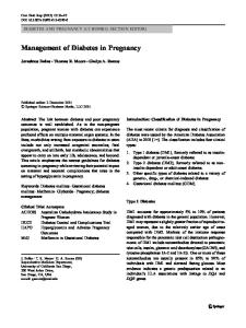
1530 FIG. 1 Melanoma-in-situ (MIS) of the nose excised with contoured excisions. The extent of the tumor is demarcated with a Wood’s lamp (a). The 5-mm margins are drawn beyond the visible extent of the tumor and 2- to 3-mm strips of tissue are excised following the contour of the tip and alae of the nose (b). The defect is sutured closed, leaving the central portion intact (c). Permanent histologic analysis of the excised tissue revealed no evidence of MIS in any of the margins, so 1 week later, the central portion is excised (d) and immediate reconstruction is performed (e). Final cosmetic outcome at 12 months (f)
next stage, which consists of excising the central lesion and performing a final reconstruction with a graft or flap under general anesthesia (Figs. 1d, e and 2f). If any of the margins are positive, the first stage is repeated until negative peripheral margins are confirmed. Histological Assessment Evaluation of Margins Sections are taken parallel to the true surgical margin and embedded en face in permanent paraffin blocks with the surgical margin (i.e., the side of the strip of tissue furthest from the MIS) face up. Specimens are processed overnight, embedded in paraffin, and made available for interpretation the day after surgery. The actual margin is considered the first section taken off the block. The sections are stained with standard hematoxylin and eosin (H&E). If these are equivocal for MIS, then immunohistochemical stains for melanoma antigens are used. These typically include S-100, and may also include MART-1/Melan-A and/or panmelanoma cocktail (MART1, HMB 45, tyrosinase). These stains are known to aid in visualization of the features necessary for diagnosis of MIS, such as broad size, confluent growth of melanocytes, upward epidermal migration of cells, and growth of melanocytes within follicular epithelium.
Evaluation of Central Specimen Once removed, the central specimen also undergoes thorough pathologic analysis. Margins are evaluated by examining parallel sections cut from the entire peripheral border of the specimen and embedded en face, measuring approximately 1–2 mm thick. The remainder of the specimen is sectioned with transverse cuts. If the central specimen is \2 cm in size, the entire specimen is submitted for pathologic analysis, allowing evaluation of the entire deep margin. If the specimen is larger than this, multiple representative sections, including any dark or nodular portions, are sampled to include the deep margin. The tissue is processed in the same fashion, stained with H&E, and when necessary examined with the aid of immunohistochemical stains. Collection and Analysis of Data The data collected included patient demographics and tumor characteristics such as lesion size and location. Lesion size was defined as the product of the largest diameter of the lesion multiplied by the largest perpendicular width of the lesion in square centimeters. Surgical data included the number of marginal excisions and the final margin width required for histological clearance, the size of the final surgical defect, and the
Surgical Management of Melanoma-In-Situ
1531
FIG. 2 Melanoma-in-situ (MIS) at the nasolabial fold. The extent of the tumor’s perimeter is demarcated with a Wood’s lamp (a). The 5mm margins are drawn beyond the visible extent of the tumor following the contour of the nose and parallel to the nasolabial fold and philtrum (b). Two- to 3-mm strips of tissue are excised (c) and oriented
for permanent histologic analysis (d). The defect is sutured closed, leaving the central portion intact (e). After permanent pathologic analysis confirming no evidence of MIS in any of the margins, central excision and reconstruction with an advancement rotational flap are performed (f). Final cosmetic outcome at 5 months (g)
type of reconstruction used. Pathology data gathered included number of patients in whom immunohistochemical stains were used, and final pathologic diagnosis with Breslow thickness if unsuspected invasive melanoma was found. Clinical data included time to recurrence (local vs. distant) or death from disease versus other causes and follow-up date. Follow-up duration was calculated from the time of central tumor excision, once all contoured margins were confirmed to be negative, to the last documented clinical visit or last patient contact obtained from the tumor
registry database. We also calculated the time (in months) elapsing from initial diagnosis to first marginal excision and to final excision and definitive reconstruction. Statistical analysis was performed by mostly descriptive summary statistics. Continuous variables are presented with median and range as well as mean and standard deviation (SD) where appropriate, while discrete variables are reported by using frequencies and percentages. For exploratory comparisons of a continuous variable (e.g., lesion size or age) between two subgroups of patients, such as the margin-positive versus margin-negative patients or
M. G. Mo¨ller et al.
1532
number of reexcisions and age or lesion size were studied by Spearman correlation coefficients. RESULTS Sixty-one patients with a diagnosis of MIS or LMM who underwent the staged contoured excision technique at our institution were identified. Twelve patients were excluded from this analysis; 11 patients had invasive melanoma (LMM) at initial diagnosis and 1 patient had MIS located on the hand. Forty-nine patients met the inclusion criteria for this study, 28 (57%) male and 21 (43%) female. The median age was 72 (range 42–88) years. All patients were white. All tumors were located on the head or neck, with 20 (41%) on the cheek, 8 (16%) on the nose, 6 (12%) on the scalp, and 5 (10%) on the temple or forehead. The median clinical lesion size was 5 cm2 (range 0.3–45 cm2; mean [SD] 7.2 [8.5] cm2) (Table 2). Marginal Excision Thirty-seven (76%) of 49 patients obtained histologic clearance of all peripheral margins at first excision performed with 5-mm margins from the visible and Wood’s lamp–defined edge of the lesion. Twelve patients (24%) required reexcision of at least one margin. Of these, six patients required one margin reexcised at the second procedure, and three and two patients each required two and three margins reexcised, respectively, and one patient required four margins reexcised at the second procedure. In addition, three patients (6% of the total) needed a third staged excision procedure to fully clear the margins. TABLE 2 Patient demographics and lesion characteristicsa
FIG. 3 Periorbital melanoma-in-situ. The lesion and surrounding 5-mm margins are demarcated and identified (left, inferior, medial, superior), following the contour of the canthus and lower eyelid to facilitate reconstruction and prevent eyelid contracture (a). Two- to 3-mm strips of tissue are excised (b). The defect is sutured closed, leaving the central portion intact pending permanent pathologic analysis (c)
the association between the margin status or final diagnosis and some variable of interest, simple descriptive statistics and the Wilcoxon two-sample rank test were used. To explore an association between a categorical variable and binary outcome (e.g., between margin status or final diagnosis and tumor subgroups defined by the greatest dimension), the Fisher exact or v2 test was performed. The correlations between total number of margins excised or
Variable
Value (n = 49)
Age (y)
72 (42–88); 70 (11)
Lesion size (cm2) Largest diameter (cm)
5 (.3–45); 7.2 (8.5) 2.5 (.5–9); 2.8 (1.6)
Sex Male
28 (57%)
Female
21 (43%)
Location of lesion
a
Cheek
20 (40.8%)
Ear
4 (8.2%)
Neck
3 (6.1%)
Nose
8 (16.3%)
Periorbital
3 (6.1%)
Scalp
6 (12.2%)
Temple
5 (10.2%)
Values are expressed as median (range); mean (SD) for continuous variables; and n (%) for discrete variables
Surgical Management of Melanoma-In-Situ
1533
TABLE 3 Characteristics of patients with negative and positive excision marginsa Variable
Negative (n = 37)
Positive (n = 12)
Age, years
71 (42–87); 70 (11)
73 (44–88); 71 (12)
Lesion size (cm2)
4 (.3–30); 5.9 (6.2)
6.9 (2.6–45); 11.5 (12.8)
Largest diameter (cm)
2.5 (.5–6); 2.6 (1.3)
3.0 (2–9); 3.6 (2.0)
2
Defect size (cm )
15.8 (1.5–42); 16.3 (10.8)
27.5 (6.3–130); 34.7 (32.9)
Time from first marginal to central excision (d)
7 (7–23); 8 (3)
14 (7–63); 17 (16)
Unsuspected invasive melanoma found in central specimen
6/37 (16%)
0/12 (0%)
a
Values are expressed as median (range); mean (SD) for continuous variables; and n (%) for discrete variables
Immunohistochemical stains were used in assessing the margins in nine patients (18%), and positive margins were identified in three patients that otherwise might have been missed with the standard H&E technique. When comparing patients with negative margins (n = 37) after the initial 5-mm marginal excision with patients having at least one margin positive (n = 12), larger lesion size did seem to be positively associated with margin status: median (mean) lesion size for negative- and positivemargin patients was 4.0 (5.9) cm2 and 6.9 (11.5) cm2, respectively (Wilcoxon p value = .03; Table 3). There seemed to be a positive association between lesion size and total number of positive margins as well as number of excisions needed to clear the margin (Spearman correlation coefficient .34 and .32, p = .02 and .03, respectively). When divided into groups on the basis of the largest single diameter of the lesion (B2 cm vs. [2 cm), positive margins were seen in 2 (11%) of 19 smaller lesions and 10 (33%) of 30 larger lesions, respectively (p = .09, Fisher’s exact test).
melanoma, respectively. Interestingly, none of these six patients with invasive melanoma required extra excisions to fully clear their margins. When divided into groups on the basis of the largest single diameter of the lesion (B2 cm vs. [2 cm), invasive cancer was seen in 4 (21%) of 19 smaller lesions and 2 (7%) of 30 larger lesions, respectively (p = .19, Fisher’s exact test).
Central Excision
The median time elapsed from the initial biopsy to first marginal excision was 53 days (range 6–172 days; mean [SD] 53 [29] days) and from initial biopsy to the time of final central excision was 60 days (range 13–179 days; mean [SD] 63 [32] days). The median time for completion of the procedure (first marginal excision to final central tumor excision and reconstruction) was 7 days (range 7– 63 days; mean [SD] 10 [9] days).
The final pathology diagnosis obtained from the resected central specimen was residual MIS in 32 patients (65%), no residual MIS in 8 patients (16%), residual atypical melanocytic proliferation in 3 patients (6%), and unsuspected invasive melanoma in 6 patients (12%), with a Breslow thickness ranging from .18 to 1.5 mm (median .33 mm; mean .52 mm). There was no statistically significant difference in lesion size between those with or without invasive melanoma in the final specimen, possibly as a result of the small number of patients with unsuspected invasive melanoma (n = 6). In fact, patients harboring unsuspected invasive melanoma in the central excision specimens had a slightly smaller lesion size compared with patients without invasive melanoma in their specimens, with a mean (median) of 5.4 (3.0) cm2 vs. 7.5 (5.0) cm2, respectively. A similar relationship existed between the largest preoperatively measured diameter of the lesion: mean (median) 2.5 (1.8) cm vs. 2.9 (2.5) cm for those with and without unsuspected invasive
Reconstruction of the Defect The median surgical defect size was 16 cm2 (range 1.5– 130 cm2, mean [SD] 21 [20] cm2). Surgical defects were reconstructed with primary closure in 1 patient (2%), rotational advancement flaps in 18 patients (37%), fullthickness grafts in 20 patients (41%), and split-thickness grafts in 10 patients (20%). Time from Biopsy to Definitive Reconstruction
Outcomes No local recurrences or disease-related deaths have been reported to date, after a median follow-up of 14 months (range 1–36 months). One patient with MIS developed brain metastases; however, this particular patient also had a synchronous invasive melanoma in a different location. DISCUSSION Complete tumor clearance is the fundamental oncologic goal for MIS and is associated with the highest local control
1534
rates, with cure rates of [91%.25 Over the past 20 years, there have been reports of several surgical methods of removal for MIS by evaluation and clearance of the perimeter margins. This was first described by Dhawan et al. in 1990, who used Mohs micrographic surgery followed by ‘‘rush’’ permanent sections.50 This was subsequently modified and simplified by Johnson and colleagues in 1997 and termed the ‘‘square’’ technique.35 With the use of staged permanent en face histologic sectioning, clearance of the entire peripheral margin can be obtained before the excision of the lesion itself. The main advantages of this multidisciplinary approach to tumor control are avoidance of open wounds, pathologic review of highquality permanent sections that are easier to process and interpret than frozen sections, maximal tissue conservation in cosmetically sensitive areas, and technical and surgical simplicity that does not require extensive training. Review of the literature shows that Agarwal-Antal et al., Mahoney et al., and Johnson et al. prefer a geometric design with angled corners and flat edges to facilitate histological processing and orientation.23,27,35 But because the clinically observed lesions of MIS on the face, scalp, and neck often have an irregular pattern and may be adjacent to functionally and aesthetically important areas, we have modified this approach by initially excising a contoured rim of tissue configured around the central lesion or anatomical structures, rather than a square or sharply angled geometric shape. This contoured excision maintains the ability to conduct careful margin analysis, but subsequently ensures optimal cosmetic reconstruction of the final defect. We have found that adequate and complete analysis of the specimen can be performed on the contoured rim of tissue. This method allows the dermatopathologist to assess the margins in the best way with permanent sections, with immunostains when needed and in an unhurried setting. Our study demonstrates that a 5-mm margin was adequate in only 76% of cases with MIS on the head and neck, and that 24% required wider excisions. In 18% of the cases, immunohistochemical stains were used by the pathologist to assess the margin status. Our findings are consistent with reported data in other studies.5,21,23,26,27,29,51,53 These findings underscore the histological challenge of subclinical extension presented by MIS in sun-damaged skin. En face histologic analysis is considered one of the pathologic standards of care in the evaluation of margins from cutaneous melanoma excision specimens. It has been in routine use at our center for all melanoma cases, even for large (2 cm), wide excisions, for over 10 years. It is the most efficient method of enabling the pathologist to view the maximum amount possible of the peripheral skin and soft tissue margin of these excisions. When conducted under a well-defined protocol for gross dissection, with marginal sections of uniform thickness and size,
M. G. Mo¨ller et al.
embedding and sectioning are relatively simple. In most excision specimens for MIS, an average of four but no more than eight en face sections are required to analyze the entire peripheral margin. An argument could be made about the increased length of time before complete tumor removal and reconstruction imposed by this staged procedure. Most of our patients were referred by community dermatologists from central Florida, and sometimes it took up to 4 to 6 weeks from the time of initial biopsy to the time of the first marginal excision (median and mean 53 days). However, the median (mean) time for completion of the entire staged procedure was 1 week (10 days). This is not clinically significantly different from patients undergoing the conventional surgical technique, in which the frequent finding of a positive margin also requires a return to surgery and delay or revision of the final reconstruction. We believe that most patients prefer a sutured, closed tumor perimeter to an open wound. Although many authors have reported on recurrence rates, only a few have obtained long-term follow-up.29,51,53 In the series by Johnson et al., no recurrences were observed in 35 patients on short-term follow-up.35 We have had approximately 2 years’ experience with this methodology at our institution, and thus the length of follow-up in our study is short (median 14 months and maximum 36 months), but no local or regional recurrences have been reported. Moreover, there is no reason to believe that once negative margins are achieved, the precise surgical technique used to gain those clear margins should influence outcome. Still, long-term follow-up data are required to substantiate the effectiveness of this treatment approach. In addition, our data emphasize that invasive melanoma frequently coexists with MIS on the head and neck: it was found in 12% of our patients, all of whom undergone a prior biopsy that indicated only MIS. If the initial evaluation reveals any suspicious areas within the residual lesion present after biopsy, full-thickness biopsies (e.g., punch biopsies) can be performed at the time of or before the marginal excision. The staged approach can be used for invasive melanomas of the head and neck as well, although we excluded these cases from our review because margins of [5 mm around the edge of the lesion were routinely taken. Lymphatic mapping and sentinel lymph node biopsy, if clinically indicated, can be carried out at the same time as marginal excision with at least a 1-cm margin for cases of invasive melanoma. The main advantages of staged, marginal excision approaches are: (1) complete excision of the tumor in aesthetically important areas while following natural anatomic lines; (2) avoidance of open wounds during the time required for detailed histopathologic examination of margins; and (3) use of high-quality permanent sections with or without immunohistochemical stains. This methodology
Surgical Management of Melanoma-In-Situ
provides margin control with equivalent results to Mohs micrographic surgery, avoiding some of the pitfalls associated with this technique, and without the requirement for specialized personnel and a laboratory dedicated to frozen section examination. Our contoured modification of the ‘‘square’’ procedure for excising MIS of aesthetically important areas of the face and neck is technically reproducible and provides a complete histological evaluation of the specimen with optimal peripheral and deep margin control, and maximal tissue preservation. It involves a multidisciplinary approach with close communication between the dermatopathologists, surgical oncologists, and reconstructive surgeons. Definitive reconstruction is facilitated with confirmation of tumor-free margins, allowing preservation of function and aesthetics. With short follow-up, local control has been achieved with this technique in 100% of cases. REFERENCES 1. Davis T, Zembowicz A. Histological evolution of lentiginous melanoma: a report of five new cases. J Cutan Pathol. 2007; 34:296–300. 2. Tannous ZS, Lerner LH, Duncan LM, et al. Progression to invasive melanoma from malignant melanoma in situ, lentigo maligna type. Hum Pathol. 2000;31:705–8. 3. Cohen LM. Lentigo maligna and lentigo maligna melanoma. J Am Acad Dermatol. 1995;33:923–36. 4. Wayte DM, Helwig EB. Melanotic freckle of Hutchinson. Cancer. 1968;21:893–911. 5. Bub JL, Berg D, Slee A, et al. Management of lentigo maligna and lentigo maligna melanoma with staged excision: a 5-year follow-up. Arch Dermatol. 2004;140:552–8. 6. Kelly JW. Following lentigo maligna may not prevent the development of life-threatening melanoma. Arch Dermatol. 1992; 128:657–60. 7. Breuninger H, Schlagenhauff B, Stroebel W, et al. Patterns of local horizontal spread of melanomas: consequences for surgery and histopathologic investigation. Am J Surg Pathol. 1999;23: 1493–8. 8. Cohen LM. Lentigo maligna and lentigo maligna melanoma. J Am Acad Dermatol. 1997;36(6 Pt 1):913. 9. Weinstock MA, Sober AJ. The risk of progression of lentigo maligna to lentigo maligna melanoma. Br J Dermatol. 1987;116: 303–10. 10. Jones WO, Harman CR, Ng AK, et al. Incidence of malignant melanoma in Auckland, New Zealand: highest rates in the world. World J Surg. 1999;23:732–5. 11. Newell GR, Sider JG, Bergfelt L, et al. Incidence of cutaneous melanoma in the United States by histology with special reference to the face. Cancer Res. 1988;48:5036–41. 12. Swetter SM, Boldrick JC, Jung SY, et al. Increasing incidence of lentigo maligna melanoma subtypes: northern California and national trends 1990–2000. J Invest Dermatol. 2005;125:685–91. 13. NIH Consensus conference. Diagnosis and treatment of early melanoma. JAMA. 1992 ;268:1314–9. 14. National Comprehensive Cancer Network clinical practice guidelines in oncology: melanoma. v.2.2007. Available at: http:// www.nccn.org/professionals/physician_gls/PDF/melanoma.pdf. Accessed September 1, 2007.
1535 15. Balch CM, Urist MM, Karakousis CP, et al. Efficacy of 2-cm surgical margins for intermediate-thickness melanomas (1 to 4 mm). Results of a multi-institutional randomized surgical trial. Ann Surg. 1993;218:262–7. 16. Veronesi U, Cascinelli N. Narrow excision (1-cm margin). A safe procedure for thin cutaneous melanoma. Arch Surg. 1991;126: 438–41. 17. Grande DJ, Koranda FC, Whitaker DC. Surgery of extensive, subclinical lentigo maligna. J Dermatol Surg Oncol. 1982;8: 493–6. 18. Albertini JG, Elston DM, Libow LF, et al. Mohs micrographic surgery for melanoma: a case series, a comparative study of immunostains, an informative case report, and a unique mapping technique. Dermatol Surg. 2002;28:656–65. 19. Osborne JE, Hutchinson PE. A follow-up study to investigate the efficacy of initial treatment of lentigo maligna with surgical excision. Br J Plast Surg. 2002;55:611–5. 20. Bricca GM, Brodland DG, Zitelli JA. Immunostaining melanoma frozen sections: the 1-hour protocol. Dermatol Surg. 2004;30: 403–8. 21. Huilgol SC, Selva D, Chen C, et al. Surgical margins for lentigo maligna and lentigo maligna melanoma: the technique of mapped serial excision. Arch Dermatol. 2004;140:1087–92. 22. Zalla MJ, Lim KK, Dicaudo DJ, et al. Mohs micrographic excision of melanoma using immunostains. Dermatol Surg. 2000;2 6:771–84. 23. Agarwal-Antal N, Bowen GM, Gerwels JW. Histologic evaluation of lentigo maligna with permanent sections: implications regarding current guidelines. J Am Acad Dermatol. 2002;47: 743–8. 24. Pitman GH, Kopf AW, Bart RS, et al. Treatment of lentigo maligna and lentigo maligna melanoma. J Dermatol Surg Oncol. 1979;5:727–37. 25. Coleman WP 3rd, Davis RS, Reed RJ, et al. Treatment of lentigo maligna and lentigo maligna melanoma. J Dermatol Surg Oncol. 1980;6:476–9. 26. Anderson KW, Baker SR, Lowe L, et al. Treatment of head and neck melanoma, lentigo maligna subtype: a practical surgical technique. Arch Facial Plast Surg. 2001;3:202–6. 27. Mahoney MH, Joseph M, Temple CL. The perimeter technique for lentigo maligna: an alternative to Mohs micrographic surgery. J Surg Oncol. 2005;91:120–5. 28. Somach SC, Taira JW, Pitha JV, et al. Pigmented lesions in actinically damaged skin. Histopathologic comparison of biopsy and excisional specimens. Arch Dermatol. 1996;132:1297–302. 29. Cohen LM, McCall MW, Zax RH. Mohs micrographic surgery for lentigo maligna and lentigo maligna melanoma. A follow-up study. Dermatol Surg. 1998;24:673–7. 30. Dawn ME, Dawn AG, Miller SJ. Mohs surgery for the treatment of melanoma in situ: a review. Dermatol Surg. 2007;33:395–402. 31. Kelley LC, Starkus L. Immunohistochemical staining of lentigo maligna during Mohs micrographic surgery using MART-1. J Am Acad Dermatol. 2002;46:78–84. 32. Davis DA, Kurtz KA, Robinson RA. Ultrarapid staining for cutaneous melanoma: study and protocol. Dermatol Surg 2005;31(7 Pt 1):753–6. 33. Hendi A, Brodland DG, Zitelli JA. Melanocytes in long-standing sun-exposed skin: quantitative analysis using the MART-1 immunostain. Arch Dermatol. 2006;142:871–6. 34. Megahed M, Schon M, Selimovic D, et al. Reliability of diagnosis of melanoma in situ. Lancet. 2002;359(9321):1921–2. 35. Johnson TM, Headington JT, Baker SR, et al. Usefulness of the staged excision for lentigo maligna and lentigo maligna melanoma: the ‘‘square’’ procedure. J Am Acad Dermatol. 1997; 37(5 Pt 1):758–64.
1536 36. McKenna JK, Florell SR, Goldman GD, et al. Lentigo maligna/ lentigo maligna melanoma: current state of diagnosis and treatment. Dermatol Surg. 2006;32:493–504. 37. Hill DC, Gramp AA. Surgical treatment of lentigo maligna and lentigo maligna melanoma. Australas J Dermatol. 1999;40: 25–30. 38. Jejurikar SS, Borschel GH, Johnson TM, et al. Immediate, optimal reconstruction of facial lentigo maligna and melanoma following total peripheral margin control. Plast Reconstr Surg. 2007;120:1249–55. 39. Mahoney EJ, Dolan RW, Choi EE, et al. Surgical reconstruction of lentigo maligna defects. Arch Facial Plast Surg. 2005;7: 342–6. 40. Temple CL, Arlette JP. Mohs micrographic surgery in the treatment of lentigo maligna and melanoma. J Surg Oncol. 2006;94:287–92. 41. Zitelli JA, Brown C, Hanusa BH. Mohs micrographic surgery for the treatment of primary cutaneous melanoma. J Am Acad Dermatol 1997;37(2 Pt 1):236–45. 42. Zalaudek I, Horn M, Richtig E, et al. Local recurrence in melanoma in situ: influence of sex, age, site of involvement and therapeutic modalities. Br J Dermatol. 2003;148:703–8. 43. Stevenson O, Ahmed I. Lentigo maligna: prognosis and treatment options. Am J Clin Dermatol. 2005;6:151–64. 44. Malhotra R, Chen C, Huilgol SC, et al. Mapped serial excision for periocular lentigo maligna and lentigo maligna melanoma. Ophthalmology. 2003;110:2011–8.
M. G. Mo¨ller et al. 45. Walling HW, Scupham RK, Bean AK, et al. Staged excision versus Mohs micrographic surgery for lentigo maligna and lentigo maligna melanoma. J Am Acad Dermatol. 2007;57:659–64. 46. Bhardwaj SS, Tope WD, Lee PK. Mohs micrographic surgery for lentigo maligna and lentigo maligna melanoma using Mel-5 immunostaining: University of Minnesota experience. Dermatol Surg. 2006;32:690–6. 47. Bricca GM, Brodland DG, Ren D, et al. Cutaneous head and neck melanoma treated with Mohs micrographic surgery. J Am Acad Dermatol. 2005;52:92–100. 48. Bienert TN, Trotter MJ, Arlette JP. Treatment of cutaneous melanoma of the face by Mohs micrographic surgery. J Cutan Med Surg. 2003;7:25–30. 49. Zitelli JA, Brown CD, Hanusa BH. Surgical margins for excision of primary cutaneous melanoma. J Am Acad Dermatol. 1997;37(3 Pt 1):422–9. 50. Dhawan SS, Wolf DJ, Rabinovitz HS, et al. Lentigo maligna. The use of rush permanent sections in therapy. Arch Dermatol. 1990;126:928–30. 51. Robinson JK. Margin control for lentigo maligna. J Am Acad Dermatol. 1994;31:79–85. 52. Cohen LM, McCall MW, Hodge SJ, et al. Successful treatment of lentigo maligna and lentigo maligna melanoma with Mohs’ micrographic surgery aided by rush permanent sections. Cancer. 1994;73:2964–70. 53. Zitelli JA. Surgical margins for lentigo maligna, 2004. Arch Dermatol. 2004;140:607–8.