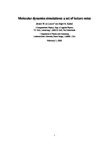
Microscopy: Science, Technology, Applications and Education A. Méndez-Vilas and J. Díaz (Eds.) ______________________________________________
Study of molecular dynamics using fluorescently tagged molecules in live cells C. N. Goulbourne 1,*, A. N. Malhas 1,*, H. Doolittle 2, A. T. Ismail 3 and D. J. Vaux 1 1 Sir William Dunn School of Pathology, University of Oxford, South Parks Road, Oxford OX1 3RE, UK. 2 Hammersmith Hospital, Du Cane Road, London W12 0HS, UK. 3 Trinity College, University of Oxford, Oxford OX1 3BH, UK. * Equal contribution. Microscopic studies of fluorescently-tagged molecules or those detected using fluorescently-tagged antibodies in fixed cells have provided insight into the localization and function of various proteins for a very long time. Such studies however do not provide information about the dynamics of labeled proteins over time. In order to assess the dynamic characteristics of fluorescently-tagged proteins within cells at the light microscope level several techniques have been developed. These include fluorescence loss in photobleaching (FLIP), fluorescence recovery after photobleaching (FRAP), inverse FRAP (iFRAP) and the use of photoactivatable fluorescent proteins (PAFPs) and caged molecules. FLIP monitors loss of fluorescence in an area distant to the bleached region while FRAP follows the recovery of fluorescently tagged proteins into the photobleached region of interest. iFRAP involves the entire fluorescently labelled area being bleached apart from a small region where the loss of fluorescence from this point is monitored over time. PAFPs are proteins that change spectral properties upon exposure to specific wavelengths of light. They can be classified into photoactivatable and photoswitchable or photoconvertible. Upon activation photactivatable fluorescent proteins change from non-fluorescent into fluorescent, while photoswitchable fluorescent proteins change their spectral properties. Application of these techniques has allowed biological questions to be answered surrounding the stability and motility of tagged proteins. However, these methods have limitations and the use of more than one of these techniques is often required to fully elucidate protein behaviour. This review will look at several recent and diverse examples of the use of these techniques to understand protein, organelle and cell dynamics. Keywords FLIP; FRAP, iFRAP, photoactivation; GFP; PAGFP.
Introduction The isolation of Green Fluorescent Protein (GFP) from the jellyfish Aequorea victoria [1, 2] and its application to molecular cell biology has revolutionized the understanding of protein dynamics inside cells. Contained within the barrel shape of GFP is a spontaneously modified tripeptide, which fluoresces when exposed to blue light causing the emission of green light [3, 4]. The shell surrounding the chromophore prevents the generation of phototoxic species, which would otherwise harm the cell. The sequence of the initial isolate of GFP has been widely mutated to produce different versions of the fluorescent protein with various applications [5-8]. Enhanced GFP (EGFP) was generated by a single point mutation that increased the fluorescence intensity and photo stability of the protein. Cyan and yellow fluorescent proteins were again produced by mutations and can be used in Fluorescence Resonance Energy Transfer (FRET) [9]. Fusion of the GFP tag is often at the C or N terminus of the protein of interest and yields a GFP chimera which can be followed in live or fixed cells.
1.Use of GFP photobleaching to study protein dynamics As the fluorescence of GFP can be irreversibly photobleached by a laser of sufficient intensity, GFP chimeras enable the study of molecular dynamics as the movement into bleached regions or behavior adjacent to it can be monitored over time to reveal the motility of the GFP tagged molecule. This section will describe how Fluorescence Loss in Photobleaching (FLIP), Fluorescence Recovery after Photobleaching (FRAP) and inverse FRAP (iFRAP) have been used on commercially available confocal laser scanning microscopes to address biological questions surrounding molecular dynamics. Suitability of each of the techniques is summarized in Table 1 while Figure 1 schematically shows how each one of these techniques works.
670
©FORMATEX 2010
Microscopy: Science, Technology, Applications and Education A. Méndez-Vilas and J. Díaz (Eds.) ______________________________________________
Figure 1. FRAP, FLIP and iFRAP are various photobleaching techniques used to assess the dynamics of fluorescently tagged
molecules FRAP bleaches a region of interest (ROI represented by the small white circle) and measures the fluorescent recovery. A very mobile molecule will rapidly enter the photobleached region as indicated by the highly mobile chart. Molecules tethered to their position will show no recovery as illustrated by the immobile chart. However, the molecular dynamics often contain a pool of both immobile and mobile fractions producing a profile shown in the intermediate chart. FLIP involves continuously photobleaching the ROI and assessing changes of fluorescent intensity within adjacent areas (black rectangle). iFRAP involves all of the fluorescently labeled molecule outside of the ROI being photobleached and assessing fluorescence changes over time. Neighboring cells are used to measure the average total nuclear fluorescence signal in both FLIP and iFRAP as a means of normalizing the intensity data. Additionally a random area outside of the cell (B) should be measured for background correction for all three techniques.
Table 1. Summary table of FLIP, FRAP and iFRAP suitability for determining various parameters of fluorescently tagged proteins. Adapted from [10]
Molecular Mobility
Diffusion Constant
Immobile Fraction
Distinct pools
Continuity of Compartments
FRAP
Optimal
Optimal
Optimal
Suitable
Suitable
FLIP
Optimal
Suitable
Suitable
Optimal
Optimal
iFRAP
Unsuitable
Unsuitable
Suitable
Optimal
Unsuitable
1.1 FLIP FLIP works on the principle of repetitive partial photobleaching of an area within a cell expressing a fluorescently tagged protein, and continuous monitoring of one or more adjacent region of interest for changes in fluorescent intensity. The greater the mobility of the tagged protein, the faster it will exchange between the bleach area and the analysis area, resulting in a faster loss in fluorescent signal in the monitored ROI. This technique has been used to monitor transcription factor behavior [11], nuclear lamina stability [12] and to elucidate the continuity of cellular structures such as the Golgi apparatus, nucleolus, endoplasmic reticulum and compartments for splicing factors [13-16]. FLIP can be used to confirm FRAP data, as the photobleached area in FRAP may have sustained photo damage to the protein of interest and altered its dynamic characteristics, whereas FLIP just assesses mobility of fluorescent proteins adjacent to the bleached region. If all the proteins have similar kinetic characteristics then the fluorescence signal from the region of interest will be lost in a simple monotonic decay. However, if there are multiple pools of protein with different dynamic properties, such as some of the proteins binding to chromatin, then the loss of fluorescence will be multiphasic. Dynamics of proteins involved in the various steps of ribosome biogenesis during interphase were examined using FLIP as a means to determine if these components, within the nucleolus, rapidly exchange with the nucleoplasm or remain tethered within the nucleolus [17]. This was achieved by photobleaching one part of the nucleus and monitoring the change in fluorescent intensity of the various GFP tagged proteins associated with ribosome assembly. The study
©FORMATEX 2010
671
Microscopy: Science, Technology, Applications and Education A. Méndez-Vilas and J. Díaz (Eds.) ______________________________________________
revealed that the proteins involved in rDNA transcription, pre-rRNA processing and ribosome assembly exchange rapidly between the nucleolus and nucleoplasm whereas ribosomal proteins move relatively slowly. The authors were concerned that prolonged photobleaching may damage the cells due to the production of phototoxic species. However, they used a low laser intensity, which has been cited as generating minimal phototoxic effects [15, 18, 19]. Additionally they monitored cells for 24 hours after they received similar and higher doses of laser irradiation and showed cells were healthy and even underwent mitosis. The understanding of the functional importance of the nuclear lamina is rapidly expanding with roles being assigned in DNA replication, chromatin organization, control of gene expression and maintenance of the structural integrity of the nucleus [20-23]. Much attention has been focused on the lamin A protein, encoded by the LMNA gene [24]. Mutations within this gene produce a diverse range of symptoms depending on where the mutation resides and the diseases are collectively called nuclear laminopathies [25]. The protein responsible for causing the segmental accelerated aging laminopathy, Hutchinson-Gilford Progeria Syndrome, is called progerin [26]. It is an internally truncated form of lamin A, lacking fifty internal amino acids but has retained fifteen C-terminal amino acids that are usually cleaved off by the protease enzyme FACE1/Zmpste24 [27]. These terminal amino acids include a farnesylated cysteine and this farnesyl tail is believed to anchor the protein into the nuclear membrane and reduce its mobility [28]. FLIP experiments on GFP tagged progerin [12] revealed that the GFP signal dropped to 80% of the starting intensity over 80 seconds compared to the wild type GFP lamin A, which dropped to 20%. This means that mature wild type lamin A is quite a dynamic protein and will move into the photobleached area, while the retention of the farnesylated tail in progerin seems to severely reduce the dynamic characteristic of this protein. Lamin A rod and tail domain missense mutants, which have been associated with a wide range of laminopathies, have been examined for differences in their dynamic characteristics [29]. H222P, G232E, Q249P and R386K mutants are linked to autosomal dominant Emery-Dreifuss muscular dystrophy. The mutation E203G has been identified in cardiomyopathy with conduction system disease and the R482L mutation is the only one located in the globular tail domain and is found in patients with familial partial lipodystrophy. The study revealed that mutants H222P and R48L were significantly less dynamic than wild type GFP lamin A while the remaining mutants were a lot more mobile. Increased mobility is thought to be due to failure of higher-order intermediate filament assembly by mutated lamin A, resulting in failure of filament assembly or lateral association and instead the production of aggregates in the nucleoplasm. Lamin B1 is another component of the nuclear lamina and has been shown to control oxidative stress responses via sequestration of the transcription factor Oct-1 [11]. Malhas et al revealed by FLIP that one of the consequences of expression of severely truncated lamin B1 in mouse embryonic fibroblasts (MEFs) was a dramatic increase in the exchange of peripheral EGFP tagged Oct-1 (43% in the lamin B1 hypomorphs compared to only 8% in the wild types). The reason for this is because full-length lamin B1 is not present and cannot hold the EGFP-Oct-1 at the periphery, which allows the transcription factor to be released into the nucleoplasm and into the photobleached region resulting in a greater decrease in peripheral fluorescent intensity.
672
©FORMATEX 2010
Microscopy: Science, Technology, Applications and Education A. Méndez-Vilas and J. Díaz (Eds.) ______________________________________________
Figure 2. (a) Lamin B1 wild type MEFs transfected with EGFP tagged Oct-1 are constantly photobleached (blue circle). Lamin B1 tethers Oct-1 at the nuclear periphery and as a result there is minimal movement of the tagged protein outside the ROI near the nuclear periphery. (b) In Lamin B1 knock out MEFs the Oct-1 EGFP is released from the nuclear periphery and can migrate into the nucleoplasm where photobleaching occurs, resulting in a decrease in relative fluorescence intensity at the ROI.
1.2 FRAP FRAP was first used in the 1970s to look at translational diffusion of fluorescein isothiocyanate labeled proteins in erythrocyte membranes [30]. It took until 1996 for the resurrection of this technique and its application in live cells transfected to express fluorescently tagged proteins [13]. Since then FRAP in conjunction with GFP tagged proteins has answered many biological questions concerning molecular dynamics within the cell. The technique involves rapid, complete photobleaching of a selected region of interest (ROI) using an intense laser beam, followed by measurement of the rate of fluorescence recovery into the photobleached region. The more dynamic the tagged molecule is the greater the rate of recovery. Movement of fluorescently tagged molecules can be considered in two dimensions as everything above and below the focal plane has been photobleached by a large cone of out of focus light when using objectives with high numerical apertures (N.A. 1.2-1.45). FRAP has been used to study the dynamics of various proteins within the nucleus and nuclear envelope. An example of this is GFP tagged components of the linker of nucleoskeleton and cytoskeleton (LINC) complex, such as Sun1, Sun2 and nesprin 2 [31]. In MEFs that have had lamin A knocked out GFP tagged LINC complex proteins showed increased mobility. The LINC protein dynamics were returned to wild type mobility when the knockouts were cotransfected to express RFP-lamin A. FRAP illustrated the importance of lamin A in forming a stable LINC complex between the nuclear and cytosolic environments and how failure of these proteins to associate with lamin A may have effects on migration, differentiation, polarization, meiosis and mitosis. It is clear that although FRAP has shed light on protein behavior it has also produced conflicting mobilities for some proteins. For example, polymerase II has been suggested to have up to three kinetic states, corresponding to promoter binding, initiation and elongation. However, different studies of the polymerase conclude variously that they detect one (elongation), two (initiation and elongation) or all three of these states [32-34]. Transcription factor residence times at chromatin have also been shown to vary by up to four orders of magnitude, which range from a few milliseconds up to several seconds [35-38]. These very different dynamics may be explained by technical variables between the different experiments or assumptions used to generate the kinetic models. However, it is clear that a degree of standardization is necessary when collecting and analyzing FRAP data so as to minimize conflicting reports of dynamic behavior. 1.3 iFRAP iFRAP involves bleaching the fluorescent signal in the entire cell apart from one ROI where the loss of signal is monitored over time. It is a useful technique for studying the rate of movement out of a region rather than the rate of recovery as in FRAP. However, a major limitation of this technique is the fact that it takes time to photobleach everywhere outside of the non-bleached region and consequently monitoring fast translocations is very difficult as the signal is lost during the bleaching phase.
©FORMATEX 2010
673
Microscopy: Science, Technology, Applications and Education A. Méndez-Vilas and J. Díaz (Eds.) ______________________________________________
Nonetheless, this method proved particularly useful for illustrating how transport carrier proteins on the Golgi membrane eventually move to the plasma membrane by vesicular transport [39]. It was achieved by bleaching everywhere outside of a section of the Golgi membrane labeled with GFP tagged vesicular stomatitis virus G protein (VSV-G–GFP). After the photobleaching had stopped fluorescent signal of the GFP tagged protein was visualised moving towards and eventually residing in the plasma membrane. Studying protein dynamics within the nucleus has also been aided by iFRAP. The mammalian nucleotide excision repair (NER) system is a multi-protein complex that binds to sites of chromatin that have been damaged by UV radiation. The complex forms at these sites with the XPC protein initiating binding to the DNA helix and recruiting other proteins to cause it to be unwound before the damaged region becomes exposed for repair. Cells expressing XPCmOrange (a mutant fluorescent protein derived from the Discosoma sp. Red fluorescent protein, DsRed) were irradiated with UV to induce DNA damage and cause recruitment of the XPC-mOrange [40]. 30 minutes after irradiation everywhere, outside a region where the XPC was locally concentrated, was photobleached. It revealed that the XPC unit rapidly dissociates from repair complexes, which contrasts previous reports that an NER factors binds irreversibly to sites of DNA damage. FLIP experiments were conducted to verify these findings and found that XPC rapidly dissociated with a half life of 25 seconds, as observed in iFRAP. This reversibility of protein binding is believed to be advantageous, as it increases the specificity of recognizing lesions without compromising efficiency. The transcription of genes and subsequent splicing out of introns in pre-mRNA was believed to occur before a completed transcript was released from the site of transcription. However, there is increasing evidence that intron containing transcripts, as a result of a slow splicing intron or mutation that affects splicing, move towards and are tethered at nuclear speckles [41]. These sites are located at the interchromatin region and contain processing factors with mature mRNA being observed to diffuse away from these sites, suggesting that they contain splicing machinery. A Drosophila Fushitarazu (ftz) pre mRNA, which contained one intron between two exons and had a forty nucleotide poly A tail, was fluorescently tagged with Alexa Fluor 546 [42]. This was micro-injected into the nuclei of COS7 cells, which showed foci formation at speckles after 1 minute; a control probe without the intron did not accumulate at the speckles. iFRAP was used to determine the stability at the foci of the fluorescently tagged pre-mRNA, which showed movement into the cytoplasm and total RNA analysis revealed that 50% of the micro injected pre-mRNA was being processed. It also revealed that movement from the foci was dependent on ATP as the dynamic nature of the tagged molecule was reduced in the absence of ATP, suggesting an energy dependent process.
2. Photoactivatable fluorescent proteins Photoactivatable fluorescent proteins (PAFPs) are proteins that change spectral properties upon exposure to specific wavelengths of light. They can be classified into photoactivatable and photoswitchable or photoconvertible. Upon activation photactivatable fluorescent proteins change from non-fluorescent into fluorescent, while photoswitcheable fluorescent proteins change their spectral properties. Table 2 summarises the spectral properties of some PAFPs. The reader is referred to original references for more detailed spectral properties as well as the exact derivitisation methods for each of these FPs. In this section we will focus more on some recent applications of PAFPs. Examples of potoactivatable proteins are PA-GFP and PA-mCherry which upon activation with UV light become green and red FPs respectively. Kaede and PS-CFP are examples of photoswitchable FPs which upon activation with UV change from green to red and blue to green FPs respectively. mOrange and mKate are also photoswitchable FPs which convert from orange to far red and red to green respectively [43]. Dronpa on the other hand is a green FP which after being bleached can be reversibly reactivated by UV. The newly reported photoactivatable green cherry (GPAC) probe [44] is unique in that it enables photoactivation of a specific pool while still being able to follow the entire protein population. In addition to PAFPs, caged molecules photoactivation or switching have also been utilized in spatial and temporal studies. Activation or conversion makes PAFPs an ideal tool to study protein dynamics in living cells. This part of the review will discuss recent advances in photoactivation and its applications in tracking labeled proteins to understand protein, organelle and cell dynamics. There are advantages of PAFPs over FPs in that the activation of a pool of PAFP tagged protein follows that specific pool. A technique like FLIP involves continuous bleaching of a specific cellular location with usually high power which might be toxic to cells whereas the PAFP activation is usually shorter and hence less toxic. 2.1 Use of PA-GFP to study chromosome positioning following mitosis Although we and others had previously shown that positioning of chromosomes in the interphase nucleus is nonrandom [21] the fate of these positions during mitosis is unclear. Fluorescent in situ hybridization (FISH) experiments suggests that the arrangements of centromeres and chromosome territories show significant symmetry in daughter cells [54]. In 2003, Walter et al and Gerlich et al performed live cell-imaging experiments to follow chromatin organization through mitosis [55, 56]. Both groups used photobleaching of fluorescently labeled histone H2B and used it as a chromatin marker. Gerlich et al reported that chromosome positions were maintained after mitosis and the distribution in the daughter cells was very similar to that of the mother cell [56]. Walter et al, on the other hand, reported that,
674
©FORMATEX 2010
Microscopy: Science, Technology, Applications and Education A. Méndez-Vilas and J. Díaz (Eds.) ______________________________________________
although chromosome position was maintained during interphase, major changes in chromatin organization are seen from one cell cycle to the next [55]. We addressed this apparent discrepancy by investigating the inheritance of nuclear domains using each of the histones H2B, H3 and H4 labeled with PA-GFP. Photoactivation was achieved by two-photon excitation using a Mira titanium-sapphire pulsed laser tuned to 830nm. Multiphoton excitation avoided the problems of photo damage, which have previously prevented this very useful label being used to investigate chromatin. The tagged histones acted as chromatin markers. A region of the nucleus was photoactivated and the nuclear domains were followed through the cell cycle. Our results showed that chromosome positions are stable during interphase and that they are heritable after mitosis since the photoactivated region remained definable all through interphase and was also visible in a similar pattern in daughter cells (Figure 3).
Figure 3: Inheritance of nuclear domains using H4-PAGFP as a chromatin marker NRK cells were transfected to express H4-PAGFP and DsRed ER. At each time point, a Z-stack has been merged into a single plane. The time indicated on each image represents the number of hours since photoactivation.
2.2 Use of PA-GFP to investigate nuclear rotation Nuclear rotation is a cellular phenomenon of unknown function that has been observed in a number of cell types and organisms since its discovery in the nineteenth century. However, very little is known about which nuclear components are involved in nuclear rotation, what drives and regulates the process and what its function is. We utilized PA-GFP to characterize the shear plane of nuclear rotation since it is not clear whether this rotation takes place at the level of the nuclear lamina or whether the nuclear membranes also rotate. In these experiments, NRK cells were transfected with lamin B receptor (LBR) fused to PA-GFP (LBR-PA-GFP) as a marker for the inner nuclear membrane and DsRed-ER as an ER marker. Following photoactivation of a region of interest within nuclei cells were imaged over time (Figure 4). We found that LBR rotated as the nuclei rotated indicating that the rotation includes at least the inner nuclear membrane.
t=0
t = 60
Figure 4 Investigating the level of nuclear rotation NRK cells were transfected to express PA-GFP-LBR and DsRed-ER. An ROI of PAGFP-LBR was photo-activated and followed over time. The nucleus of the bottom cell rotated after 60 min indicating that the rotation includes the inner nuclear membrane. The white line indicates the edge of the photo-activated ROI and hence the extent of rotation.
©FORMATEX 2010
675
Microscopy: Science, Technology, Applications and Education A. Méndez-Vilas and J. Díaz (Eds.) ______________________________________________
2.3 Applications using other PAFPs The wide variety of available FPs and the range of wavelengths that can be used to activate them is particularly useful when a specific wavelength is required for an aspect of the experiment that is distinct from photoactivation. An example is the use of the recently introduced mOrange which converts from orange to far red when activated with a 488nm laser. This has been recently exploited by Luijsterburg et al. [40] in a study of the kinetics of the nucleotide excision repair (NER) machinery. Cells expressing low levels of XPC-mOrange were subjected to localized DNA damage in a specific region of interest using a UV laser. This resulted in the accumulation of XPC-mOrange that was visible in the orange channel at the site of damage. This visible pool of XPC-mOrange was then converted to the far red form using a 488nm laser in the same ROI. This allowed the tracking of both the non-converted pool of XPC-mOrange into the damaged area and the converted pool of XPC-mOrange out of the area in order for the dissociation kinetics to be calculated. Dronpa has been used to study the real-time dynamics of the nuclear factor of activated T cells (NF-AT) nucleocytoplasmic shuttling [57]. The nuclear import of NF-ATc1-Dronpa was investigated in COS7 cells following stimulation with ionomycin and calcium. In this experiment, NF-ATc1-Dronpa was “erased”, a cytoplasmic pool was activated and its import into the nucleus was followed over time. In order to investigate nuclear export dynamics, calcium signaling was terminated using cyclosporine A, NF-ATc1-Dronpa was “erased” again and the nuclear pool was activated and its export was followed over time. PAFPs have also been used to study dynamics of cell populations within animals. PA-GFP-tubulin has been used to monitor mesoderm spreading [58] while GPAC has been used to study hemocyte migration patterns in Drosophila [44]. Activated regulatory T cell trafficking has also been followed using Kaede transgenic mice [59]. Fluorescent timers (FTs) change fluorescent properties over time. Examples are E5, which changes from green to red [60] and three monomeric mCherry-derived variants which change from blue to red over periods of time ranging from 0.25 to 9.8 hours [52]. The latter FTs have been used to track the lysosome-associated membrane protein type 2A (LAMP-2A) within cells. More recently Chen et al. [61] utilized the different maturation times of the well characterized GFP (t1/2=30 min) and tdimer2 (t1/2=2 hours). The bicistronic expression of the two proteins enabled tracking of time dependent activity of the myo-3 promoter in C. elegans.
Table 2: Summary of PAFPs and their spectral properties
Fluorescent protein
Activation or switching
PA-GFP PA-mRFP1 PA-mCherry1 PS-CFP Kaede Dronpa
UV UV UV UV UV Bleach: 488nm Activation: 405nm UV or 488nm UV Over time
Dendra2 mKikGR Fluorescent timers (FTs) mEosFP mOrange GPAC
UV 488nm UV
Excitation/Emission before activation
Excitation/Emission
Reference
after activation 488/520nm 578/605nm 564/595nm 490/511nm 572/580nm 503/518nm
45 46 47 48 49 50
490/507nm 505/517nm 401/464nm
553/573 580/591nm 579/600nm
48 51 52
505/516nm 543/600nm 587/610nm
569/581nm 610/640nm 587/610nm and 504/517nm
53 43 44
402/468nm 508/518nm
3. Conclusion and Future directions The use of fluorescently tagged proteins has proved invaluable for studying protein, organelle and cell dynamics. Each of the techniques described above has its own advantages and allows multiple biological questions concerning intracellular mobility to be answered. All of the techniques described here are now relatively easy to implement on most commercially available confocal laser scanning microscopes, broadening the availability of these methods to researchers. These techniques use skills not only from molecular and biochemical fields but also implement methods from bio-physical and mathematical disciplines, enabling the analysis of kinetic visual data. Clearly the discrepancy in some of the reported dynamic properties for various proteins is a concern, as it produces an uncertain picture of what is happening and how reproducible these methods are. A greater degree of standardization in analysis of data and
676
©FORMATEX 2010
Microscopy: Science, Technology, Applications and Education A. Méndez-Vilas and J. Díaz (Eds.) ______________________________________________
experimental procedure should pave the way to resolving this issue and providing a greater understanding of molecular dynamics within the living cell. Acknowledgements Work in the author’s lab is funded by the Medical Research Council (grant number G0801917), the Tim and Kit Kemp Fellowship and the EP Abraham Trust.
References [1] Shimomura O, Johnson F, Saiga Y. Extraction, purification and properties of aequorin, a bioluminescent protein from the luminous hydromedusan, Aequorea. J Cell Comp Physiol 1962; 59: 223–39. [2] Prendergast F, Mann K. Chemical and physical properties of aequorin and the green fluorescent protein isolated from Aequorea forskalea. Biochemistry. 1978; 17: 3448–53. [3] Morise, H, Shimomura, O, Johnson, FH, Winant, J. Intermolecular energy transfer in the bioluminescent system of Aequorea. Biochemistry.1974; 13: 2656-2662. [4] Patterson, GH, Lippincott-Schwartz, J. A photoactivatable GFP for selective photolabeling of proteins and cells. Science. 2002; 297, 1873-1877. [5] Creemers, TMH, Lock, AJ, Subramaniam, V, Jovin, TM, Volker, S. Photophysics and optical switching in green fluorescent protein mutants. Proc. Natl. Acad. Sci. USA. 2000; 97: 2974–2978. [6] Dickson, RM, Cubitt, AB, Tsien, RY, Moerner, WE. On/off blinking and switching behavior of single molecules of green fluorescent protein. Nature. 1997; 388, 355–358. [7] Heim, R, Prasher, DC, Tsien, RY. Wavelength mutations and posttranslational autoxidation of green fluorescent protein. Proc. Natl Acad. Sci. USA. 1994; 91, 12501–12504. [8] Prendergast, FG. Biophysics of the green fluorescent protein. Methods Cell Biol.1999; 58, 1–18. [9] Tsien, RY. The green fluorescent protein. Annu. Rev.Biochem.1998; 67, 509–544. [10] Dundr, M, Misteli, T. Measuring Dynamics of Nuclear Proteins by Photobleaching. Current Protocols in Cell Biology. 2003; 18, 1-18. [11] Malhas, AN, Lee, CF, Vaux, DJ. Lamin B1 controls oxidative stress responses via Oct-1. Journal of Cell Biology. 2009; 184, 45-55. [12] Cao, K, Capell, BC, Erdos, MR, Djabali, K, Collins, FS. A lamin A protein isoform overexpressed in Hutchinson–Gilford progeria syndrome interferes with mitosis in progeria and normal cells. Proc. Natl Acad. Sci. USA. 2006; 104, 4949-4954. [13] Cole, NB, Smith, CL, Sciaky, N, Terasaki, M, Edidin, M, Lippincott-Schwartz, J. Diffusional mobility of Golgi proteins in membranes of living cells. Science. 1996; 273, 797–801. [14] Nehls, S, Snapp, EL, Cole, NB, Zaal, KJM, Kenworthy, AK, Roberts, TH, Ellenberg, J Presley, JF, Siggia, E, LippincottSchwartz, J. Dynamics and retention of misfolded proteins in native ER membranes. Nat Cell Biol. 2000; 2, 288–295. [15] Phair, RD, Misteli, T. High mobility of proteins in the mammalian cell nucleus. Nature. 2000; 404, 604–609. [16] Goldman, RD, Spector, DL. Live Cell Imaging: A Laboratory Manual. In: Rabut, G, Ellenberg, J. Photobleaching Techniques to Study Mobility and Molecular Dynamics of Proteins in Live Cells:FRAP, iFRAP and FLIP. Cold Spring Harbor Laboratory Press; 2005: 101-126. [17] Chena, D, Huang, S. Nucleolar Components Involved in Ribosome Biogenesis Cycle between the Nucleolus and Nucleoplasm in Interphase Cells. Journal of Cell Biology. 2001; 153, 169-176. [18] White, J, Stelzer, E. Photobleaching GFP reveals protein dynamics inside live cells. Trends Cell Biol. 1999; 9, 61-65. [19] Krulak, MJ, Lever, MA, Fischle, W, Verdin, E, Bazett-Jones, DP, Hendzel, MJ. Reduced mobility of the alternate splicing factor (ASF) through the nucleoplasm and steady state speckle compartments. Journal of Cell Biology. 2000; 150, 41-51. [20] Capco, DG, Penman, S. Mitotic architecture of the cell: the filament networks of the nucleus and cytoplasm. Journal of Cell Biology. 1983; 96, 896-906. [21] Malhas, A, Lee, CF, Sanders, R, Saunders, N, Vaux, DJ. Defects in lamin B1 expression or processing affect interphase chromosome position and gene expression. Journal of Cell Biology. 2007; 176, 593-603. [22] Moir, RD, Spann, TP, Herrmann, H, Goldman, RD. Disruption of nuclear lamin organization blocks the elongation phase of DNA replication. Journal of Cell Biology. 2000; 149, 1179-1192. [23] Steen, RL, Collas, P. Mistargeting of B-type lamins at the end of mitosis: implications on cell survival and regulation of lamins A/C expression. Journal of Cell Biology. 2001; 153, 621-626. [24] Fisher, DZ, Chaudhary, N, Blobel, G. cDNA sequencing of nuclear lamins A and C reveals primary and secondary structural homology to intermediate filament proteins. Proc. Natl Acad. Sci. USA. 1986; 83, 6450-6454. [25] Worman, HJ, Fong, LG, Muchir, A, Young, SG. Laminopathies and the long strange trip from basic cell biology to therapy. Journal of Clinical Investigation. 2009; 119, 1825-1836. [26] Eriksson, M, Brown, WT, Gordon, LB. Recurrent de novo point mutations in lamin A cause Hutchinson-Gilford progeria syndrome. Nature. 2003; 423, 293-298. [27] Young, SG, Fong, L, Michaelis, S. Prelamin A, Zmpste24, misshapen cell nuclei, and progeria--new evidence suggesting that protein farnesylation could be important for disease pathogenesis. Journal of Lipid Research. 2005; 46, 2531-2558. [28] Fong, LG, Frost, D, Meta, M, Qiao, X, Yang, SH, Coffinier, C, Young, SG. A Protein Farnesyltransferase Inhibitor Ameliorates Disease in a Mouse Model of Progeria. Science. 2006; 311, 1621-1623. [29] Tripathi, K, Muralikrishna, B, Parnaik, VK. Differential dynamics and stability of lamin A rod domain mutants. International Journal of Integrative Biology. 2009; 5, 1-8. [30] Peters, R, Peters, J, Tews, KH, Bahr, W. A microfluorimetric study of translational diffusion in erythrocyte membranes. Biochim. Biophys. Acta. 1974; 367, 282–294.
©FORMATEX 2010
677
Microscopy: Science, Technology, Applications and Education A. Méndez-Vilas and J. Díaz (Eds.) ______________________________________________
[31] Östlund, C, Folker, ES, Choi, JC, Gomes, ER, Gundersen, GG, Worman, HJ. Dynamics and molecular interactions of linker of nucleoskeleton and cytoskeleton (LINC) complex proteins. Journal of Cell Science. 2009; 122, 4099-4108. [32] Kimura, H, Sugaya, K, Cook, PR. The transcription cycle of RNA polymerase II in living cells. Journal of Cell Biology. 2002; 159, 777-782. [33] Darzacq, X, Shav-Tal, Y, de Turris, V, Brody, Y, Shenoy, SM, Phair, RD, Singer, RH. In vivo dynamics of RNA polymerase II transcription. Nat Struct Mol Biol. 2007; 14, 796-806. [34] Boireau, S, Maiuri, P, Basyuk, E, de la, MM, Knezevich, A, Pradet-Balade, B, Backer, V, Kornblihtt, A, Marcello, A, Bertrand, E. The transcriptional cycle of HIV-1 in real-time and live cells. Journal of Cell Biology. 2007; 179, 291-304. [35] Sprague, BL, Pego, RL, Stavreva, DA, McNally, JG. Analysis of binding reactions by fluorescence recovery after photobleaching. Biophys J. 2004; 86, 3473-3495. [36] Phair, RD, Scaffidi, P, Elbi, C, Vecerova, J, Dey, A, Ozato, K, Brown, DT, Hager, G, Bustin, M, Misteli, T. Global nature of dynamic protein–chromatin interactions in vivo: three-dimensional genome scanning and dynamic interaction networks of chromatin proteins. Mol Cell Biol. 2004; 24, 6393-6402. [37] Hinow, P, Rogers, CE, Barbieri, CE, Pietenpol, JA, Kenworthy, AK,DiBenedetto, E. The DNA binding activity of p53 displays reaction-diffusion kinetics. Biophys J. 2006; 91,330-342. [38] Farla, P, Hersmus, R, Trapman, J, Houtsmuller, AB. Antiandrogens prevent stable DNA-binding of the androgen receptor. J Cell Sci. 2005; 118, 4187-4198. [39] Lippincott-Schwartz, J. The secretory membrane system studied in real-time. Histochem Cell Biol. 2001; 97-107. [40] Luijsterburg, MS, von Bornstaedt, G, Gourdin, AM, Politi, AZ, Moné, MJ, Warmerdam, DO, Goedhart, J, Vermeulen, W, van Driel, R, Höfer, T. Stochastic and reversible assembly of a multiprotein DNA repair complex ensures accurate target site recognition and efficient repair. Journal of Cell Biology. 2010; 189, 445-463. [41] Johnson, C, Primorac, D, McKinstry, M, McNeil, J, Rowe, D, Lawrence, JB. Tracking COL1A1 RNA in osteogenesis imperfecta. Splice-defective transcripts initiate transport from the gene but are retained within the SC35 domain. Journal of Cell Biology. 2000; 150, 417–432. [42] Ishihama, Y, Tadakuma, H, Tani, T, Funatsu, T. The dynamics of pre-mRNAs and poly(A)+ RNA at speckles in living cells revealed by iFRAP studies. Exp Cell Res. 2008; 314, 748-762. [43] Kremers GJ, Hazelwood KL, Murphy CS, Davidson MW, Piston DW. Photoconversion in orange and red fluorescent proteins. Nat Methods. 2009;6:355-358. [44] Welman A, Serrels A, Brunton VG, Ditzel M, Frame MC. Two-color photoactivatable probe for selective tracking of proteins and cells. J Biol Chem. 2010 285:11607-11616. [45] Patterson GH, Lippincott-Schwartz J. A photo-activatable GFP for selective photolabeling of proteins and cells. Science. 2002;297:1873-1877. [46] Verkhusha VV, Sorkin A. Conversion of the monomeric red fluorescent protein into a photoactivatable probe. Chem Biol. 2005;12:279-285. [47] Subach FV, Patterson GH, Manley S, Gillette JM, Lippincott-Schwartz J, Verkhusha VV. Photoactivatable mCherry for highresolution two-color fluorescence microscopy. Nat Methods. 2009;6:153-159. [48] Chudakov DM, Verkhusha VV, Staroverov DB, Souslova EA, Lukyanov S, Lukyanov KA. Photoswitchable cyan fluorescent protein for protein tracking. Nat Biotechnol. 2004;22:1435-1439. [49] Ando R, Hama H, Yamamoto-Hino M, Mizuno H, Miyawaki A. An optical marker based on the UV-induced green-to-red photoconversion of a fluorescent protein. Proc Natl Acad Sci U S A. 2002; 99:12651-12656. [50] Ando R, Mizuno H, Miyawaki A. Regulated fast nucleocytoplasmic shuttling observed by reversible protein highlighting. Science. 2004;306:1370-1373. [51] Habuchi S, Tsutsui H, Kochaniak AB, Miyawaki A, van Oijen AM. mKikGR, a monomeric photoswitchable fluorescent protein. PLoS One. 2008;3:e3944. [52] Subach FV, Subach OM, Gundorov IS, Morozova KS, Piatkevich KD, Cuervo AM, Verkhusha VV. Monomeric fluorescent timers that change color from blue to red report on cellular trafficking. Nat Chem Biol. 2009;5:118-126. [53] Wiedenmann J, Ivanchenko S, Oswald F, Schmitt F, Rocker C, Salih A, Spindler KD, Nienhaus GU. EosFP, a fluorescent marker protein with UV-inducible green-to-red fluorescence conversion. Proc Natl Acad Sci U S A. 2004;101:15905-15910. [54] Habermann FA, Cremer M, Walter J, Kreth G, von Hase J, Bauer K, Wienberg J, Cremer C, Cremer T,Solovei I. Arrangements of macro- and micro-chromosomes in chicken cells. Chromosome Res. 2001;9:569-584. [55] Walter J, Schermelleh L, Cremer M, Tashiro S, Cremer T. Chromosome order in HeLa cells changes during mitosis and early G1, but is stably maintained during subsequent interphase stages. J Cell Biol. 2003;160:685-697. [56] Gerlich D, Beaudouin J, Kalbfuss B, Daigle N, Eils R, Ellenberg J. Global chromosome positions are transmitted through mitosis in mammalian cells. Cell. 2003;112:751-764. [57] Kwon OY, Kwon IC, Song HK, Jeon H. Real-time imaging of NF-AT nucleocytoplasmic shuttling with a photoswitchable fluorescence protein in live cells. Biochim Biophys Acta. 2008;1780:1403-1407. [58] Murray MJ, Saint R. Photoactivatable GFP resolves Drosophila mesoderm migration behaviour. Development. 2007;134:39753983. [59] Tomura M, Honda T, Tanizaki H, Otsuka A, Egawa G, Tokura Y, Waldmann H, Hori S, Cyster JG, Watanabe T, Miyachi Y, Kanagawa O, Kabashima K. Activated regulatory T cells are the major T cell type emigrating from the skin during a cutaneous immune response in mice. J Clin Invest. 2010; 120:883-893. [60] Terskikh A, Fradkov A, Ermakova G, Zaraisky A, Tan P, Kajava AV, Zhao X, Lukyanov S, Matz M, Kim S, Weissman I, Siebert P. "Fluorescent timer": protein that changes color with time. Science. 2000;290:1585-1588. [61] Chen MR, Yang S, Niu WP, Li ZY, Meng LF, Wu ZX. A novel fluorescent timer based on bicistronic expression strategy in Caenorhabditis elegans. Biochem Biophys Res Commun. 2010;395:82-86.
678
©FORMATEX 2010