ARTHRITIS & RHEUMATISM Vol. 48, No. 2, February 2003, pp 370–377 DOI 10.1002/art.10777 © 2003, American College of Rheumatology
Safety and Efficacy of Long-Term Intraarticular Steroid Injections in Osteoarthritis of the Knee A Randomized, Double-Blind, Placebo-Controlled Trial Jean-Pierre Raynauld,1 Chris Buckland-Wright,2 Rupert Ward,2 Denis Choquette,1 Boulos Haraoui,1 Johanne Martel-Pelletier,1 Imad Uthman,3 Visithan Khy,4 Jean-Luc Tremblay,1 Carole Bertrand,5 and Jean-Pierre Pelletier1 tient’s assessment of pain, range of motion (ROM) of the affected knee, and 50-foot walking time. Clinical symptoms were assessed just before each injection. Results. At the 1-year and 2-year followup evaluations, no difference was noted between the two treatment groups with respect to loss of joint space over time. The steroid-injected knees showed a trend toward greater symptom improvement, especially at 1 year, for the WOMAC pain subscale, night pain, and ROM values (P ⴝ 0.05) compared with the saline-injected knees. Using area under the curve analyses, knee pain and stiffness were significantly improved throughout the 2-year study by repeated injections of triamcinolone acetonide, but not saline (P < 0.05). Conclusion. Our findings support the long-term safety of IA steroid injections for patients with symptomatic knee OA. No deleterious effects of the long-term administration of IA steroids on the anatomical structure of the knee were noted. Moreover, long-term treatment of knee OA with repeated steroid injections appears to be clinically effective for the relief of symptoms of the disease.
Objective. To evaluate the safety and efficacy of long-term intraarticular (IA) steroid injections for knee pain related to osteoarthritis (OA). Methods. In a randomized, double-blind trial, 68 patients with OA of the knee received IA injections of triamcinolone acetonide 40 mg (34 patients) or saline (34 patients) into the study knee every 3 months for up to 2 years. The primary outcome variable was radiologic progression of joint space narrowing of the injected knee after 2 years. Measurements of minimum joint space width were performed by an automated computerized method on standardized fluoroscopically guided radiographs taken with the patient standing and with the knee in a semiflexed position. The clinical efficacy measure of primary interest was the pain subscale from the Western Ontario and McMaster Universities OA Index (WOMAC). Efficacy measures of secondary interest were the total score on the WOMAC, physician’s global assessment, patient’s global assessment, paSupported in part by a grant from the Fonds de la Recherche en Sante´ du Que´bec. 1 Jean-Pierre Raynauld, MD, Denis Choquette, MD, Boulos Haraoui, MD, Johanne Martel-Pelletier, PhD, Jean-Luc Tremblay, MD, Jean-Pierre Pelletier, MD: Ho ˆpital Notre-Dame, Centre Hospitalier de l’Universite´ de Montre´al, Montreal, Quebec, Canada; 2Chris Buckland-Wright, PhD, DSc, Rupert Ward, MPhil: King’s College, London, UK; 3Imad Uthman, MD: American University of Beirut, Beirut, Lebanon; 4Visithan Khy, MD: Centre Hospitalier Ho ˆtel-Dieu d’Amos, Amos, Quebec, Canada; 5Carole Bertrand, MD: Ho ˆpital Maisonneuve-Rosemont, Montreal, Quebec, Canada. Address correspondence and reprint requests to Jean-Pierre Raynauld, MD, Unite´ de Recherche en Arthrose, Centre Hospitalier de l’Universite´ de Montre´al, Ho ˆpital Notre-Dame, 1560 rue Sherbrooke est, Montreal, Quebec H2L 4M1, Canada. E-mail: jp.
[email protected]. Submitted for publication May 13, 2002; accepted in revised form October 23, 2002.
Knee osteoarthritis (OA) is a disease characterized mainly by cartilage degradation, which is reflected clinically by a gradual development of pain, stiffness, and loss of motion of the joint. Pain relief is still a primary goal in treating patients who have knee OA. However, pain may have a protective role for the affected knee by causing a reduction in weight bearing. Therefore, simply alleviating pain may lead to further joint and cartilage damage (1–3). Intraarticular (IA) corticosteroid injections have been used for decades in clinical practice for pain relief and control of local inflammation in OA 370
SAFETY AND EFFICACY OF LONG-TERM IA STEROIDS IN KNEE OA
(4–10), although in two recent studies, no predictors of response could be identified (11,12). IA corticosteroid injections are part of the treatment paradigm suggested in the American College of Rheumatology (ACR) practice guidelines for the treatment of knee OA (13). However, this practice is still controversial because there is fear that these injections, especially when used repeatedly as long-term treatment, could promote joint destruction and tissue atrophy (14–16). Conversely, studies both in vitro and in vivo in experimental models have shown that corticosteroid injections can, in fact, reduce progression of structural changes (17–21). Published studies of the long-term effect of IA steroid injections on knee OA are very scarce. There are no long-term (i.e., more than a few months) studies on efficacy. Moreover, there are no studies on the functional impact of these injections based on validated instruments, such as the Western Ontario and McMaster Universities OA Index (WOMAC) (22). Finally, no structural or anatomical evaluation, which is now feasible in OA, as suggested by many investigators (23–29), has yet been performed to directly address the question of whether steroid injections induce joint structure damage. We conducted a 2-year, double-blind, salinecontrolled trial to evaluate the long-term effect of IA steroid injections on the cartilage structure of the knee and clinical outcome, as well as the safety of these injections, in patients with knee OA. Our findings are presented herein. PATIENTS AND METHODS The study was conducted at the outpatient clinic of the Ho ˆpital Notre-Dame, Centre Hospitalier de l’Universite´ de Montre´al (Montreal, Quebec, Canada). The protocol was approved by both the scientific and ethics committees of our institution, and all patients gave their informed consent. Information about the trial was sent to more than 20 specialists treating musculoskeletal diseases. Patient population. Patients were recruited from outpatient rheumatology clinics affiliated with the University of Montreal. Most of the patients were provided by the 10 rheumatologists at the Arthritis Division, Ho ˆpital Notre-Dame in Montreal. Male and female patients were eligible for the study if they were between 40 and 80 years old, fulfilled the ACR criteria for knee OA (30), had symptomatic knee OA requiring treatment, and had not responded adequately to treatment with acetaminophen or a traditional nonsteroidal antiinflammatory drug (NSAID). Eligible patients were required to have radiologic evidence of OA of the affected knee on a radiograph obtained within 6 months of the start of the study. In addition, they were required to have a severity grade of 2 or 3 on the Kellgren/
371
Lawrence scale (31) for joint space narrowing, osteophytes, or sclerosis. Patients with chondrocalcinosis were excluded. Patients were also excluded if they had isolated patellofemoral OA, if their knee OA was secondary to other conditions (including inflammation, sepsis, metabolic abnormalities, and trauma), or if they had acute or chronic infection (including tuberculosis). Additional exclusion factors consisted of a history (past or present) of gastrointestinal ulceration, IA corticosteroid injection in the study knee within the previous 6 months, and radiologic grade 4 OA (Kellgren/Lawrence scale). Patients with severe functional disability, candidates for imminent knee joint surgery, and patients with contralateral total joint replacement were also excluded. Patients in whom both knees were symptomatic chose the more symptomatic knee for injection or, when symptoms were similar bilaterally, tossed a coin to determine which knee would be studied. Treatment. Patients were randomly assigned to the IA steroid or IA saline treatment group based on a table of randomly assorted digits. Patients assigned to the IA steroid group received IA injections of triamcinolone acetonide (Kenalog; Westwood-Squibb, Montreal, Quebec, Canada) 40 mg (1 cc) in the affected knee every 3 months. This dosage was chosen based on the dosage used in rheumatology practice as well as the dosage that yielded a sustained level of efficacy in previous studies (7,8,11). Patients assigned to the IA saline group received IA injections of saline (1 cc) in the affected knee every 3 months. Patients in both groups received a total of 8 injections during the 2-year study. In order to preserve the blind, the injections were given by a rheumatologist (DC or BH) other than the evaluators. Lidocaine 2% without epinephrine (Astra Pharma, Mississauga, Ontario, Canada) was used to anesthetize the skin, but was not injected into the joint. Additional injections in the study knee were not permitted between study injections. Patients in both treatment groups were permitted to receive simple analgesics and NSAIDs, and analgesic regimens could be changed according to the rheumatologist’s preferences and the patient’s clinical course. The patients’ treatment regimens were closely monitored and any changes were noted. The use of indomethacin was not permitted because of its potential to promote the progression of OA (32–34). Patients due for an injection were contacted by mail or, if necessary, by telephone. Compliance was assessed by regular review of all medical charts. Patients were withdrawn from the study if a severe reaction to the injections occurred or if there was evidence of an active infection in the injected joint at any time during the study. Followup and assessment of outcome. Clinical evaluations. Patients underwent clinical evaluation at baseline and every 3 months thereafter just prior to their injections. Investigators performed these evaluations in a blinded manner using validated measures. Patients were evaluated using the WOMAC VA 3.0 (visual analog version). This measure, developed by Bellamy et al (22), is a tri-dimensional selfadministered questionnaire probing pain (5 items), stiffness (2 items), and physical function (17 items). It has been fully validated and established as reliable (35). In addition, physicians made a subjective judgment of the patient’s disease activity based on the patient’s symptoms, functional capacity, physical examination, and, if applicable, laboratory parameters, using a visual analog scale (VAS; 0 ⫽ best, 100 ⫽
372
RAYNAULD ET AL
Table 1. Baseline characteristics of patients with OA of the knee assigned to receive IA injections of steroids or saline* Baseline characteristic Age, years Sex, % Females Males Education, years Duration of knee OA, years Weight, kg Kellgren/Lawrence grade, % Grade 2 Grade 3 Concomitant medication use, % Analgesics NSAIDs 50-foot walking time, seconds Range of motion, degrees Physician’s global assessment, 100-mm VAS Patient’s global assessment, 100-mm VAS Patient’s assessment of pain, 100-mm VAS Patient’s assessment of pain at night, 100-mm VAS WOMAC (VA 3.0) score Pain Stiffness Function Total index Minimum JSW, mm (Buckland-Wright protocol)
IA steroid (n ⫽ 34)
IA saline (n ⫽ 34)
63.1 ⫾ 9.1
63.3 ⫾ 9.0
74 26 13.0 ⫾ 3.6 9.8 ⫾ 7.2 84.1 ⫾ 15.1
61 39 12.7 ⫾ 3.2 8.7 ⫾ 6.8 85.4 ⫾ 16.6
64.7 35.3
67.6 31.2
47.1 50.0 11.6 ⫾ 3.6 126.9 ⫾ 12.2 45.4 ⫾ 20.2 44.6 ⫾ 27.5 42.5 ⫾ 24.1 26.2 ⫾ 28.8
45.9 52.9 11.6 ⫾ 2.1 129.9 ⫾ 14.1 40.3 ⫾ 21.3 45.2 ⫾ 29.5 48.3 ⫾ 27.7 23.2 ⫾ 23.5
40.1 ⫾ 25.6 45.3 ⫾ 32.7 32.9 ⫾ 21.7 35.3 ⫾ 26.7 4.07 ⫾ 1.01
47.7 ⫾ 28.2 55.0 ⫾ 33.6 39.3 ⫾ 26.8 42.3 ⫾ 25.0 3.93 ⫾ 0.91
* Except where indicated otherwise, values are the mean ⫾ SD. OA ⫽ osteoarthritis; IA ⫽ intraarticular; NSAIDs ⫽ nonsteroidal antiinflammatory drugs; VAS ⫽ visual analog scale (0 ⫽ best; 100 ⫽ worst); WOMAC ⫽ Western Ontario and McMaster Universities Osteoarthritis Index (visual analog version); JSW ⫽ joint space width (see refs. 36 and 37 for an explanation of the Buckland-Wright protocol).
worst). The patients themselves also used a VAS to make a global assessment of their condition (0 ⫽ very good, 100 ⫽ very bad) and to rate the pain they were having that day (0 ⫽ no pain, 100 ⫽ most severe pain). The range (flexion minus extension) of motion (ROM) of the affected knee was mea-
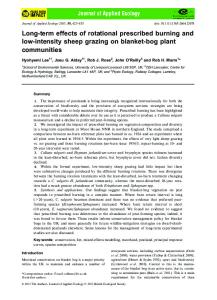
Figure 1. Changes over time in the medial compartment joint space width (JSW) of the knee in patients with knee osteoarthritis treated for 2 years with intraarticular (IA) steroids (solid bars) or IA saline (open bars), as measured according to the Buckland-Wright protocol (36,37). Values are the mean and SEM. The difference between the 2 treatment groups was not significant (P ⫽ 0.57).
sured with a long-arm goniometer, and the time required to walk a distance of 50 feet “as fast as possible” was measured with a stop-watch and reported in seconds. “Flares” of knee pain and effusions during and between patient visits were also recorded. Analysis of synovial fluid. Patients in both groups underwent joint taps before the injections. The presence and volume of synovial fluid were noted. Radiologic evaluation. The progression of joint space narrowing in the injected knee over 2 years was evaluated at the narrowest point according to the published protocol (36,37). This protocol permits standardization of radiographs by positioning the knee in a semiflexed position aided by fluoroscopy and appending a metal sphere to the head of the fibula to correct for the effect of radiographic magnification. The films were then digitized by scanning and integrating the images in a computer, which automatically computes the narrowest joint space width (36). Safety evaluations. Adverse events reported by patients or observed by the investigator were recorded in the patients’ medical charts and, if necessary, were reported to an independent external committee for evaluation. Laboratory evaluations, including a complete blood cell count, determination of the erythrocyte sedimentation rate and rheumatoid factor titer, and levels of blood urea nitrogen, serum creatinine, alkaline phosphatase, alanine aminotransferase, aspartate aminotrans-
SAFETY AND EFFICACY OF LONG-TERM IA STEROIDS IN KNEE OA
373
Table 2. Change in clinical outcome measures after 1 year of treatment* IA steroid (n ⫽ 33)
IA saline (n ⫽ 33)
Difference†
P
⫺10.3 ⫾ 20.9‡ ⫺5.8 ⫾ 17.4 ⫺10.7 ⫾ 18.3 ⫺0.20 ⫾ 3.0 4.40 ⫾ 3.6
⫺14.1 ⫾ 21.1‡ ⫺6.0 ⫾ 20.8 2.6 ⫾ 21.2 ⫺0.00 ⫾ 3.1 2.70 ⫾ 3.3
⫺3.8 ⫾ 21.0 0.2 ⫾ 19.1 7.1 ⫾ 20.5 0.20 ⫾ 3.0 1.70 ⫾ 3.5
0.46 0.96 0.08 0.79 0.05
⫺17.4 ⫾ 19.3‡ ⫺13.2 ⫾ 27.4‡ ⫺12.7 ⫾ 20.6 ⫺13.7 ⫾ 19.7‡
⫺11.2 ⫾ 21.1‡ ⫺16.2 ⫾ 21.6‡ ⫺13.1 ⫾ 21.8‡ ⫺12.9 ⫾ 19.2‡
6.2 ⫾ 20.4 ⫺3.0 ⫾ 24.5 ⫺0.4 ⫾ 20.9 0.8 ⫾ 19.5
0.22 0.62 0.94 0.87
Outcome measure Physician’s global assessment, 100-mm VAS Patient’s global assessment, 100-mm VAS Patient’s assessment of pain at night, 100-mm VAS 50-foot walking time, seconds Range of motion, degrees WOMAC score Pain Stiffness Function Total index
* Values are the mean ⫾ SD. Negative values signify improvement for all measures except for range of motion. P values were determined by 2-sample t-test. IA ⫽ intraarticular; VAS ⫽ visual analog scale (0 ⫽ best; 100 ⫽ worst); WOMAC ⫽ Western Ontario and McMaster Universities Osteoarthritis Index. † Difference between the two treatment groups with regard to change in outcome measures from baseline. A positive difference favors the IA steroid group. ‡ P ⬍ 0.05 versus baseline within the treatment group.
ferase, gamma glutamyl transferase, calcium, phosphorus, albumin, and thyroid-stimulating hormone, were performed at baseline and periodically throughout the study. Statistical analysis. Power calculation. The original sample size calculation for this study was determined based on the number of patients needed to show a difference between the treatment groups with respect to the primary anatomical outcome variable: joint space narrowing, as estimated according to measurements of plain radiographs. The alpha level and power level were chosen as 0.05 and 0.80, respectively. The joint space narrowing of an OA knee was initially estimated based on published data available at the time the study was started (26,38) to be ⬃0.25 mm/year, or 0.50 mm at 2 years. The aim was to demonstrate a therapeutic effect of the IA steroid injections of 25% (0.125 mm less progression) on the progression of joint space narrowing between the two treatment groups at 2 years. The standard deviation for measuring the narrowest point of the joint space width using a standard radiograph and a semiflexed view was ⬃0.25 mm (38). Therefore, according to a standardized effect size of 0.5, with the alpha level at 0.05 and the beta level at 0.20, 27 patients were needed for each treatment group. A dropout rate of 20% was expected at 2 years of followup, so ⬃68 patients were needed for this study. Data analyses. All analyses were based on an intent-totreat principle. The primary outcome variable was the radiologic progression of joint space narrowing of the injected knee after 2 years of treatment. The efficacy measure of primary interest was the pain subscale from the WOMAC. Efficacy measures of secondary interest included the total score on the WOMAC, the physician’s global assessment, patient’s global assessment, patient’s assessment of pain, ROM of the affected knee, and 50-foot walking time. All of the data (clinical, radiologic, and laboratory) were systematically entered into a computerized database using a double-entry procedure. For continuous variables, the mean changes from baseline at the 1- and 2- year followup visits were estimated for each group, and the mean changes in the two groups were compared with the use of a 2-sample t-test
at 1 year, and an analysis of variance for repeated measurements at 2 years. The clinical efficacy measures were also analyzed using the normalized area under the curve (AUC) for the difference in scores from baseline to month 24, and a 2-sample t-test was used to detect significant differences of the AUCs between treatment groups. All tests were 2-sided. P values less than or equal to 0.05 were considered statistically significant. P values were not corrected for multiple comparisons.
RESULTS Patient characteristics. Sixty-eight patients, 34 in the IA steroid group and 34 in the IA saline group, were enrolled in the study. The treatment groups were generally similar at baseline with respect to demographic and disease characteristics (Table 1). The percentage of female patients and the duration of knee OA were slightly higher in the IA steroid group. A total of 66 patients completed the 2-year study. One patient in the IA steroid group received a corticosteroid injection between the second and third study injections because of an acute flare of knee pain; this was considered a protocol violation. One patient in the IA saline group refused to continue after the second injection because of lack of pain relief. These 2 patients were excluded from the analyses. Radiologic progression between treatment groups. Examination of radiographs obtained at baseline and after 1 or 2 years of treatment revealed no differences between the treatment groups with respect to the mean joint space width. There was no indication of disease progression in either group at either followup (P ⫽ 0.57) (Figure 1). Moreover, changes in the mean
374
RAYNAULD ET AL
Table 3. Change in clinical outcome measures after 2 years of treatment* IA steroid (n ⫽ 33)
IA saline (n ⫽ 33)
Difference†
P
⫺9.2 ⫾ 27.2 ⫺7.6 ⫾ 23.8 ⫺2.8 ⫾ 19.1 ⫺0.4 ⫾ 3.0 13.3 ⫾ 4.0
⫺9.5 ⫾ 27.7 ⫺7.1 ⫾ 21.3 ⫺1.0 ⫾ 24.1 ⫹0.03 ⫾ 3.1 ⫺0.10 ⫾ 4.1
⫺0.3 ⫾ 27.5 0.5 ⫾ 22.5 1.8 ⫾ 23.0 0.07 ⫾ 3.0 1.43 ⫾ 2.7
0.96 0.92 0.74 0.92 0.16
⫺11.4 ⫾ 19.2‡ ⫺12.1 ⫾ 25.1‡ ⫺10.9 ⫾ 32.6 ⫺11.1 ⫾ 28.3‡
⫺13.8 ⫾ 21.5‡ ⫺13.2 ⫾ 25.4‡ ⫺13.1 ⫾ 34.1‡ ⫺13.5 ⫾ 24.3‡
⫺2.4 ⫾ 20.5 ⫺1.1 ⫾ 25.2 ⫺2.2 ⫾ 33.3 ⫺2.4 ⫾ 22.7
0.63 0.86 0.79 0.71
Outcome measure Physician’s global assessment, 100-mm VAS Patient’s global assessment, 100-mm VAS Patient’s assessment of pain at night, 100-mm VAS 50-foot walking time, seconds Range of motion, degrees WOMAC score Pain Stiffness Function Total Index
* Values are the mean ⫾ SD. Negative values signify improvement for all measures except for range of motion. P values were determined by analysis of variance for repeated measurements. IA ⫽ intraarticular; VAS ⫽ visual analog scale (0 ⫽ best; 100 ⫽ worst); WOMAC ⫽ Western Ontario and McMaster Universities Osteoarthritis Index. † Difference between the two treatment groups with regard to change in outcome measures from baseline. A positive difference favors the IA steroid group. ‡ P ⬍ 0.05 versus baseline within the treatment group.
joint space width within treatment groups was not significant (P ⫽ 0.81 in the steroid group and P ⫽ 0.74 in the saline group for 2-year data versus baseline, by 1-sample t-test). Clinical changes between treatment groups. After 1 year of treatment (4 systematic injections), the change in ROM from baseline was significantly greater in the IA steroid group than in the IA saline group (P ⫽ 0.05) (Table 2). In addition, trends toward greater improvement in the IA steroid group were noted for the WOMAC pain subscale (P ⫽ 0.24) and night pain (P ⫽ 0.39) versus baseline. Improvement was greater for the patients in the IA steroid group for all other variables except the physician’s global assessment and the WOMAC stiffness subscale. After 2 years of treatment (8 systematic injections), there were no statistically significant differences
between the treatment groups with respect to any of the primary or secondary efficacy variables (Table 3). The trends favoring the IA steroid group that were apparent after 1 year were less evident after 2 years. Knee pain and stiffness, as assessed by the WOMAC questionnaire, were significantly improved (P ⬍ 0.05) within each group at 1 and 2 years of followup compared with baseline (Tables 2 and 3). The AUC analyses revealed statistically significant differences between the treatment groups, favoring the steroid group for night pain (Figure 2) and stiffness (Figure 3) in the study knee (P ⬍ 0.05). All the other variables showed no significant differences, although trends favored the steroid-treated group. Moreover, none of the variables we evaluated gave any indication of significant worsening in either treatment group over the 2-year course of treatment.
Figure 2. Changes over time in levels of knee pain at night in patients with knee osteoarthritis treated for 2 years with intraarticular (IA) steroids or IA saline, as assessed by visual analog scales (see Patients and Methods for details). The area under the curve (AUC) of the normalized values for night pain is shown for each group. The difference between the 2 treatment groups was significant (P ⫽ 0.0047).
Figure 3. Changes over time in levels of knee stiffness in patients with knee osteoarthritis treated for 2 years with intraarticular (IA) steroids or IA saline, as assessed by visual analog scales (see Patients and Methods for details). The area under the curve (AUC) of the normalized values for knee stiffness is shown for each group. The difference between the 2 treatment groups was significant (P ⫽ 0.0511).
SAFETY AND EFFICACY OF LONG-TERM IA STEROIDS IN KNEE OA
Evaluation of the clinical course of the study patients demonstrated no differences between the two groups with regard to the number of OA flares or the magnitude of the effusions (data not shown). There were also no interactions between the primary clinical outcomes and age, sex, weight, NSAID use, or the other covariates analyzed. Safety. No infections (local or systemic) or acute flares were associated with the injections during the study. Findings of laboratory tests performed during the study were within normal limits, and there were no differences between the treatment groups. DISCUSSION The present study demonstrated that OA patients who received long-term IA injections of triamcinolone acetonide had a significantly greater change in ROM and a slightly greater improvement in pain compared with baseline than did the saline control patients, especially at the first year of followup. Moreover, the AUC analyses revealed significant improvements in knee pain and stiffness during the repeated triamcinolone injection regimen. Measurement of the radiographic joint space width after 1 and 2 years of treatment revealed no differences in the anatomical progression of OA between the two treatment groups. Osteoarthritis is a slowly evolving articular disease characterized by a gradual development of joint pain, stiffness, and loss of full range of motion. Degeneration of cartilage is among the most prominent pathologic changes. Clinical OA is a complex interaction of degradation and repair of the cartilage, bone, and synovium with secondary components of inflammation (39–42). Chondrocytes from OA patients have been demonstrated to be deficient in glucocorticoid receptors (43). The resulting decreased responsiveness of OA cells to circulating glucocorticoid may be among the factors that lead to an increased level of cytokine and metalloprotease synthesis in degrading cartilage (44). It is therefore logical to consider interventions that could control inflammation in OA patients. The antiinflammatory effects of synthetic glucocorticoids serve as a basis for such a therapeutic approach. Although these steroids are administered chiefly for relief of symptoms, there is some evidence in experimental animal models of OA to suggest that they also exert protective effects, in that they may reduce the severity of cartilage lesions and the size of osteophytes (17,18,21). These effects are presumably the result of the suppressive action of glucocorticoids on the synthesis by connective tissue cells of a
375
number of the cytokines and metalloproteases associated with cartilage degradation (20). Physicians have treated patients with IA injections of corticosteroids for more than 4 decades. Despite extensive clinical experience, certain aspects of local corticosteroid treatment remain controversial, such as its relative efficacy, its possible deleterious effects, and its potential chondroprotective properties. Dieppe et al (7) reported that IA steroid injections caused a significantly greater reduction in pain and tenderness than did placebo injections and were preferred by patients, but the benefit was transient. A 6-month, randomized, singleblind clinical trial conducted in patients with symptomatic knee OA in Finland (10) showed that IA injections of triamcinolone hexacetonide might have a long-term beneficial effect on symptoms. One retrospective observational study evaluated IA steroid injections that were repeated over a period of 4–15 years in 65 patients (4). Based on findings from radiologic assessments, those investigators concluded that, if used judiciously, IA corticosteroid therapy plays an important role in the management of chronic arthritis. They found no real evidence to support the suggestion that repeated IA injections inevitably led to rapid destruction of the injected joint. Our study is the first long-term, prospective, double-blind, and placebo-controlled trial to examine the safety and efficacy of IA steroid injections. Our results showed that long-term injections of IA steroids over 2 years did not have a deleterious effect on the anatomical structure of the knee. Our findings expand and enhance observations made in previous short-term studies in which a strong trend toward greater clinical improvement in the IA steroid group was shown, this difference being statistically significant for key variables such as knee pain and stiffness using the AUC analysis strategy. The apparent lack of difference, using mean change compared with baseline, between the IA steroid and IA saline treatment groups for the other variables in the present study can be explained in several ways. First, our protocol did not include a washout period for NSAIDs and other analgesics used pretreatment. It is possible that the patients in our study had better pain control and function at baseline than was expected, resulting in an underestimation of the effect of the study treatments. Second, our sample size was based on the number of patients needed to show differences in radiographic data between the treatment groups. The sample size may not have been large enough to reveal significant between-group differences in clinical efficacy data.
376
Third, the efficacy of steroid injections is usually rapid but short-lived. Because the primary focus of the study was to determine the effect of steroid injections on knee structure, data were not collected on the days or weeks immediately after the injections. As a result, the shortterm effect of the injections on pain and function could not be assessed. The AUC analysis is therefore more appropriate for capturing the long-term efficacy difference between the treatment groups. Fourth, arthrocentesis and injection of saline may benefit patients by washing away mediators of inflammation and IA debris from the joint, as has been suggested (45). The statistically significant improvement in both the steroid and the saline groups between baseline and years 1 and 2 of followup, as shown for knee pain and stiffness, tends to favor that hypothesis. Finally, the study did not include a washout period for NSAIDs and other analgesics prior to each clinical evaluation, as is now suggested by regulatory agencies for studies of OA treatments (26). Consequently, the clinical effect of the steroid injections on knee pain and function in our study may have been underreported as a result of the influence of concomitant medications at each evaluation. Although our study did not demonstrate that steroid injections may prevent cartilage degradation, it is reassuring to note that after 2 years of injections, no significant difference between the two experimental groups was noted. This finding addresses the safety issue of repeated IA injections of steroids for pain relief and suggests that these injections are not associated with accelerated disease progression. The methods we used to determine radiologic disease progression have been refined since they were first described (26,38). According to a publication that followed the start of our study (36), a loss of joint space width of 0.18 mm per year in the study knee would be expected in patients who meet the following criteria: radiographs showing disease in the internal compartment of the knee that is worse than that in the lateral compartment and the presence of osteophytes on the anteroposterior view. Without these 2 radiographic elements, disease progression would be expected to be significantly slower than originally thought. Therefore, our original power calculations were incorrect. A recent study using glucosamine sulfate and a good study sample size demonstrated that narrowing of the joint space width on standardized radiographs can be used as a primary outcome variable in trials of knee OA (46). In our own study, IA injections of saline combined with insufficient statistical power may explain the lack of radiologic progression in our OA “control” group.
RAYNAULD ET AL
In conclusion, no significant deleterious effects of the steroids on the anatomical joint structure were seen in this study. This finding suggests that repetitive IA steroid injections appear to be safe. Moreover, the long-term use of IA injections of triamcinolone acetonide afforded relief of some of the symptoms of knee OA, including pain and stiffness. ACKNOWLEDGMENTS The authors wish to thank Professors Michel Lequesne, Eric Vignon, and Roy Davis Altman for their valuable time and expertise in the analysis of the radiographs. The authors also wish to thank Raymonde Gre´goire, RN, for administrative and medical support and Santa Fiori for assistance in preparing the manuscript.
REFERENCES 1. Mankin HJ. The reaction of articular cartilage to injury and osteoarthritis (first of two parts). N Engl J Med 1974;291:1285–92. 2. Salter RB, Field P. The effects of continuous compression on living articular cartilage. J Bone Joint Surg Am 1960;42:31–9. 3. Wada J, Koshino T, Morii T, Sugimoto K. Natural course of osteoarthritis of the knee treated with or without intraarticular corticosteroid injections. Bull Hosp Jt Dis 1993;53:45–8. 4. Balch HW, Gibson JM, El-Ghobarey AF, Bain LS, Lynch MP. Repeated corticosteroid injections into knee joints. Rheumatol Rehabil 1977;16:137–40. 5. Blyth T, Hunter JA, Stirling A. Pain relief in the rheumatoid knee after steroid injection: a single-blind comparison of hydrocortisone succinate, and triamcinolone acetonide or hexacetonide. Br J Rheumatol 1994;33:461–3. 6. Cederlof S, Jonson G. Intraarticular prednisolone injection for osteoarthritis of the knee: a double blind test with placebo. Acta Chir Scand 1966;132:532–7. 7. Dieppe PA, Sathapatayavongs B, Jones HE, Bacon PA, Ring EF. Intra-articular steroids in osteoarthritis. Rheumatol Rehabil 1980; 19:212–7. 8. Friedman DM, Moore ME. The efficacy of intraarticular steroids in osteoarthritis: a double-blind study. J Rheumatol 1980;7:850–6. 9. Gray RG, Gottlieb NL. Intra-articular corticosteroids: an updated assessment. Clin Orthop 1983;235–63. 10. Valtonen EJ. Clinical comparison of triamcinolonehexacetonide and betamethasone in the treatment of osteoarthrosis of the knee-joint. Scand J Rheumatol Suppl 1981;41:1–7. 11. Gaffney K, Ledingham J, Perry JD. Intra-articular triamcinolone hexacetonide in knee osteoarthritis: factors influencing the clinical response. Ann Rheum Dis 1995;54:379–81. 12. Jones A, Doherty M. Intra-articular corticosteroids are effective in osteoarthritis but there are no clinical predictors of response. Ann Rheum Dis 1996;55:829–32. 13. American College of Rheumatology Subcommittee on Osteoarthritis Guidelines. Recommendations for the medical management of osteoarthritis of the hip and knee: 2000 update. Arthritis Rheum 2000;43:1905–15. 14. Behrens F, Shepard N, Mitchell N. Alterations of rabbit articular cartilage by intra-articular injections of glucocorticoids. J Bone Joint Surg Am 1975;57:70–6. 15. Creamer P. Intra-articular corticosteroid treatment in osteoarthritis. Curr Opin Rheumatol 1999;11:417–21. 16. Papachristou G, Anagnostou S, Katsorhis T. The effect of intra-
SAFETY AND EFFICACY OF LONG-TERM IA STEROIDS IN KNEE OA
17.
18.
19.
20.
21.
22.
23.
24.
25. 26. 27.
28.
29. 30.
articular hydrocortisone injection on the articular cartilage of rabbits. Acta Orthop Scand 1997;132–4. Pelletier J-P, Martel-Pelletier J, Cloutier J-M, Woessner JF Jr. Proteoglycan-degrading acid metalloprotease activity in human osteoarthritic cartilage, and the effect of intraarticular steroid injections. Arthritis Rheum 1987;30:541–8. Pelletier J-P, Martel-Pelletier J. Protective effects of corticosteroids on cartilage lesions and osteophyte formation in the Pond-Nuki dog model of osteoarthritis. Arthritis Rheum 1989;32: 181–93. Pelletier J-P, Mineau F, Raynauld J-P, Woessner JF Jr, GunjaSmith Z, Martel-Pelletier J. Intraarticular injections with methylprednisolone acetate reduce osteoarthritic lesions in parallel with chondrocyte stromelysin synthesis in experimental osteoarthritis. Arthritis Rheum 1994;37:414–23. Pelletier J-P, Di Battista JA, Raynauld J-P, Wilhelm S, MartelPelletier J. The in vivo effects of intraarticular corticosteroid injections on cartilage lesions, stromelysin, interleukin-1 and oncogene protein synthesis in experimental osteoarthritis. Lab Invest 1995;72:578–86. Williams JM, Brandt KD. Triamcinolone hexacetonide protects against fibrillation and osteophyte formation following chemically induced articular cartilage damage. Arthritis Rheum 1985;28: 1267–74. Bellamy N, Buchanan WW, Goldsmith CH, Campbell J, Stitt LW. Validation study of WOMAC: a health status instrument for measuring clinically important patient relevant outcomes to antirheumatic drug therapy in patients with osteoarthritis of the hip or knee. J Rheumatol 1988;15:1833–40. Altman RD, Fries JF, Bloch DA, Carstens J, Cooke TD, Genant H, et al. Radiographic assessment of progression in osteoarthritis. Arthritis Rheum 1987;30:1214–25. Brandt KD, Fife RS, Braunstein EM, Katz B. Radiographic grading of the severity of knee osteoarthritis: relation of the Kellgren and Lawrence grade to a grade based on joint space narrowing, and correlation with arthroscopic evidence of articular cartilage degeneration. Arthritis Rheum 1991;34:1381–6. Ravaud P, Ayral X, Dougados M. Radiologic progression of hip and knee osteoarthritis. Osteoarthritis Cartilage 1999;7:222–9. Lequesne M, Brandt K, Bellamy N, Moskowitz R, Menkes CJ, Pelletier J-P, et al. Guidelines for testing slow acting drugs in osteoarthritis. J Rheumatol Suppl 1994;41:65–73. Scott WW Jr, Lethbridge-Cejku M, Reichle R, Wigley FM, Tobin JD, Hochberg MC. Reliability of grading scales for individual radiographic features of osteoarthritis of the knee: the Baltimore longitudinal study of aging atlas of knee osteoarthritis. Invest Radiol 1993;28:497–501. Schouten JS, Van den Ouweland FA, Valkenburg HA. A 12 year follow up study in the general population on prognostic factors of cartilage loss in osteoarthritis of the knee. Ann Rheum Dis 1992;51:932–7. Spector TD, Dacre JE, Harris PA, Huskisson EC. Radiological progression of osteoarthritis: an 11 year follow up study of the knee. Ann Rheum Dis 1992;51:1107–10. Altman R, Asch E, Bloch D, Bole G, Borenstein D, Brandt K, et al. Development of criteria for the classification and reporting of osteoarthritis: classification of osteoarthritis of the knee. Arthritis Rheum 1986;29:1039–49.
377
31. Kellgren JH, Lawrence JS. Radiological assessment of osteoarthrosis. Ann Rheum Dis 1957;16:494–501. 32. Huskisson EC, Berry H, Gishen P, Jubb RW, Whitehead J, and the Longitudinal Investigation of Nonsteroidal Antiinflammatory Drugs in Knee Osteoarthritis Study Group. Effects of antiinflammatory drugs on the progression of osteoarthritis of the knee. J Rheumatol 1995;22:1941–6. 33. Palmoski MJ, Brandt KD. Effects of some nonsteroidal antiinflammatory drugs on proteoglycan metabolism and organization in canine articular cartilage. Arthritis Rheum 1980;23:1010–20. 34. Rashad S, Revell P, Hemingway A, Low F, Rainsford K, Walker F. Effect of non-steroidal anti-inflammatory drugs on the course of osteoarthritis. Lancet 1989;2:519–22. 35. Choquette D, Bellamy N, Raynauld JP. A French-Canadian version of the WOMAC Osteoarthritis Index [abstract]. Arthritis Rheum 1994;37 Suppl 9:S226. 36. Buckland-Wright C. Protocols for precise radio-anatomical positioning of the tibiofemoral and patellofemoral compartments of the knee. Osteoarthritis Cartilage 1995;3:71–80. 37. Buckland-Wright JC, Macfarlane DG, Williams SA, Ward RJ. Accuracy and precision of joint space width measurements in standard and macroradiographs of osteoarthritic knees. Ann Rheum Dis 1995;54:872–80. 38. Lequesne M, Nguyen NJH, Rodriguez P, Masse JP, Glimet T. Vitesse du pincement de l’interligne au cours de la gonarthrose fe´moro-tibiale interne: validite´ de la mesure [abstract]. Rev Rhum Mal Osteoartic 1990;57:A14. 39. Revell PA, Mayston V, Lalor P, Mapp P. The synovial membrane in osteoarthritis: a histological study including the characterization of the cellular infiltrate present in inflammatory osteoarthritis using monoclonal antibodies. Ann Rheum Dis 1988;47:300–7. 40. Haraoui B, Pelletier J-P, Cloutier J-M, Faure M-P, MartelPelletier J. Synovial membrane histology and immunopathology in rheumatoid arthritis and osteoarthritis: in vivo effects of antirheumatic drugs. Arthritis Rheum 1991;34:153–63. 41. Lindblad S, Hedfors E. Arthroscopic and immunohistologic characterization of knee joint synovitis in osteoarthritis. Arthritis Rheum 1987;30:1081—8. 42. Pelletier J-P, Howell DS. Etiopathogenesis of osteoarthritis. In: McCarty DJ, Koopman WJ, editors. Arthritis and allied conditions: a textbook of rheumatology. 12th ed. Philadelphia: Lea & Febiger; 1993. p. 1723–34. 43. Di Battista JA, Martel-Pelletier J, Wosu LO, Sandor T, Antakly T, Pelletier J-P. Glucocorticoid receptor mediated inhibition of interleukin-1 stimulated neutral metalloprotease synthesis in normal human chondrocytes. J Clin Endocrinol Metab 1991;72:316–26. 44. Di Battista JA, Martel-Pelletier J, Antakly T, Tardif G, Cloutier J-M, Pelletier J-P. Reduced expression of glucocorticoid receptor levels in human osteoarthritic chondrocytes: role in the suppression of metalloprotease synthesis. J Clin Endocrinol Metab 1993; 76:1128–34. 45. Ravaud P, Moulinier L, Giraudeau B, Ayral X, Guerin C, Noel E, et al. Effects of joint lavage and steroid injection in patients with osteoarthritis of the knee: results of a multicenter, randomized, controlled trial. Arthritis Rheum 1999;42:475–82. 46. Reginster JY, Deroisy R, Rovati LC, Lee RL, Lejeune E, Bruyere O, et al. Long-term effects of glucosamine sulphate on osteoarthritis progression: a randomised, placebo-controlled clinical trial. Lancet 2001;357:251–6.