J. Phys. Chem. C 2007, 111, 14113-14124
14113
Role of Different Phospholipids in the Synthesis of Pearl-Necklace-Type Gold-Silver Bimetallic Nanoparticles as Bioconjugate Materials Mandeep Singh Bakshi,*,†,‡,§,⊥ Fred Possmayer,†,‡ and Nils O. Petersen*,†,‡,| Department of Obstetrics and Gynaecology, Department of Biochemistry, and Department of Chemistry, UniVersity of Western Ontario, 339 Windermere Road, London, Ontario, Canada N6A 5A5, National Institute for Nanotechnology, Edmonton, Alberta, Canada, and Department of Chemistry, Guru Nanak DeV UniVersity, Amritsar 143005, Punjab, India ReceiVed: April 12, 2007; In Final Form: July 12, 2007
A seed-growth (S-G) method has been used to synthesize gold (Au) and Au-silver (Ag) bimetallic pearnecklace-type nanoparticles (NP) as bioconjugate materials by using a series of phospho-glycerol (PG) and phospho-choline (PC) lipids as capping agents. All PG lipids produce a fine pear-necklace arrangement of Au NP with dimensions between 10 and 20 nm. Addition of Ag converts this arrangement into Au-Ag bimetallic NP with a diameter essentially close to that of Au NP. Use of PC lipids does not show this arrangement but promotes a significant anisotropic growth especially in the presence of Ag. This difference has been attributed to a difference in the capping ability of PG and PC lipids because of their anionic and zwitterionic nature, respectively. XPS results have demonstrated the presence of adsorbed PG and PC lipids on Au or Au-Ag bimetallic surfaces in their respective samples. The results also indicate a decrease in the capping amount of a lipid with an increase in the growth of Au-Ag bimetallic NP. The growth of Au-Ag bimetallic NP from Au NP has been ascribed to the nucleation of Ag atoms at the {111} facets of Au NP in the presence of PG lipids, while anisotropic growth is occurring mainly at all other planes of fcc crystal geometry of Au-Ag bimetallic NP in the presence of PC lipids. It has been concluded that in order to get fine Au-Ag bimetallic bioconjugate materials capped with PG lipids, one has to use ascorbic acid (AA) as a weak reducing agent at the end of the S-G reaction sequence so as to give the required time for lipid molecules to adsorb at the liquid-solid interface.
Introduction Phospholipids are the essential constituents of pulmonary surfactants.1 A collective contribution of phospho-glycerol (PG) and phospho-choline (PC) lipids along with surfactant proteins helps the lung to acquire minimum surface tension during a normal breathing process.1 High surface activity is achieved when lipid molecules form a fluid film of lipid bilayers at airalveolar interfaces. An uninterrupted bilayer is highly surfaceactive and helps the air-alveolar interface to achieve a surface tension value close to 0 mNm-1 during the lung compression. Conventional surfactants, however, are also known to have a high surface activity but surface tension never drops to a value close to 0 mNm-1 even for a micellar solution.2 The main difference lies in the membrane-forming ability of lipids. Phospholipids are known to form a continuous membrane at an interface,3 whereas conventional surfactants like sodium dodecyl sulfate or cetyltrimethyl ammonium bromide adsorb individually. Thus, a continuous membrane of phospholipids is more surface-active than individually adsorbed conventional surfactant molecules. Recent reports4 have suggested that phospholipids also act as excellent capping agents for the synthesis of noble metal NP. * Corresponding authors. E-mail:
[email protected] and
[email protected]. † Department of Obstetrics and Gynaecology, University of Western Ontario. ‡ Department of Biochemistry, University of Western Ontario. § Department of Chemistry, University of Western Ontario. | National Institute for Nanotechnology. ⊥ Guru Nanak Dev University.
They provide both steric and charge stabilization to colloidal metal NP by forming a lipid bilayer on the surface of NP. The presence of a bilayer thus prevents anisotropic growth and leads to the formation of 1D monodisperse NP. Not all lipids act as good capping agents. The capping ability is generated by electrostatic interactions between the lipid polar head group and charged NP surface.5 It depends on the nature of the polar head group, whether it is zwitterionic (PC) or ionic (PG). Anionic PG lipids are generally sodium salts, whereas PC lipids are zwitterionic with phospho-choline head groups. An anionic PG head group is expected to have much-stronger interactions with a charged metal NP surface in comparison to the zwitterionic head group of PC. Thus, weak electrostatic interactions show poor capping ability and consequently leave some crystal planes uncapped, which become active sites for further nucleation. This secondary nucleation is very hard to control and consequently leads to anisotropic growth.6 Bioengineering7 is a fast developing area of fine synergism between biological molecules and nanometallic surfaces and has great implications to produce macromolecular machines. The basis of this arises from a suitable combination between the two at molecular scale. We have attempted this by synthesizing Au and Au-Ag bimetallic NP in the presence of bioactive phospholipids. Recent advances have proven to be a remarkable success in achieving the formation of nanorods or nanowires. Murphy and co-workers6a-h have synthesized 1D nanorods successfully by using a simple and convenient S-G method. Other groups6i,j as well as our group4f have also reported the synthesis of Au nanowires. In the present work, we have used
10.1021/jp072862t CCC: $37.00 © 2007 American Chemical Society Published on Web 09/01/2007
14114 J. Phys. Chem. C, Vol. 111, No. 38, 2007
Bakshi et al.
SCHEME 1 : Molecular Structures of Different PG and PC Phospholipids
a similar S-G method to synthesize lipid-capped Au and AuAg bimetallic NP. Interestingly, this method works very well for our systems too. A series of PG and PC phospholipids (Scheme 1) have been selected to evaluate their capping ability on Au NP and Au-Ag bimetallic NP in order to form bioconjugate materials. Phospholipid-NP conjugate materials are foreseen as the basic components for bionanometallic devices with potential applications in sensors, gene sequencing, and biocatalysis. Experimental Section Materials. Tetrachloroauric acid (HAuCl4), silver nitrate (AgNO3), sodium borohydride (NaBH4), and trisodium citrate (Na3Cit) were obtained from Aldrich. 1-palmitoyl-2-oleoyl-snglycero-3-[phospho-rac-(1-glycerol)] (sodium salt) (POPG) (16: 0-18:1), 1,2-dipalmitoyl-sn-glycero-3-[phospho-rac-(1-glycerol)] (sodium salt) (DPPG) (16:0), 1,2-dimyristoyl-sn-glycero3-[phospho-rac-(1-glycerol)] (sodium salt) (DMPG) (14:0), 1-palmitoyl-2-oleoyl-sn-glycero-3-phosphocholine (POPC) (16: 0-18:1), 1,2-dipalmitoyl-sn-glycero-3-phosphocholine (DPPC) (16:0), and 1,2-dimyristoyl-sn-glycero-3-phosphocholine (DMPC) (14:0) were procured from Avati Polar Lipids. Ultrapure water (18 MΩcm) was used for all aqueous preparations. Preparation of Lipid-Capped Gold Nanoparticles by the Seed-Growth (S-G) Method. In the S-G method,6a the first step includes the preparation of a seed solution. First of all, 25 mL of HAuCl4 aqueous solution ([HAuCl4] ) 0.5 mM) was taken in a screw-capped glass bottle, and Na3Cit was added in it so as to make a final concentration of [Na3Cit] ) 0.5 mM. Then, 0.6 mL of aqueous NaBH4 ([NaBH4] ) 0.1 mol dm-3) solution was added under constant stirring, giving rise to a rubyred color to the final solution, which acts as a seed solution. The growth solution was prepared by taking 120 µL of lipid (1 mg/mL) in chloroform, in a glass screw-capped tube. The chloroform was evaporated under the flux of pure N2 leaving a dried lipid film at the bottom of the tube. In this tube, 5 mL of pure water was added along with 2-3 small glass beads and it was vortexed for couple of minutes to completely disperse the lipid in the aqueous phase. This was followed by the addition of HAuCl4 so to make [HAuCl4] ) 0.5 mM, which gave a lightyellow color to the solution. Then, 0.5 mL of previously made seed solution was added in it, and, finally, 0.2 mL of freshly prepared ascorbic acid (AA) aqueous solution ([AA] ) 0.1 M) was added. This gave an instant deep-ruby-red color to the
solution. The solution was mixed couple of times by inverting the bottle and kept in the dark without disturbing it at least for 2 days. The pH of the aqueous NP solution was always close to neutral in each case. A similar procedure was adopted to synthesize Au-Ag bimetallic NP. Here, the addition of AgNO3 was carried out immediately after the addition of HAuCl4 in the growth solution, while the rest of the reaction sequence was the same as that mentioned in the previous paragraph. Three concentrations of AgNO3, namely, 0.12, 0.25, and 0.5 mM, were used. Purification of each aqueous sample of lipid-capped NP was carried out by repeated washing (at least 3 to 4 times) with distilled water after performing centrifugation at 10 000 rpm for 10 min each time to remove the maximum uncapped lipid vesicles from the aqueous phase. It is to be mentioned that no vesicles or liposomes were observed from TEM studies after the purification process. In all of these reactions, an important point to be noted is to choose whether to use AA or seed at the end of a reaction sequence because entirely different morphologies of NP appear in either case. Use of AA at the end only produces a fine pearlnecklace-type arrangement. This point has been discussed in detail in the Discussion section by taking the example of the POPG reaction. Methods. UV-visible spectra of as-prepared solutions were taken by a UV spectrophotometer (Multiskan Spectrum, model no. 1500) in the wavelength range of 200-900 nm to determine the absorbance due to surface plasmon resonance (SPR). The formation of Au and Ag NP was monitored in the visible absorption range of ∼530 and ∼410 nm, respectively. The shape and size of Au and Au-Ag bimetallic NP were characterized by transmission electron microscopy (TEM). The samples were prepared by mounting a drop of aqueous NP solution on a carbon-coated Cu grid and allowed it to dry in air. They were observed with the help of a Philips CM10 transmission electron microscope operating at 100 kV. To understand the pearlnecklace morphology of Au-Ag bimetallic NP, high-angle annular dark-field (HAADF) scanning TEM (with CM20) was also done. The X-ray diffraction (XRD) patterns were characterized by using Bruker-AXS D8-GADDS with Tsec ) 480. The chemical composition of some samples containing Au and AuAg bimetallic NP was confirmed with the help of X-ray photonelectron spectroscopic (XPS) measurements. A portion of an aqueous sample was placed onto a clean silicon wafer,
Pearl-Necklace-Type Bimetallic Nanoparticles
Figure 1. (a) UV-visible spectra of aqueous gold and gold-silver bimetallic nanoparticle solutions in the presence of POPG ([POPG] ) 0.027 mM). Black arrows indicate the absorbance due to surface plasmon resonance of nanometallic silver (see details in the text). (b) Absorbance spectra of identical reactions mentioned in part a except the addition of seed at the end of the seed-growth reaction. Inset shows two weak shoulders due to two different kinds of morphologies of Au NPs (see details in the text).
and then it was put into the introduction chamber of the XPS instrument. The liquid was then pumped away. The sample was analyzed by using a Kratos Axis Ultra X-ray photoelectron spectrometer. XPS can detect all elements except hydrogen and helium and can probe the surface of the sample to a depth of 7-10 nm. Survey scan analyses were carried out with an analysis area of 300 × 700 µm. Results UV-Vis Measurements. PG-Capped NP. Nanosized Au particles show absorbance in the UV-visible region due to SPR, which originates due to the resonance of collective conduction electrons with incident electromagnetic radiations.8 It provides a sharp absorbance in the visible region around 520 nm. The shape of the resonance peak can be qualitatively related to the nature of NP. Small and uniform-sized NP with a narrow size distribution gives a sharp absorbance, whereas NP with a wide size distribution or any kind of aggregation shows a broad absorbance.8 Ag NP demonstrates this behavior around 410 nm. Figure 1 shows the UV-visible absorbance of POPG-capped Au NP in the absence and presence of Ag. The POPG-capped Au NP in the absence of Ag shows a sharp absorbance at ∼520
J. Phys. Chem. C, Vol. 111, No. 38, 2007 14115 nm (Figure 1a). Addition of AgNO3 at constant lipid concentration has a significant effect on the SPR of Au NP. Increase in [AgNO3] from 0.12 to 0.5 mM causes a blue shift of ∼40 nm in the absorbance of Au NP from 520 to 480 nm, while a shoulder due to the SPR of Ag NP starts appearing at ∼350 nm (shown by arrows). Further increase in the amount of AgNO3 ([AgNO3] ) 0.5 mM) makes it quite prominent and red shifts to 380 nm. It appears that the increase in the amount of AgNO3 induces nucleation of Ag° atoms on the surface of already available Au NP, thus reducing their SPR.9 This might cause a blue shift in the absorbance of Au NP and consequently produces a red shift in the absorbance of Ag NP. It can most probably be due to some kind of self-association among both kinds of NP10 in order to form Au-Ag bimetallic NP (we will come to this point along with TEM images) at the expense of smaller Ag NP through the Ostwald ripening process.11 Similar UV-visible spectra were obtained for Au and Au-Ag bimetallic NP when DMPG and DPPG were used as the capping agents (see the Supporting Information, Figure S1). A drastic change in the absorbance behavior is observed when the seed solution is added at the end of the reaction sequence instead of the AA (see the Experimental Section). Figure 1b shows the absorbance of an aqueous NP solution obtained from the identical reactions except adding seed at the end of the reaction sequence. For pure Au NP, a broad absorbance is obtained in comparison to the sharp one obtained in Figure 1a for the same reaction. A close up (inset) indicates that this broad maximum in fact consists of two shoulders around 535 and 570 nm (indicated by arrows). Both shoulders take the form of broad maxima around 520 and 620 nm, respectively, in the presence of [AgNO3] ) 0.25 and 0.5 mM. The first one shows a blue shift of 15 nm, and the second one red shifts with 50 nm in comparison to that in the absence of Ag. A blue shift from 535 to 520 nm can be due to the formation of Au-Ag bimetallic NP9 as demonstrated by a similar NP in Figure 1a, whereas the red shift in the latter case indicates the presence of some ordered arrangement.11 Additional rise in the amount of Ag ([AgNO3] ) 0.5 mM) produces only one prominent broad maximum at ∼540 nm with a very-weak hump at ∼420 nm, suggesting the presence of a large Au-Ag bimetallic NP in an aggregated state. TEM images will differentiate between the NP obtained by using different reaction sequences in the next section. PC-Capped NP. Interestingly, when instead of anionic PG, corresponding zwitterionic PC lipids (Scheme 1) such as POPC, DMPC, and DPPC were used, an entirely different behavior of SPR was observed (see the Supporting Information, Figure S2). The Au NP synthesized in the presence of all PC lipids show mainly broad absorbance between 500 and 700 nm when [AgNO3] ) 0-0.5 mM is used, which indicated the presence of a large NP with unspecified growth.8e,f Thus, a marked difference between the SPR of PG- and PCcapped Au or Au-Ag bimetallic NP indicates a fundamental difference between the capping ability of lipids during their synthesis. This difference one can easily speculate from the ionic and zwitterionic nature of PC and PG lipid head groups, which have to provide charge and steric stabilization10b of colloidal NP during crystal growth. TEM Studies. As observed in the previous section that the capping behavior of PG (Figure 1a) and PC (see the Supporting Information, Figure S2) lipids produce entirely different kinds of absorption spectra of Au or Au-Ag bimetallic NP, we have divided this section into two parts in order to understand that how the shape and structure of NP is different in the presence of PG and PC lipids. The first section explains the shape and
14116 J. Phys. Chem. C, Vol. 111, No. 38, 2007
Bakshi et al.
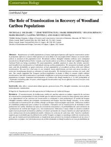
Figure 2. (a) TEM micrographs of POPG-capped gold nanoparticles ([POPG] ) 0.029 mM). Note the POPG bilayer around individual as well as groups of nanoparticles shown by block arrows. Inset shows a high-resolution image of a single Au NP covered by a 4-nm POPG bilayer. Part b shows the pearl-necklace type gold-silver bimetallic NP made up of several spindle-shaped particles (indicated by block arrows). Part c shows a much-smoother arrangement with a coating of POPG bilayer.
Figure 3. Mechanistic point of view of bilayer formation on a citrate-stabilized gold nanoparticle surface.
structure of NP in the presence of PG lipids, and the second section explains the same in the presence of PC lipids. Apart from this, the UV-visible spectra also indicate a dramatic
change when AA (Figure 1a) or seed (Figure 1b) is used at the end of the reaction sequence; therefore, this aspect has also been explained in detail with TEM studies.
Pearl-Necklace-Type Bimetallic Nanoparticles
J. Phys. Chem. C, Vol. 111, No. 38, 2007 14117
Figure 4. (a) High-angle annular dark-field (HAADF) scanning transmission electron microscopic image of a POPG-capped pearl-necklace-type Au-Ag bimetallic. (b) The corresponding energy-dispersive X-ray spectrum (see details in the text).
PG-Capped NP. POPG as Capping Agent (AA Added Last). TEM image in the absence of Ag (Figure 2a) mainly shows spherical POPG capped Au NP with an average size distribution of 13.9 ( 4.5 nm. The Au NP seems to arrange in a typical pearl-necklace model.4f A high-resolution image (inset) shows the presence of a lipid bilayer of ∼4 nm thickness around each or group of Au NPs (indicated by block arrows). The corresponding lipid membrane studies12 have also reported the thickness of the lipid bilayer around 4 nm. The bilayer formation is thought to be achieved due to the following steps (Figure 3). In the first step, the Na+ counterions of PG lipids interact with the citrate stabilized negatively charged Au NP surface. This is followed by electrostatic interactions with anionic lipid molecules to form a monolayer. Because hydrophobic tails cannot survive in the aqueous phase, they interact with the tails of other lipid molecules and thus lead to a bilayer arrangement. The origin of the pearl-necklace arrangement is thought to arise from the fusion of lipid bilayers of adjoining POPG-capped Au NPs.13,4f In the presence of [AgNO3] ) 0.12 mM (Figure 2b), the spherical NPs (15.3 ( 4.9 nm) merge with each other in a clear pearl-necklace-type arrangement with an average thickness of 14.5 nm and become much smoother as the amount of AgNO3 reaches 0.5 mM (Figure 2c). Figure 2c also shows that each arrangement is completely covered with a lipid bilayer. Interestingly, the average size distribution of the Au NPs (Figure 2a) and the thickness of this arrangement (Figure 2c) are very close to each other, which probably suggests that fusion among Au NPs in the presence of Ag forms Au-Ag bimetallic NPs. This is further proved by the HAADF STEM image (Figure 4a) as well as its corresponding energy-dispersive X-ray microanalysis
(EDAX) (Figure 4b). One would clearly see many bright discshaped connections (shown by block arrows) made up of Ag NP probably bound to [111] facets of roughly spherical NP (will be explained further by XRD later in the Discussion section). Similarly, because we use DMPG as a capping agent, the observed morphologies of Au and Au-Ag bimetallic NPs (Figure 5) are almost identical to those obtained in the presence of POPG. The spherical Au NPs (11.7 ( 4.7 nm) capped with DMPG bilayers are arranged in a perfect pearl-necklace model (Figure 5a) in the absence of Ag and that converts into a fine arrangement as the amount of Ag increases (Figure 5b and c). In the case of DPPG (Figure 6), the overall transformation of Au NPs to a pearl-necklace arrangement essentially follows the same order as mentioned in the case of POPG and DMPG, but there are some significant differences as far as the size and shape of the NPs are concerned. First, the average size distribution of DPPG-capped spherical Au NPs is equal to 20.3 ( 7.3 nm (Figure 6a), which is much larger than that obtained for POPG (13.9 ( 4.5 nm) and DMPG (11.7 ( 4.7 nm) capped Au NP. Second, the addition of [AgNO3] ) 0.12 mM clearly changes the spherical Au NPs into polyhedral geometries with size distributions of 23.7 ( 9.5 nm (Figure 6b). No clear DPPG bilayer is observed in Figure 6a, whereas broken bilayer is indeed present around polyhedral NPs (Figure 6b). Increase in the amount of [AgNO3] ) 0.5 mM transfers the polyhedral NPs into a pearl-necklace arrangement but with less continuity and smoothness (Figure 6c). The difference in the morphology of Au or Au-Ag bimetallic NPs in the presence of DPPG from POPG and DMPG will be explained in the Discussion section.
14118 J. Phys. Chem. C, Vol. 111, No. 38, 2007
Bakshi et al.
Figure 5. (a) TEM micrographs of DMPG-capped gold nanoparticles ([DMPG] ) 0.030 mM). (b) Pearl-necklace-type gold-silver bimetallic NP made up of several spindle-shaped particles. (c) Much-smoother arrangement.
POPG as Capping Agent (Seed Added Last). Entirely different morphologies of Au and Au-Ag bimetallic NPs are observed when the seed solution is added at the end of the reaction sequence. In the absence of Ag, two different kinds of Au NP morphologies arranged in a clear pearl-necklace model are observed in the same sample as shown in Figure 7a-c. Figure 7a shows large flower-like Au NP with an average size distribution of 68.0 ( 11.1 nm (see the Supporting Information, Figure S3a), whereas Figure 7c contains several groups of mainly spherical Au NPs with size distributions of 31.9 ( 6.9 nm (see the Supporting Information, Figure S3c). UV-visible absorbance with two weak shoulders in fact corresponds to these two different morphologies (Figure 1b, inset). It appears that the absorbance at ∼570 nm is due to large flower-like NPs, whereas at ∼535 nm it is due to relatively smaller spherical NPs. Figure 7b may be an intermediate state between the two. A close inspection of Figure 7a-c (see the NP in circle in each case) indicates that there is some fundamental difference between the capping ability of POPG from one case to another. Figure 7a suggests anisotropic growth in comparison to predominantly uniform growth in Figure 7c. Recently, some studies14a,b have reported the synthesis of multibranched starshaped Ag NPs, but a flower-like morphology has never been observed according to our knowledge. However, almost identical hyperbranched flower-like Au NPs have been reported recently by Lou et al.14c They attributed such growth to shape-directing citrate salt.15 Murphy et al.16 have mentioned that when AA is added at the end of the S-G reaction the growth of NPs becomes too slow, but the reaction completes immediately if seed solution is added at the end. In relevance to a time-dependent adsorption of phospholipids at immiscible interfaces,17 it seems that the slow adsorption of POPG on the Au surface is surpassed by
the fast reduction in the later case. This leaves some of the crystal planes uncapped and consequently leads to anisotropic growth. On the contrary, slow reduction provides enough time for POPG to cap the Au surface successfully and thus prevents any secondary nucleation process. As we add [AgNO3] ) 0.12 mM, even larger flower-like NPs (101.2 ( 42.5 nm, see the Supporting Information, Figure S3d) with greater anisotropic growth are obtained (Figure 7d), which are connected to other much-smaller dendritic-type NPs (42.7 ( 8.3 nm, see the Supporting Information, Figure S3e) (Figure 7e). This sample also shows the presence of small NPs (17.0 ( 5.1 nm, see the Supporting Information, Figure S3f) in the form of the pearl-necklace model (Figure 7f). Two distinct UVvisible absorbances in Figure 1b for this sample are in fact originating from large and small NPs. Increase in the size of flower-like NPs from 68.0 (Figure 7a) to 101.2 nm (Figure 7d) causes a red shift from 570 to 620 nm, whereas the formation of Au-Ag bimetallic NPs (Figure 7f) brings a blue shift from 535 to 520 nm as observed in Figure 1a. At [AgNO3] ) 0.25 mM, both large flower-like (120.4 ( 46.8 nm, see the Supporting Information, Figure S3ga) and small dendritic NPs (50.7 ( 9.0 nm, see the Supporting Information, Figure S3gb) are present in almost equal amounts (Figure 7ga,b) along with the presence of a fine pearl-necklace arrangement (Figure 7h). Further increase in the amount of [AgNO3] ) 0.5 mM reduces the number of flower-like NPs dramatically. One could see them converting into groups of small dendritic Au-Ag bimetallic NPs as shown in encircled regions of Figure 7i. A complete conversion would produce a clear pear-necklace arrangement (Figure 7j) of such NPs (58.0 ( 12.4 nm, see the Supporting Information, Figure S3j) and thus could be the cause of only one broad absorbance in Figure 1c for this sample.
Pearl-Necklace-Type Bimetallic Nanoparticles
J. Phys. Chem. C, Vol. 111, No. 38, 2007 14119
Figure 6. (a) TEM micrographs of DPPG-capped gold nanoparticles ([DPPG] ) 0.028 mM). (b) Pearl-necklace-type gold-silver bimetallic NP polyhedral nanoparticles. (c) Much-smoother arrangement.
TABLE 1: Shape, Size, and Arrangement of Au and Au-Ag Bimetallic NPs Synthesized in the Presence of Different PG and PC Phospholipids (AA Added Last)a POPG
POPC
DMPG
DMPC
DPPG
DPPC
Au + Ag ) 0 mM
S NP 13.9 ( 6.5 nm P-N
S NP 10.3 ( 2.4 nm
S NP 11.7 ( 4.7 nm P-N
P NP 15.3 ( 8.4 nm
S NP 20.3 ( 9.3 nm P-N
P NP 9.07.3 nm
Au + Ag ) 0.12 mM
P NP 15.3 ( 7.9 nm fused P-N
P NP fused
P NP 13.7 ( 6.7 nm fused P-N
P NP fused
P NP 23.7 ( 9.5 nm fused P-N
P NP fused
Au + Ag ) 0.25 mM
P NP 15.8 ( 6.7 nm fused P-N
P NP fused
P NP 14.9 ( 7.3 nm fused P-N
P NP fused
P NP 24.9 ( 9.1 nm fused P-N
P NP fused
Au + Ag ) 0.5 mM
fused P-N Th )14.5 6.5 nm
clumps
fused P-N Th )13.7 5.9 nm
P NP fused
fused P-N Th )23.7 ( 8.1 nm
P NP fused
a
S, spherical; P, polyhedral; P-N, pearl-necklace; Th, thickness.
PC-Capped NP. When POPC is used as a capping agent for the synthesis of Au NPs; the shape, size, and aggregation morphology (Figure 8) change dramatically in comparison to that obtained in the presence of POPG (Figure 2). Figure 8a shows spherical Au NPs with a size distribution of 10.3 ( 2.4 nm but without any clear pearl-necklace arrangement. Even no lipid bilayer was observed in this case. As the amount of Ag
increases ([AgNO3] ) 0.25 mM, Figure 8b), the NPs start merging with each other in a random fashion that subsequently converts them into large clumpy aggregates at [AgNO3] ) 0.5 mM (Figure 8c). The presence of large aggregates is mainly responsible for broad UV-vis absorbance (see the Supporting Information, Figure S2). Using seed at the end of this reaction sequence gives no flower-like or dendritic NPs. Large polyhedral
14120 J. Phys. Chem. C, Vol. 111, No. 38, 2007
Bakshi et al.
Figure 7. TEM micrographs of flower-shaped (a) and polyhedral (c) gold nanoparticles arranged in a typical pearl-necklace model in the presence of POPG ([POPG] ) 0.027 mM). The image shown in b is considered to be intermediate between a and c. TEM images of flower-shaped (d), predominantly dendritic nanoparticle (e), and smoother pearl-necklace-type gold-silver bimetallic NP arrangements in the presence of [AgNO3] ) 0.12 mM. Parts g and h show flower-like/dendritic nanoparticles in the presence of [AgNO3] ) 0.25 mM. Part i demonstrates how flower-like nanoparticles convert into dendritic nanoparticles (j) in the presence of [AgNO3] ) 0.5 mM. All NPs are obtained by using seed at the end of the reaction sequence (see details in the text for all images).
Au NPs are obtained in the absence of Ag (see the Supporting Information, Figure S4a) while micrometer-sized Au-Ag bimetallic plates are observed in the presence of [AgNO3] ) 0.5 mM (see the Supporting Information, Figure S4b). Similarly, when DMPC is used as a capping agent (see the Supporting Information, Figure S5), the overall morphology of Au and AuAg bimetallic NPs is very much similar to that explained for POPC. Use of DPPC does not show any ordered morphologies of Au NPs (see the Supporting Information, Figure S6a),
whereas the addition of Ag produces significant anisotropic growth (see the Supporting Information, Figure S6b). Thus, the TEM images fully support the UV-visible results and demonstrate that all PG lipids used as capping agents herein produce ordered shapes and structures of Au or Au-Ag bimetallic NPs when AA is added at the end of the seed-growth reaction, whereas this is not so when PC lipids are used as capping agents. All results of TEM studies have been summarized in Table 1 where one can clearly differentiate between
Pearl-Necklace-Type Bimetallic Nanoparticles
J. Phys. Chem. C, Vol. 111, No. 38, 2007 14121
Figure 8. (a) TEM micrographs of polyhedral POPC-capped gold nanoparticles ([POPC] ) 0.028 mM). (b) Polyhedral POPC-capped gold-silver bimetallic nanoparticles with anisotropic growth in the presence of [AgNO3] ) 0.12 mM, and (c) [AgNO3] ) 0.5 mM.
TABLE 2: Binding Energies/eV (I) and Results of XPS Analysis in Atomic Percent (II) sample
Au + (Ag ) 0mM) Au + (Ag ) 0.25mM) Au + (Ag ) 0.5mM) Au + (Ag ) 0mM) Au + (Ag ) 0.25mM) Au + (Ag ) 0.5mM) a
Au 4f (I)
(II)
84.15
2.0
84.15
9.6
84.15
7.6
83.95
0.7
83.95
1.4
84.25
2.1
Ag 3d (I)
368.35 (0.64)a 368.35 (1.05)a
368.05 (1.14)a 368.35 (1.95)a
C 1s (II)
6.1 8.0
1.6 4.1
(I)
P 2p (II)
(I)
285.05 (26.2)b 285.75 (4.7)b 285.75 (4.2)b
POPG 52.5 133.15 (0.15)c 45.0 135.25 (0.09)c 31.8 135.25 (0.04)c
283.75 (115)b 283.15 (51.0)b 284.95 (32.0)b
POPC 80.9 131.95 (2.6)c 71.4 131.95 (1.0)c 67.2 134.05 (0.76)c
O 1s (II) 0.3 0.9 0.3
1.8 1.4 1.6
(I)
Na 1s (II)
531.5 (19.5)d 532.85 (3.5)d 532.85 (3.25)d
39.1
530.95 (20.0)d 530.35 (13.1)d 531.55 (8.76)d
14.0
33.8 24.7
18.4 18.4
(I)
N 1s (II)
1069.7 (1.45)e 1071.8 (0.08)e 1071.8 (0.12)e
2.9
1068.5 (0.29)e 1068.8 0.4 (0.29)e 1069.4 (0.19)e
0.2
(I)
(II)
0.8 0.9
400.75 (2.57)f 399.85 (0.71)f
1.8 1.0
0.4
(Ag/Au). b (C/Au). c (P/Au). d (O/Au). e (Na/Au),
the shape and structure of Au or Au-Ag bimetallic NPs obtained when PG and corresponding PC lipids are used as capping agents. XPS Studies. The surface composition of Au NPs is examined by XPS measurements. Figure 9 shows the XPS spectra of Au and Au-Ag bimetallic NPs capped with POPG, and similar spectra for POPC are shown in the Supporting Information, Figure S7. Table 2 lists the binding energies and atomic percent values. Each spectrum has been divided into two ranges of binding energies for the sake of clarity. Figure 9a shows strong emission peaks due to Au 4f, Au 4d, and Ag 3d electrons, whereas weak peaks due to Au 4p, Au 4s, Ag 3p, and Ag 3s are present in Figure 9b. Apart from this, other strong peaks due to C 1s and O 1s, and weak peaks due to P 2p, are also
visible in each case. As mentioned in the previous sections that the nature of the lipids significantly influences the morphology of the NPs, we expect the presence of both kinds of lipids, that is, PG as well as PC adsorb on the nanometallic surface as capping agents. In the case of POPG, the emission peaks due to P 2p (Figure 9a) along with O 1s (Figure 9b) suggest that PO4- group appears to interact with the positively charged Au surface (due to the presence of adsorbed Na+ ions on citrate stabilized Au NP, Figure 3). In the case of POPC, the presence of both P 2p as well as N 1s emission peaks (see the Supporting Information, Figure S7) indicates that POPC is interacting with the Au surface through zwitterionic head groups. Strong C 1s emission can therefore be due to the presence of hydrocarbon tails of adsorbed POPG/POPC molecules in each case. To make
14122 J. Phys. Chem. C, Vol. 111, No. 38, 2007
Figure 9. XPS spectra of gold and gold-silver bimetallic nanoparticles in the presence of POPG.
a qualitative comparison among different samples in the absence and presence of Ag, we have computed the amount of each element adsorbed on the Au surface from the respective ratios, that is, Ag/Au, C/Au, P/Au, O/Au, and Na/Au (Table 2). The Ag/Au ratio increases as the amount of Ag increases in AuAg bimetallic NPs, but the ratios of rest of the elements decrease in a similar fashion. This demonstrates that the amount of POPG/ POPC adsorbed on the Au surface actually decreases when Au NPs turn into Au-Ag bimetallic NPs. This is readily understood on the basis of two main reasons. First, the addition of Ag would obviously lead to an increase in the amount of nanometallic surfaces against the constant amount of POPG/POPC. Second, the Au-Ag bimetallic surface is not expected to be positively charged because citrate-stabilized Au NPs would only welcome the Na+ ions (or ammonium groups in the case of zwitterionic POPC), whereas neutral Au-Ag bimetallic surfaces should not have any affinity with charged groups. Thus, the XPS results clearly demonstrate that Au and Au-Ag bimetallic NPs are capped with POPG or POPC in their respective samples. Discussion All results clearly indicate that two main characteristic properties of present lipids strongly influence the morphologies of NPs. The first one is the time-dependence adsorption of lipid molecules at the liquid-solid interface and the second is the polarity. A uniform morphology of NPs capped with PG lipids can be achieved if sufficient time is given for the adsorption of lipid molecules at the liquid-solid interface under slow growth
Bakshi et al. conditions. A slow reduction by a weak reducing agent like AA provides enough time for lipid molecules to adsorb at a freshly synthesized nanometallic surface. On the contrary, if the reaction is accelerated by using seed at the end, many crystal planes remain uncapped and vulnerable for secondary nucleation. This proves that the secondary nucleation process is much faster than the time taken by lipid molecules to adsorb at the liquid-solid interface. Therefore, slow reduction by AA is the most-ideal situation to synthesize Au-Ag bimetallic NPs, which takes nearly 1 h (see the Supporting Information, Figure S8) to complete the reaction in comparison to fast reduction. This provides enough time for lipid molecules to cap the nanometallic surface and reduce the maximum chances of secondary nucleation. That is why a fine pearl-necklace-type Au-Ag bimetallic arrangement is obtained for all PG lipids under slow growth conditions. Alternatively, all PC lipids promote greater anisotropic growth rather than PG. This point is directly related to the capping ability of a lipid molecule. PG lipids are ionic in nature and are expected to have stronger interactions with electropositive gold surfaces (because of the adsorbed Na+ ions on the citratestabilized gold surface)4f in comparison to zwitterionic PC lipids (Scheme 2). This provides relatively less probability for anisotropic growth in the former case rather than in the latter case. The stronger adsorption of POPG is very much evident from the bilayer formation in Figure 2a, whereas no bilayer is observed in the case of POPC-capped Au NPs (Figure 8a). Because the amphiphilic property of a stable bilayer is expected to be the driving force for the formation of pearl-necklace arrangement due to bilayer fusion,13,4f such an arrangement is always observed when PG lipids are used as capping agents. The absence of this arrangement in the case of PC-capped NPs is simply due to its insufficient capping amount. Apart from this, the adsorption of POPG molecules are expected mainly on {100} facets just like that of conventional surfactants such as SDS and CTAB, and hence would promote the growth on {111} facets in the presence of Ag18-22 (see the XRD patterns of Au or Au-Ag bimetallic NP and the variation of relative intensity in the Supporting Information, Figure S9). On the contrary, a close-packed bilayer formation in the case of POPC may not be similar to that of POPG for the following reasons. First, POPGs have relatively smaller head groups than POPCs and can therefore better conform to a high curvature where the head groups occupy smaller areas than the tails.23 Thus, zwitterionic PC head groups would have much-weaker interactions with charged Au surfaces in comparison to ionic PG head groups. Second, such interactions may lead to a highly unstable arrangement on the basis of charge stabilization if the bilayer arrangement is presumed to be identical to that of the POPGs (see arrangements a and b in Scheme 2). The zwitterionic head group is expected to interact with the citrate-stabilized gold surface through the electropositive ammonium group, but the same charge repulsions between the adjoining PO4- groups will not allow an arrangement similar to that of POPGs. Therefore, the most stable could be the one shown in part c of Scheme 2 where effective packing is possible because the positive charge on one lipid associates with the negative charge on the neighboring lipid. This arrangement is mainly responsible for very-clear crystalline arrays that give spiral structures on airwater interfaces in the case of DPPC.23 This arrangement would therefore leave some of crystal planes uncapped with PC molecules and consequently result in anisotropic growth. As far as a comparison among various PG lipids is concerned, both POPG and DMPG exhibit almost similar capping abilities
Pearl-Necklace-Type Bimetallic Nanoparticles
J. Phys. Chem. C, Vol. 111, No. 38, 2007 14123
SCHEME 2 : Schematic Arrangement of POPG/POPC Lipid Molecules on the Gold Surface
as evident from the identical morphology of Au and Au-Ag bimetallic NPs in both cases (Figures 2 and 5, respectively). A significant change in the shape of DPPG-capped Au NPs in the presence of [AgNO3] ) 0.12 mM (Figure 6b) indicates a drastic reduction in the capping ability of DPPG in comparison to similar cases of POPG- (Figure 2b) and DMPG-capped NP (Figure 5b). No appropriate explanation can be given at this point and needs further investigation. But one can speculate on the basis of the fluidity behavior of the present PG lipids. Both POPG and DMPG are more-fluid in comparison to DPPG24 because of the presence of unsaturation (one double bond in the hydrocarbon chain) in the former case and less hydrophobicity (C14) in the latter case. A complete saturation and greater hydrophobicity (C16) of DPPG with respect to POPG and DMPG will make it less-fluid. Thus, greater fluidity would provide easy access for lipid molecules to leave the vesicular structures for a capping process in comparison to the lesserfluid vesicular structures of DPPG. Conclusions The present study concludes that a fine synergism between phospholipids and noble metal surfaces at the nanoscale level can be achieved by appropriately selecting both the polarity of a phospholipid and the reaction conditions. Because of a timedependence adsorption of lipid molecules at the liquid-solid interface, a proper capping ability can only be achieved under slow reducing conditions. It has been observed that the addition of ascorbic acid at the end of the S-G method reduces the probability of secondary nucleation and thus produces PGcapped fine pearl-necklace-type Au-Ag bimetallic arrangements. The results also conclude that because of the anionic nature of PG lipids, they act as wonderful capping agents just like that of conventional surfactants. Because of their appropriate capping of {100} crystal planes, the growth is directed at {111} facets with the results of Au-Ag bimetallic NPs are obtained in each
case. This is not achieved in the case of all PC lipids under identical growth conditions because of their poor capping ability that resulted in anisotropic growth of Au-Ag bimetallic NPs. Thus, the present study is a step forward in synthesizing organized assemblies of bioconjugate materials made up of bioactive phospholipids and noble metals (Au or Au-Ag bimetallic) at the nanoscale level. Acknowledgment. These studies were supported by Grants MOP 66406 and FRN 15462 from the Canadian Institutes of Health Research. Supporting Information Available: This material is available free of charge via the Internet at http://pubs.acs.org. References and Notes (1) (a) Nag, K.; Rodriguez-Capote, K.; Panda, A. K.; Frederick, L.; Hearn, S. A.; Petersen, N. O.; Schurch, S.; Possmayer, F. Am. J. Physiol.Lung Cell. Mol. Physiol. 2004, 287, L1145. (b) Rodriguez-Capote, K.; Nag, K.; Schurch, S.; Possmayer, F. Am. J. Physiol.-Lung Cell. Mol. Physiol. 2001, 281, L231. (c) Schurch, S.; Bachofen, H.; Possmayer, F. Comp. Biochem. Physiol., Part A: Mol. Integr. Physiol. 2001, 129, 195. (d) Possmayer, F.; Nag, K.; Rodriguez, K.; Qanbar, R.; Schurch, S. Comp. Biochem. Physiol., Part A: Mol. Integr. Physiol. 2001, 129, 209. (e) Veldhuizen, E. J. A.; Batenburg, J. J.; van Golde, L. M. G.; Haagsman, H. P. Biophys. J. 2000, 79, 3164. (f) Possmayer, F.; Hutzal, J.; Inchley, K.; Schurch, S.; Petersen, N. O. Biophys. J. 1998, 74, A375. (2) (a) Bakshi, M. S.; Singh, J.; Kaur, G. Chem. Phys. Lipids 2005, 138, 81. (b) Bakshi, M. S. J. Colloid Interface Sci. 2000, 227, 78. (c) Ko, J. S.; Oh, S. W.; Kim, K. W.; Nakashima, N.; Nagadome, S.; Sugihara, G. Colloids Surf., B 2005, 45, 90. (d) Okano, T.; Tamura, T.; Abe, Y.; Tsuchida, T.; Lee, S.; Sugihara, G. Langmuir 2000, 16, 1508. (e) Hisatomi, M.; Abe, M.; Yoshino, N.; Lee, S.; Nagadome, S.; Sugihara, G. Langmuir 2000, 16, 1515. (f) Ranganathan, R.; Vautier-Giongo, C.; Bakshi, M. S.; Bales, B. L.; Hajdu, J. Chem. Phys. Lipids 2005, 135, 93. (g) Riske, K. A.; Nascimento, O. R.; Peric, M.; Bales, B. L.; Lamy-Freund, M. T. Biochim. Biophys. Acta-Biomembranes 1999, 1418, 133. (h) Riske, K. A.; Fernandez, R. M.; Nascimento, O. R.; Bales, B. L.; Lamy-Freund, M. T. Chem. Phys. Lipids 2003, 124, 69. (3) (a) Ridsdale, R. A.; Palaniyar, N.; Possmayer, F.; Harauz, G. J. Membr. Biol. 2001, 180, 21. (b) Schurch, S.; Green, F. H. Y.; Bachofen,
14124 J. Phys. Chem. C, Vol. 111, No. 38, 2007 H. Biochim. Biophys. Acta-Molecular Basis of Disease 1998, 1408, 180. (c) Kim, S. H.; Franses, E. I. Colloids Surf., B 2005, 43, 256. (d) Biswas, S. C.; Rananavare, S. B.; Hall, S. B. Biochim. Biophys. Acta-Biomembranes 2005, 1717, 41. (e) Schram, V.; Anyan, W. R.; Hall, S. B.; Biochim. Biophys. Acta-Biomembranes 2003, 1616, 165. (4) (a) He, P.; Urban, M. W. Biomacromolecules 2005, 6, 1224. (b) Zhu, H. F.; Tao, C.; Zheng, S. P.; Li, J. B. Colloids Surf., A 2005, 257258, 411. (c) Ibano, D.; Yokota, Y.; Tominaga, T. Chem. Lett. 2003, 32, 574. (d) Chow, G. M.; Markowitz, M. A.; Rayne, R.; Dunn, D. N.; Singh, A. J. Colloid Interface Sci. 1996, 183, 135. (e) Markowitz, M. A.; Chow, G. M.; Singh, A. Abstr. Pap. Am. Chem. Soc. 1995, 210, 137-MSE. (f) Bakshi, M. S.; Possmayer, F.; Petersen, N. O. Chem. Mater. 2007, 19, 1257. (5) (a) Zhang, L. X.; Sun, X. P.; Song, Y. H.; Jiang, X.; Dong, S. J.; Wang, E. A. Langmuir 2006, 22, 2838. (b) Sastry, M. Pure Appl. Chem. 2002, 74, 1621. (c) Sastry, M.; Rao, M.; Ganesh, K. N. Acc. Chem. Res. 2002, 35, 847. (d) Patil, V.; Malvankar, R. B.; Sastry, M. Langmuir 1999, 15, 8197. (6) (a) Murphy, C. J.; San, T. K.; Gole, A. M.; Orendorff, C. J.; Gao, J. X.; Gou, L.; Hunyadi, S. E.; Li, T. J. Phys. Chem. B 2005, 109, 13857. (b) Johnson, C. J.; Dujardin, E.; Davis, S. A.; Murphy, C. J.; Mann, S. J. Mater. Chem. 2002, 12, 1765. (c) Murphy, C. J.; Gole, A. M.; Hunyadi, S. E.; Orendorff, C. J. Inorg. Chem. 2006, 45, 7544. (d) Orendorff, C. J.; Sau, T. K.; Murphy, C. J. Small 2006, 2, 636. (e) Orendorff, C. J.; Murphy, C. J. J. Phys. Chem. B 2006, 110, 3990. (f) Orendorff, C. J.; Gearheart, L.; Jana, N. R.; Murphy, C. J. Phys. Chem. Chem. Phys. 2006, 8, 165. (g) Gou, L. F.; Murphy, C. J. Chem. Mater. 2005, 17, 3668. (h) Orendorff, C. J.; Hankins, P. L.; Murphy, C. J. Langmuir 2005, 21, 2022. (i) Vasilev, K.; Zhu, T.; Wilms, M.; Gillies, G.; Lieberwirth, I.; Mittler, S.; Knoll, W.; Kreiter, M. Langmuir 2005, 21, 12399. (j) Ji, C.; Searson, P. C. J. Phys. Chem. B 2003, 107, 4494. (7) (a) Hill, R. T.; Shear, J. B. Anal. Chem. 2006, 78, 7022. (b) Kulak, A.; Davis, S. A.; Dujardin, E.; Mann, S. Chem. Mater. 2003, 15, 528. (8) (a) Liz-Marzan, L. M. Langmuir 2006, 22, 32. (b) Schultz, D. A.; Curr. Opin. Biotechnol. 2003, 14, 13. (c) Sun, Y.; Xia, Y. Analyst 2003, 128, 686. (d) Schofield, C. L.; Haines, A. H.; Field, R. A.; Russell, D. A. Langmuir 2006, 22, 6707. (e) El-Sayed, M. A. Acc. Chem. Res. 2001, 34, 257. (f) Eustis, S.; El-Sayed, M. A. Chem. Soc. ReV. 2006, 35, 209. (9) (a) Kalsin, A. M.; Pinchuk, A. O.; Smoukov, S. K.; Paszewski, M.; Schatz, G. C.; Grzyboswski, B. A. Nano Lett. 2006, 6, 1896. (b) Sun. Y.; Wiley, B.; Li, Z-Y.; Xia, Y. J. Am. Chem. Soc. 2006, 126, 9399. (10) (a) M. S. Bakshi, A. Kaura, P. Bhandari, G. Kaur, K. Torigoe, K. Esumi. J. Nanosci. Nanotechnol. 2006, 6, 1405. (b) M. S. Bakshi, P. Sharma, T. S. Banipal, G. Kaur, and K. Torigoe, N. O. Petersen, F. Possmayer J. Nanosci. Nanotechnol. 2007, 7, 916. (c) Link, S.; El Sayed, M. A. Int. ReV. Phys. Chem. 2000, 19, 409. (d) Link, S.; El Sayed, M. A. J. Phys. Chem. B 1999, 103, 4212. (11) Sun, Y.; Mayers, B.; Herricks, T.; Xia, Y. Nano Lett. 2003, 3, 955. (12) (a) Diemel, R. V.; Snel, M. M.; Van Golde, L. M.; Putz, G.; Haagsman, H. P.; Batenburg, J. J. Biochemistry 2002, 41. 15007. (b) Saccani, J.; Castano, S.; Desbat, B.; Blaudez, D. Biophys. J. 2003, 85, 3781.
Bakshi et al. (13) (a) Bayburt, T. H.; Grinkova, Y. V.; Sligar, S. G. Nano Lett. 2002, 2, 853. (b) Brozell, A. M.; Muha, M. A.; Sanii, B.; Parikh, A. N. J. Am. Chem. Soc. 2006, 128, 62. (c) Ross, E. E.; Bondurant, B.; Spratt, T.; Conboy, J. C.; O’Brien, D. F.; Saavedra, S. S. Langmuir 2001, 17, 2305. (d) Gong, Y.; Luo, Y.; Bong, D. J. Am. Chem. Soc. 2006, 128, 14430. (14) (a) Nehl, C. L.; Liao, H.; Hafner, J. H. Nano Lett. 2006, 6, 683. (b) Bakr, O. M.; Wunsch, B. H.; Stellacci, F. Chem. Mater. 2006, 18, 3297. (c) Lou, X. W.; Yuan, C.; Archer, L. A. Chem. Mater. 2006, 18, 3921. (15) (a) Me´traux, G. S.; Mirkin, C. A. AdV. Mater. 2005, 17, 412. (b) Jin, R.; Cao, Y.; Mirkin, C. A.; Kelly, K. L.; Schatz, G. C.; Zheng, J. G. Science 2001, 294, 1901. (c) Jin, R.; Cao, Y.; Hao, E.; Me´traux, G. S.; Schatz, G. C.; Mirkin, C. A. Nature 2003, 425, 487. (16) Sau, T. K.; Murphy, C. J. Langmuir 2004, 20, 6414. (17) (a) Johansson, D.; Bergenstahl, B. J. Am. Oil Chem. Soc. 1995, 72, 205. (b) Sabra, M. C.; Jorgensen, K.; Mouritsen, O. G. Biochim. Biophys. Acta-Biomembranes 1995, 1233, 89. (c) Lockwood, N. A.; Abbott, N. L. Curr. Opin. Colloid Interface Sci. 2005, 10, 111. (d) Caseli, L.; Zaniquelli, M. E. D.; Furriel, P. R. M.; Leone, F. A. Colloids Surf., B 2003, 30, 273. (e) Ross, M.; Krol, S.; Janshoff, A.; Galla, H. J. Eur. Biophys. J. Biophys. Lett. 2002, 31, 52. (f) Cseh, R.; Hetzer, M.; Wolf, K.; Kraus, J.; Bringmann, G.; Benz, R. Eur. Biophys. J. Biophys. Lett. 2000, 29, 172. (g) Sundaram, S.; Stebe, K. J. Langmuir 1997, 13, 1729. (18) Kim, F.; Conner, S.; Song, H.; Kuykendall, T.; Yang, P. Angew. Chem., Int. Ed. 2004, 43, 3673. (19) (a) Xiang, Y.; Wu, X.; Liu, D.; Jiang, X.; Chu, W.; Li, Z.; Ma, Y.; Zhou, W.; Xie, S. Nano Lett. 2006, 6, 2290. (b) Yu, Y. Y.; Chang, S. S.; Lee, C. L.; Wang, C. R. J. Phys. Chem. B 1997, 101, 6661. (c) Z, S.-H.; Jiang, Z.-Y.; Xie, Z.-X.; Xu, X.; Huang, R.-B.; Zheng, L.-S. J. Phys. Chem. B 2005, 109, 9416. (20) (a) Ah, C. S.; Hong, S. D.; Jang, D. J. J. Phys. Chem. B 2001, 105, 7871. (b) Huang, C.; Yang, Z.; Chang, H. T. Langmuir 2004, 20, 6089. (21) Ni, C.; Hassan, P. A.; Kaler, E. W. Langmuir 2005, 21, 3334. (22) (a) Kuo, C. H.; Huang, M. H. Langmuir 2005, 21, 2012. (b) Kuo, C. H.; Chiang, T. F.; Chen, L. J.; Huang, M. H. Langmuir 2004, 20, 7820. (23) (a) Weidemann, G.; Vollhardt, D. Colloids Surf. 1995, 100, 187. (b) Zhou, Z.; Sayer, B. G.; Hughes, D. W.; Stark, R. E.; Epand, R. M. Biophys. J. 1999, 76, 387. (c) Katsaras, J.; Yang, S-C. D.; Epand, R. M. Biophys. J. 1992, 63, 1170. (24) (a) Takamoto, D. Y.; Lipp, M. M.; von Nahmen, A.; Lee, K. Y. C.; Waring, A. J.; Zasadzinski, J. A. Biophys. J. 2001, 81, 153. (b) Koppenol, S.; Tsao, F. H. C.; Yu, H.; Zografi, G. Biochim. Biophys. Acta-Biomembranes 1998, 1369, 221. (c) Pastranarios, B.; Flach, C. R.; Brauner, J. W.; Mautone, A. J.; Mendelsohn, R. Biochemistry 1994, 33, 5121. (d) Ding, J.; Doudevski, I.; Warriner, H. E.; Alig, T.; Zasadzinski, J. A.; Waring, A. J.; Sherman, M. A. Langmuir 2003, 19, 1539. (e) Davidson, S. M. K.; Regen, S. L. Chem. ReV. 1997, 97, 1269.