RETINAL BLOOD VESSEL SEGMENTATION USING GEODESIC VOTING METHODS Youssef Rouchdy and Laurent D. Cohen CEREMADE, UMR 7534 CNRS, Universit´e Paris Dauphine, 75775 Paris Cedex 16, France ABSTRACT Methods to segment retinal blood vessels are presented. Many authors have used minimal cost paths, or similarly geodesics relative to a weight potential P , to find a vessel pathway between two end points. We focus on the use of a set of such geodesic paths to extract retinal blood vessels, using minimal interaction. The approach consists in computing geodesics from a set of end points scattered in the image in such way that there is a high geodesic density on the vessels. This geodesic density is used to approximate the centerlines and walls of the vessels. The methods were applied to segment blood vessels from images of the retina. Index Terms— Geodesic voting, Fast Marching, minimal paths, tree structure segmentation, retinal blood vessel segmentation. 1. INTRODUCTION The segmentation of blood vessels is useful for the diagnosis of diseases or in the assessment of the efficacy of therapy. Diameter and branching angles of retinal blood vessels are important criteria in these tasks, [1, 2]. These measurements can be computed by segmenting retinal blood vessels. The focus of this paper is to adapt and apply our geodesic voting method [3], developed initially to segment any kind of thin tree structure in medical imaging, to the segmentation of retinal blood vessels. Retinal blood vessels can be considered as a network composed of tree structures. There have been many studies on the segmentation of blood vessels but only few of them focus on retinal blood vessels in particular. We can classify the retinal segmentation method into two categories: supervised methods which require manually labeled images [4] and unsupervised methods [5, 6]. The geodesic voting method presented in this paper belongs to the former category. For a general review on blood vessels segmentation, see [7, 8]. Minimal paths techniques were extensively used for centerlines extraction of blood vessels. These approaches are robust to the presence of local perturbations due to the presence of stenosed branches or imaging artifacts where the image information might be insufficient to guide the growing process. Several minimal path techniques have been proposed to deal with this problem [9]. These methods required the definition
by the user of a starting point (propagation source) and end points. Each end point allows to extract a minimal path from this point to the source point, the points located on the minimal path are very likely to be located on the vessel of interest. Few works have been devoted to reduce the interaction of the user in the segmentation of tree structure to the initialization of the propagation from a single point. In this paper, we present a new approach to extract blood vessels from retinal images without using any a priori information and based on the user providing only a single point on the vessels. While the first version of this method was presented in conferences [3], namely the geodesic voting, we give here a summary and adapt this method to the segmentation retinal vessels. Furthermore, we will compare here our methods with competing methods used for the segmentation of blood vessels. The geodesic voting method consists in computing geodesics from a set of end points scattered in the image to a given source point. The target structure corresponds to image points with a high geodesic density. The geodesic density is defined at each pixel of the image as the number of geodesics that pass over this pixel. A potential is defined in such a way that it takes low values on the vessels, therefore geodesics will locate preferably on the vessels and thus the geodesic density should be high. In Section 2, we present the tools needed in Section 3 to introduce geodesic voting methods for vessel segmentation. In Sections 4, we apply our methods to the segmentation of blood vessels from retinal images. 2. BACKGROUND 2.1. Minimal paths In the context of image segmentation Cohen and Kimmel proposed, in [10], a deformable model to extract contours between two points given by the user. The model is formulated as finding a geodesic for a weighted distance: min y
Z
0
L
� w + P (y(s)) ds,
(1)
the minimum is considered over all curves y(s) traced on the image domain Ω that link the two end points, that is, y(0) = x0 and y(L) = x1 . The constant w imposes regularity on the curve. P > 0 is a potential cost function computed from
the image, it takes lower values near the edges or the features. For instance P (y(s)) = I(y(s)) leads to darker lines while P (y(s)) = g(||∇I||) leads to edges, where I is the image and g is a decreasing positive function. To compute the solution associated to the source x0 of this problem, [10] proposed a Hamiltonian approach: Find the geodesic weighted distance U that solves the Eikonal equation: ||∇U(x)|| = w + P (x), ∀x ∈ Ω. The ray y is subsequently computed by back-propagation from the end point x1 by solving the Ordinary Differential Equation (ODE): y ′ (s) = −∇U(y). The idea behind the Fast Marching algorithm is to propagate the wave in only one direction, starting with the smaller values of the action map U and progressing to the larger values using the upwind property of the scheme. Therefore, the Fast Marching method permits to solve the Eikonal � in complexity O n log(n) , for details see [10]. 2.2. Geodesic voting for the segmentation of tree structures We have introduced in [3] a new concept to segment a tree structure from only one point given by the user in the tree structure. This method consists in computing the geodesic density from a set of geodesics extracted from the image. Assume you are looking for a tree structure for which a potential cost function has been defined as above and has lower values on this tree structure. First we provide a starting point x0 roughly at the root of the tree structure and we propagate a front in the whole image with the Fast Marching method, obtaining the minimal action U. Then assume you consider an end point anywhere in the image. Backtracking the minimal path from the end point you will reach the tree structure somewhere and stay on it till the start point is reached. So a part of the minimal path lies on some branches of the tree structure. The idea of this approach is to consider a large number of end points {xk }N k=1 on the image domain, and analyze the set of minimal paths yk obtained. For this we consider a voting scheme along the centerlines. When backtracking each path, you add 1 to each pixel you pass over. At the end of this process, pixels on the tree structure will have a high vote since many paths have to pass over it. On the contrary, pixels in the background will generally have a low vote since very few paths will pass over them. The result of this voting scheme is what we can call the geodesic density. This means at each pixel the density of geodesics that pass over this pixel. The tree structure corresponds to the points with high geodesic density. The set of end points for which you consider the geodesics can be defined through different choices. This could be all pixels over the image domain, random points, scattered points according to some criterion, or simply the set of points on the boundary of the image domain. We define the voting score or
the geodesic density at each pixel p of the image by µ(p) =
N X
δp (yk )
(2)
k=1
where the function δp (y) returns 1 if the path y crosses the pixel p, else 0. Once the geodesic voting is made, the tree structure is obtained by a simple thresholding of the geodesic density µ. As shown in [11], the contrast between the background and the tree is large and the threshold can be chosen easily. 3. GEODESIC VOTING METHODS FOR BLOOD VESSEL SEGMENTATION Geodesic voting method gives a good approximation of the localization of the tree branches, but it does not allow to extract the tubular aspect of the tree. Here, we use the geodesic voting method to build a shape prior to constrain the level set evolution in order to segment the boundary of the tubular structure. 3.1. Geodesic voting in an augmented space In this section we introduce a constraint that ensures that the segmented tree approximates well the centerlines of the tree and we adapt the geodesic voting method to segment the walls of the tubular tree structure. The idea is to perform the geodesic voting with a potential that integrates an extradimension used to measure the distance from the centerline to the walls of the vessels. The potential proposed by [12] incorporates this measure. More precisely, this potential is defined by P˜ : (x, r) ∈ Ω × [0, rmax ] −→ P˜ (x, r). It incorporates the full set of image values within the sphere of center x and radii r and it is designed in such a way that the whole sphere lies inside the desired object and is as large as possible so that it is tangential to the boundary of the object. The extension of the minimal path extraction model (1) to the case of a potential with an extra-dimension is achieved by minimizing the following energy Z t � ω + P˜ (c(s), r(s)) ds. (3) min c,r
0
The minimization of this energy allows simultaneous approximation of the minimal path and the radii of the spheres tangents to the boundary of the tube with centers located along the minimal path. The computation of the path is achieved with the framework presented in the Section 2. Using the potential P˜ and a set of end points (xk , rk ) (uniform grid) in the domain, we extract a set of geodesics yk from which we compute the geodesic density (x, r) −→ µ(x, r) given by the equation (2). In this case the geodesic voting map is a function of the spatial dimension and also of the radii of the spheres. There are many ways to use
this (3D+radius) geodesic density in order to extract the tree Statistics structure [11]. Here we use the following spatial density: Mean rX max Std µ ˜m (x) = µ(x, r). (4) r=0
Dice GVR GVP 0.770 0.775 0.044 0.063
Sensitivity GVR GVP 0.66 0.657 0.066 0.094
Specificity GVR GVP 0.929 0.908 0.036 0.056
Table 1. Comparison of GVR and GVP methods on the data test.
3.2. Geodesic voting prior to constrain the level set evolution Here we present a second approach to extract the walls of the vessels using the original geodesic voting method. A shape prior constraint is constructed from the geodesic voting tree to constrain the evolution of a level set active contour in order to extract the walls of the tree. A Bayesian approach is used to introduce this prior into the level set formulation. The model is formulated as a minimization problem of a global energy composed of two terms. The first term corresponds to a deformation energy for a standard region based level set method and the second term introduces the shape prior:
Eb (φ, c1 , c2 ) = V(φ, c1 , c2 ) +
γ 2σ 2
Z
˜ 2 δǫ (φ)dx, (5) (φ − φ)
Ω
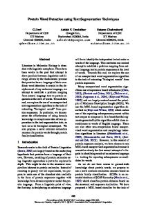
where the factor term δǫ allows us to restrict the shape prior within the region of interest, and φ˜ is the signed distance computed from the geodesic voting tree. The segmentation of vessels with this approach is achieved in two steps: (1) the geodesic voting tree is extracted using the original geodesic voting method (2) the walls of the vessels are extracted by minimization of the functional Eb . Figure 1 illustrates the segmentation process. Details about this methods are given in [13]. 4. RETINAL BLOOD VESSEL SEGMENTATION In this section, we will compare on the one hand the geodesic voting methods GVR (method presented in Section 3.1) and GVP (method presented in Section 3.2) for vessel segmentation from retinal images on the DRIVE data (Digital Retinal Images for Vessel Extraction) [4]. On the other hand we will compare GVR and GVP with other approaches used for vessels segmentation. The DRIVE data were acquired using a Canon CR5 non-mydriatic 3CCD camera with a 45 degree field of view (FOV). Each image was captured using 8 bits per color plane at 768 by 584 pixels. The FOV of each image is circular with a diameter of approximately 540 pixels. For this database, the images have been cropped around the FOV. The DRIVE data is composed of 40 images for which manual segmentations are also provided. Considering the complexity of the retinal images and the properties of our algorithm, we have cropped twelve different images from the 40 images available and evaluated our method on them.
For the GVP method, we have used the following potential P (x) = I(x)3 to run the geodesic voting segmentation, where I is the grayscale intensity of the image. For the GVR method, the augmented potential P˜ used is described in Section 3.1. In table 1, we compare the GVR and GVP results for vessel segmentation on the DRIVE database in terms of the following evaluation measures: Dice, specificity, and sensitivity. We found that the GVR and GVP gave similar results. In the following, we compare the performance of GVP method with the edge and region based level set methods and the fuzzy connectedness method in the segmentation of vessels. Figure 1 shows the segmentation result obtained with GVP. The shape prior allows us to constrain the propagation inside the tubular tree. Figure 2 (first row, right) shows that the propagation without shape constraints (γ = 0 in the Equation (5)) can leak outside of the tree structure. Figure 2-(second row,left)- shows the results obtained with the fuzzy connectedness method. The segmentation of the tree is obtained by thresholding the fuzzy connectedness map. For a small threshold the method does not allow to extract all the branches of the tree, and when the threshold is increased the propagation leaks outside of the tree. The same problems were observed with the edge based level set method when we increased the number of iterations, see Figure 2(second row, left). Figure 2 compares the results obtained with all these approaches. The GVP method gives the best results: it succeed in segmenting more tree branches without leaking outside of the tree structures.
Fig. 1. Segmentation of retinal blood vessels with the geodesic voting method presented in section 3.2. Left panel: in red the voting tree on the image is shown; center panel: the voting tree after thresholding (red); right panel: segmentation result obtained with the geodesic voting segmentation (red).
[3] Youssef Rouchdy and Laurent D. Cohen, “Image segmentation by geodesic voting. application to the extraction of tree structures from confocal microscope images,” in The 19th International Conference on Pattern Recognition, Tampa, Florida, 2008, pp. 1–5. [4] Joes Staal, Michael D. Abramoff, Meindert Niemeijer, and Max A. Viergeverand Bram van Ginneken, “Ridge based vessel segmentation in color images of the retina,” IEEE Trans. Med. Imaging, vol. 23, no. 4, pp. 501–509, 2004. [5] Koichiro Akita and Hideki Kuga, “A computer method of understanding ocular fundus images,” Pattern Recognition, vol. 15, no. 6, pp. 431 – 443, 1982. [6] Elena M. Martinez-Perez, Alun D. Hughes, Simon A. Thom, Anil A. Bharath, and Kim H. Parker, “Segmentation of blood vessels from red-free and fluorescein retinal images,” Medical Image Analysis, vol. 11, no. 1, pp. 47 – 61, 2007.
Fig. 2. Comparison of the geodesic voting approach with other methods. First row, left panel: shows in red the segmentation obtained with the edge based level set method; the right panel shows in red the segmentation results obtained with a Chan and Vese method without using the geodesic voting prior. Second row, the left panel shows the fuzzy connectedness segmentation; the right panel shows the segmentation result obtained with our approach. 5. CONCLUSION AND FUTURE WORK In this paper we have presented a new method for the segmentation retinal blood vessels. These methods are adapted to segment automatically the centerline and the walls of a tree from a single point given by the user, no a priori information. In contrast, the methods present in the literature for the segmentation of vessels are not fully automatic and require prior information about the structure to be segmented. We have applied our approach to segment retinal blood vessels from 12 images. The results are satisfying in terms of the statistics: Dice measure, sensitivity, specificity. 6. REFERENCES [1] Tien Yin Wong and Rachel McIntosh, “Hypertensive retinopathy signs as risk indicators of cardiovascular morbidity and mortality,” British Medical Bulletin, vol. 73-74, no. 1, pp. 57–70, 2005. [2] F Skovborg, A V Nielsen, E Lauritzen, and O Hartkopp, “Diameters of the retinal vessels in diabetic and normal subjects.,” Diabetes, vol. 18, no. 5, pp. 292–298, 1969.
[7] Cemil Kirbas and Francis Quek, “A review of vessel extraction techniques and algorithms,” ACM Comput. Surv., vol. 36, no. 2, pp. 81–121, 2004. [8] David Lesage, Elsa D. Angelini, Isabelle Bloch, and Gareth Funka-Lea, “A review of 3D vessel lumen segmentation techniques: Models, features and extraction schemes,” Medical image analysis, vol. 13, no. 6, pp. 819–845, 2009. [9] Thomas Deschamps and Laurent D. Cohen, “Fast extraction of minimal paths in 3D images and applications to virtual endoscopy,” Medical Image Analysis, vol. 5, no. 4, pp. 281 – 299, 2001. [10] Laurent D. Cohen and Ron Kimmel, “Global minimum for active contour models: A minimal path approach,” International Journal of Computer Vision, vol. 24, no. 1, pp. 57–78, 1997. [11] Youssef Rouchdy and Laurent D. Cohen, “A geodesic voting method for the segmentation of tubular tree and centerlines,” in Eigth IEEE International Symposium on Biomedical Imaging (ISBI’11), Chicago, Illinois, USA, 2011, pp. 979–983. [12] Hua Li and Anthony Yezzi, “Vessels as 4D curves: Global minimal 4D paths to extract 3D tubular surfaces and centerlines,” IEEE Transactions on Medical Imaging, vol. 26, pp. 1213–1223, 2007. [13] Youssef Rouchdy and Laurent D.Cohen, “A geodesic voting shape prior to constrain the level set evolution for the segmentation of tubular trees.,” in Third International Conference on Scale Space and Variational Methods in Computer Vision (SSVM), Ein-Gedi, Israel, 2011, pp. 1–12.