The Journal of Immunology
Requirements for Apoptotic Cell Contact in Regulation of Macrophage Responses1 Mark Lucas,2* Lynda M. Stuart,2* Ailiang Zhang,* Kairbaan Hodivala-Dilke,† Maria Febbraio,‡ Roy Silverstein,‡ John Savill,* and Adam Lacy-Hulbert3* An important consequence of macrophage engulfment of apoptotic cells is suppression of inflammatory responses, which was first defined by assay of TNF-␣ release stimulated by LPS. These effects are apparently mediated in part by paracrine effects of TGF- released by the subset of stimulated macrophages that ingest apoptotic cells, which suppresses neighboring cells. However, the apoptotic cell-derived signal that stimulates TGF- release, and the nature of any additional signals required for the antiinflammatory response remain poorly defined. In this study, we investigate the requirements for apoptotic cell engagement of macrophage surface receptors in these responses. We show that the apoptotic cell receptors CD36 and ␣v3 contribute to apoptotic cell phagocytosis by mouse macrophages, but are not essential for anti-inflammatory responses, suggesting that the mechanisms of response and phagocytosis are separate. In further defining requirements for response, we confirm the importance of TGF- in suppression by apoptotic cells, and identify an additional level of control of these effects. We show that LPS-stimulated mouse macrophage TNF-␣ release is only suppressed if macrophages have first contacted apoptotic cells, and hence, bystander macrophages are refractory to TGF- released by phagocytosing macrophages. We conclude that the profound suppression of LPSdriven TNF-␣ release by macrophage populations requires hitherto obscure contact-dependent licensing of macrophage responsiveness to TGF- by apoptotic cells. The Journal of Immunology, 2006, 177: 4047– 4054.
I
nflammation can be initiated by a variety of insults, including infection, trauma, and autoimmune mediated damage. Tissue macrophages, along with mast cells, are often the first components of the immune system to encounter local injury (1). Through their ability to recognize and phagocytose pathogens and dying cells, macrophages produce chemokines that recruit other inflammatory cells and secrete cytokines that condition both the local and systemic environments, orchestrating the outcome of an inflammatory process. Importantly, activated macrophages can adopt distinct states: either classic inflammatory cells able to recruit and activate bystander lymphocytes and hence amplify immune responses, or the less well-defined alternatively activated macrophage that favors tissue repair and regeneration (2). Important determinants of the macrophage phenotype are the cells and pathogens that they encounter at the outset of inflammation, and these interactions are able to program the subsequent macrophage response. Interestingly, encounter with cells dying by programmed cell death, such as apoptotic cells found during the resolution of inflammation or in remodeling tissue, has been shown to cause mac*Medical Research Council/University of Edinburgh Centre for Inflammation Research, Queen’s Medical Research Institute, Edinburgh, United Kingdom; †Cell Adhesion and Disease Laboratory, Cancer Research U.K., London, United Kingdom; and ‡Department of Cell Biology, Lerner Research Institute, Cleveland Clinic Foundation, Cleveland, OH 44195 Received for publication February 22, 2006. Accepted for publication June 27, 2006. The costs of publication of this article were defrayed in part by the payment of page charges. This article must therefore be hereby marked advertisement in accordance with 18 U.S.C. Section 1734 solely to indicate this fact. 1 This work was funded by Wellcome Trust Program Grant 064487 (to J.S.). L.M.S. was funded by Wellcome Trust Clinical Training Fellowship 056647 and Wellcome Trust Clinician Scientist Fellowship 36731. A.L.-H. is funded by a United Kingdom Research Council Fellowship. 2
rophages to adopt an alternative anti-inflammatory phenotype (3– 5). Numerous experimental systems have demonstrated that professional phagocytes such as macrophages, dendritic cells, and monocytes respond to encounter with apoptotic cells by increased production of anti-inflammatory cytokines (notably TGF- and IL10) and decreased proinflammatory cytokine response to subsequent microbial stimuli (3, 6 – 8). The mechanism of this antiinflammatory response has not been fully elucidated, but appears to require recognition of modified components of the apoptotic cell surface, such as exposed lipids or oxidized moieties (9). Several researchers have highlighted the importance of one such component, the anionic lipid phosphatidylserine (PS),4 in such responses (10, 11). Notably, PS-containing vesicles can recapitulate antiinflammatory signaling in vitro and promote resolution in vivo (4), and blockade of PS recognition inhibits inflammation resolution and shifts the balance of immune responses from tolerance to immunity (12, 13). Recognition of PS appears to occur via soluble bridging molecules, including MFG-E8 and Del1, which bind PS and engage ␣v integrins (14, 15), and Gas6, which engages the Mer tyrosine kinase (16), and both receptors have been implicated in anti-inflammatory responses. Other apoptotic cell surface moieties also promote anti-inflammatory signals, again often through soluble proteins such as thrombospondin (TSP), which engages ␣v integrins and CD36. Apoptotic cell-stimulated anti-inflammatory responses have been suggested to be an important checkpoint favoring resolution of inflammation and promoting immune tolerance (5). Furthermore, mice lacking certain apoptotic cell receptors (17) or soluble apoptotic cell-binding molecules (18, 19) show increased numbers of uncleared apoptotic cells and autoimmunity, prompting the assertion that failed apoptotic cell phagocytosis results in autoimmunity. It has been proposed that this is due to the subsequent accumulation of apoptotic cell-derived autoantigens
M.L. and L.M.S. contributed equally to this work.
3
Address correspondence and reprint requests to Dr. Adam Lacy-Hulbert, Center for Cancer Research, Massachusetts Institute of Technology, 40 Ames Street E17-227, Cambridge, MA 02139. E-mail address:
[email protected] Copyright © 2006 by The American Association of Immunologists, Inc.
4
Abbreviations used in this paper: PS, phosphatidylserine; TSP, thrombospondin. 0022-1767/06/$02.00
4048
APOPTOTIC CELL CONTACT AND MACROPHAGE RESPONSES
and necrotic cells increasing the risk of immunogenic self Ag presentation. However, it is also possible that loss of antiinflammatory responses to apoptotic cells leads to a failure to induce tolerance to phagocytosed apoptotic material. Necrotic cells are phagocytosed by similar mechanisms to apoptotic cells (20) and can induce similar anti-inflammatory responses (20, 21). Furthermore, recent investigations have identified knockout mice in which apoptotic cell phagocytosis is impaired and apoptotic cells accumulate without apparent inflammation or autoimmunity (22, 23), raising the possibility that phagocytosis and response are separable. In this respect, the failure to respond to apoptotic cells in a nonphlogistic manner may be more detrimental than the persistence of uncleared apoptotic cells. In this study, we investigate the mechanism for the antiinflammatory response of macrophages to apoptotic cells and focus on the early events that occur upon cell contact. By using macrophages from mice deficient for apoptotic cell receptors, we demonstrate that these molecules have important roles in phagocytosis, but are redundant for anti-inflammatory signaling. We show that TGF- release by macrophages following apoptotic cell uptake is insufficient to down-regulate cytokine responses, and that additional signals provided by apoptotic cell contact are required to license subsequent anti-inflammatory responses. Furthermore, we demonstrate that because of the requirement for contact, bystander macrophages or macrophages added later to the inflammatory conditions respond normally with production of TNF-␣ despite the presence of TGF- in the medium. These observations suggest a two-step model for response analogous to the tether and tickle model for apoptotic cell engulfment (24, 25). We propose a model in which cells must first bind phagocytes using one or more of many partially redundant receptors, which 1) stimulate phagocytosis; 2) promote release of TGF-; and 3) license the cell for response to TGF-. Importantly, all aspects of this “kiss and tell” model are required for efficient anti-inflammatory signaling to occur. Taken together, these data suggest that apoptotic cell phagocytosis and response use distinct receptor pathways, confirm an important role for TGF- in responses, and highlight an important cell surface interaction that is required for anti-inflammatory responses to occur and that therefore limits their extent.
Materials and Methods Mice BALB/c and C57BL/6 mice were purchased from B & K Universal, and mice were maintained in specific pathogen-free conditions at the University of Edinburgh. TSP-1⫺/⫺ and control mice were also obtained from Beth Israel Deaconess Medical School. For experiments using knockout mice (26 –29), controls were matched for age, sex, and genetic background, and housed in the same facility (CD36⫺/⫺ and TSP-1⫺/⫺) or were littermates (␣v3⫺/⫺, ␣v5⫺/⫺). Bone marrow-derived macrophages were prepared from 8- to 12-wk-old mice. Housing and animal procedures were approved by the appropriate local authority.
Macrophage culture Macrophages were cultured, as described previously (30 –32). Femurs were removed and cleaned, and bone marrow was flushed through and plated in DMEM supplemented with 2 mM L-glutamine, 100 U/ml penicillin, 100 g/ml streptomycin, 10% heat-inactivated FBS, and 10% conditioned supernatant from L929 cells, which was changed on day 2 and every 3 days subsequently. Macrophages were used between day 7 and 10 of culture. For TSP-1⫺/⫺ and relevant control macrophages, bone marrow cells were grown in macrophage serum-free medium (Invitrogen Life Technologies) supplemented with 2 mM L-glutamine, 100 U/ml penicillin, 100 g/ml streptomycin, and 10 ng/ml rM-CSF (R&D Systems).
Generation of apoptotic cells Human neutrophils were extracted from peripheral blood of healthy volunteers, as described previously (33). Blood was separated using dextran
sedimentation and a Percoll gradient. This yielded highly pure human neutrophils (⬎95%), which were allowed to undergo constitutive apoptosis by aging overnight in Iscove’s modified DMEM with 100 U/ml penicillin, 100 g/ml streptomycin, and 10% autologous platelet-rich plasma-derived serum. After this period, the cells were routinely 60 – 80% apoptotic, assessed by cytospin morphology. No significant necrosis was detected, as assessed by trypan blue exclusion (⬍1% positive). Aged thymocytes were used as an alternative source of apoptotic cells in experiments using TSP-1⫺/⫺ mice to avoid any potential TSP contamination and other response experiments where stated. The mouse thymus was removed and disaggregated in RPMI 1640 supplemented with 2 mM L-glutamine, 100 U/ml penicillin, and 100 g/ml streptomycin, to yield a single-cell suspension. Thymocytes were aged overnight at 2 ⫻ 106/ml, which yielded a largely annexin Vpositive (⬎60%), trypan blue-negative (⬍1%) cell population.
Apoptotic cell phagocytosis Macrophages in 24-well culture plates were washed twice with PBS and cultured with fluorescent green-labeled (8) apoptotic cells (2.5 ⫻ 106 cells/ well, except where stated otherwise; corresponds to a 1:5 ratio of macrophages to apoptotic cells) in serum-free DMEM for between 30 min and 2 h (as stated in figure legends). Plates were washed three to five times with ice-cold PBS to remove noningested cells, as verified by microscopy, and fixed with 4% paraformaldehyde for 20 min. Phagocytosis was measured by comparison of phase-contrast and fluorescent microscopy to show all cells and apoptotic cells, respectively. Fluorescent apoptotic cells completely encircled by nonfluorescent macrophages were considered phagocytosed. Phagocytosis was expressed as either the percentage of macrophages that had internalized apoptotic cells or the phagocytic index, calculated as the percentage of phagocytic macrophages multiplied by the average number of apoptotic cells ingested by phagocytic macrophages.
Macrophage cytokine production For cytokine production experiments, macrophages were cultured with combinations of apoptotic cells and LPS (500 ng/ml; Escherichia coli serotype 026:B6; Sigma-Aldrich). Exact conditions for each experiment are given in figure legends. Supernatants were harvested 24 h after LPS addition, cleared by centrifugation, and frozen at ⫺70°C until cytokine analysis was performed by ELISA (using duoset kits; R&D Systems). To measure the effects of TGF- on cytokine expression, macrophages were stimulated with both LPS and rTGF-1 (0 –1000 pg/ml; R&D Systems) for 24 h, and supernatants were harvested. To block phagocytosis, macrophages were incubated with 10 g/ml colchicine (Calbiochem) for 30 min, washed, and incubated with apoptotic cells/LPS, as above. Macrophage-macrophage coculture experiments are described in detail in figure legends, and used 0.3-m Transwell inserts (Costar plastics; Corning Life Sciences) to separate cell cultures.
Results Role of phagocytic receptors in responses to apoptotic cells The ability of apoptotic cells to inhibit macrophages has been demonstrated in numerous previous reports (3, 30), and is thought to be triggered by the same receptors that mediate binding and/or engulfment. Hence, macrophages ingest apoptotic cells through recognition of TSP bound to the apoptotic cell surface by the scavenger receptor CD36 and integrin ␣v3 and Abs to both CD36 and ␣v3 stimulate anti-inflammatory responses in monocytes or macrophages, similar to those triggered by apoptotic cells (6, 34). To define whether these molecules were required for phagocytosis and/or responses to apoptotic cells, we studied macrophages derived from CD36, TSP-1, and integrin 3/5 knockout mice. Macrophages lacking either ␣v3 or CD36 showed significantly reduced apoptotic cell phagocytosis (Fig. 1), with the proportion of phagocytic macrophages and the phagocytic index both reduced to ⬃60% of control levels (data for phagocytic index only shown). Macrophages lacking TSP-1 showed a small reduction in phagocytosis (80% of control) that was not statistically different from controls, perhaps reflecting overlapping functions of additional apoptotic cell opsonins, such as MFG-E8 (14), or some subtle effect of macrophage preparation in the absence of serum.
The Journal of Immunology
4049
FIGURE 1. Macrophage reprogramming is independent of phagocytic receptors. A, Mouse bone marrow-derived macrophages from control mice (䡺) or indicated knockout mice (u) were incubated with apoptotic cells, and phagocytosis was measured. Phagocytic indices were calculated and normalized to phagocytosis by macrophages from appropriate control mice. Data are expressed as mean relative phagocytic index ⫾ SEM for at least three independent cultures in each case. ⴱ, Significant difference from controls; p ⬍ 0.05, Student’s t test. NS, Not significantly different from control. B, Macrophages from indicated knockout and control mice were stimulated with LPS (u), or with apoptotic cells for 2 h, then with LPS (f) or unstimulated (䡺), and TNF-␣ and TGF- production was measured after 24 h. Data shown are means ⫾ SEM for three wells of macrophages (mean of two wells for TSP/TGF- results), and similar results were seen in three (CD36, integrin ␣v3/␣v5) or two (TSP) experiments. ⴱ, Significant difference between LPS-stimulated and LPS ⫹ apoptotic human neutrophil (ac)-stimulated macrophages; p ⬍ 0.002, Student’s t test.
Knockout macrophages were then stimulated with combinations of apoptotic cells and LPS to assess the contribution of these receptors on macrophage response. Interestingly, lack of CD36, TSP-1, or integrin 5 resulted in reduced (CD36) or increased (5, TSP-1) responses of macrophages to LPS, suggesting that these molecules may act as coreceptors or modulators of the LPS response of macrophages. Furthermore, macrophages from the TSP⫺/⫺ experiment, which were cultured under serum-free conditions in the presence of M-CSF, showed 10-fold lower TNF-␣ production than macrophages cultured in serum-replete conditions, probably due to lack of serum LPS-binding proteins. However, despite these differences, apoptotic cells efficiently inhibited LPS
responses in both control and knockout macrophages, and loss of any single molecule did not interfere with responses to apoptotic cells, despite previous observations that receptor ligation can recapitulate apoptotic cell binding (6, 34). These results show that CD36 and ␣v3 play important nonredundant roles in apoptotic cell phagocytosis, such that loss of either molecule reduced the efficiency of uptake by nearly 40%. The anti-inflammatory response, however, was more robust, appearing normal in the absence of CD36 or ␣v3. This suggested either that apoptotic cell response was not signaled through these molecules, in contrast to experiments with cross-linking Abs, or that responses were signaled through a redundant array of multiple receptors. Additionally, these
4050
APOPTOTIC CELL CONTACT AND MACROPHAGE RESPONSES
experiments showed that the anti-inflammatory response occurred normally under conditions in which phagocytosis was significantly inhibited. We decided therefore to investigate further the relationship between phagocytosis and response. Apparent paracrine inhibition of macrophages in apoptotic cell macrophage cultures Interestingly, in many reports of anti-inflammatory effects of apoptotic cells, 100% inhibition of proinflammatory cytokine production is seen in cultures in which only ⬃30% of macrophages have engulfed apoptotic cells. This is consistent with the observation that macrophages that internalize apoptotic cells secrete antiinflammatory mediators, such as TGF-, that suppress bystander macrophage inflammatory responses. To determine the limits of these responses and to define the degree of inhibition of phagocytosis that would be required to inhibit response, we titrated apoptotic cells into LPS-stimulated macrophage cultures and measured TNF-␣ production. Addition of apoptotic cells at a ratio of 1:1 with macrophages resulted in greater than 60% inhibition of TNF-␣, despite only 25% of macrophages demonstrating internalized dying cells (Fig. 2). Furthermore, we saw greater than 80% inhibition of TNF-␣ at higher apoptotic cell:macrophage ratios, although seldom was phagocytosis greater than 50 – 60%. Thus, low numbers of phagocytosing macrophages were sufficient to inhibit TNF-␣ production significantly, supporting a paracrine effector-mediated anti-inflammatory response. In our earlier
FIGURE 2. Dose response of apoptotic cells on macrophage cytokine production. Mouse bone marrow-derived macrophages were incubated with increasing numbers of apoptotic human neutrophils (0.5– 4 ⫻ 106 apoptotic cells/well of 0.5 ⫻ 106 macrophages). After 2 h, uningested apoptotic cells were removed by gentle washing with PBS, and response to LPS (A) or phagocytosis (B) was measured. Data shown are means ⫾ SEM for three wells of macrophages. ⴱ, Significant difference between LPSstimulated and LPS ⫹ apoptotic cell-stimulated macrophages; p ⬍ 0.002, Student’s t test.
work in this culture system, blockade of TGF- using either a TGF--soluble receptor or neutralizing Abs greatly reduced the inhibition of macrophages (8), confirming the role of this cytokine in the paracrine anti-inflammatory response, as reported by others (3). TGF- is insufficient to recapitulate fully the effects of apoptotic cells The correlation between TGF- production and inhibition of bystander cells suggested that we should determine the requirements for TGF- for inhibition. We first explored whether TGF- was sufficient to inhibit TNF-␣ production in mouse bone marrowderived macrophages. Interestingly, LPS-induced TNF-␣ production was inhibited only if rTGF- was added at levels significantly higher than those detected endogenously following apoptotic cell uptake (250 –1000 pg/ml vs ⬃100 pg/ml; Fig. 3). Furthermore, unlike apoptotic cells, which routinely reduced TNF-␣ by 90% or more, rTGF- alone never reduced TNF-␣ production as efficiently. Taken together, these data demonstrate that soluble rTGF- is inadequate to inhibit macrophages fully and was ⬃10-fold less efficient than TGF- in the context of apoptotic cells. Paracrine anti-inflammatory mediators are insufficient to inhibit bystander macrophage responses It is possible that TGF- alone is less efficient than apoptotic cells in generating anti-inflammatory responses, because other important mediators such as PGE2 and IL-10 are also released in response to apoptotic cells and these could act in concert with TGF- to reduce the threshold of TGF- response (3, 6). We therefore asked whether medium conditioned by macrophages ingesting apoptotic cells would be as efficient as apoptotic cells at inhibiting nonphagocytosing bystander cells. First, we separated two populations of macrophages by a Transwell filter. To one population of macrophages we added apoptotic cells and subsequently stimulated the cultures with LPS. Interestingly, TNF-␣ was still produced at high levels in these cocultures (Fig. 4A). Therefore, paracrine factors produced by macrophages ingesting apoptotic cells were unable to block LPS responses in macrophages separated across a Transwell system. These data did not exclude a role for certain paracrine factors that are too labile to act across a
FIGURE 3. Role of TGF- in macrophage reprogramming. Mouse bone marrow-derived macrophages were stimulated with LPS in the presence of increasing amounts of rTGF-1 (0 –1000 pg/ml). Culture supernatants were harvested 24 h after treatment, and TNF-␣ levels were determined by ELISA. Data shown are means ⫾ SEM for three wells of macrophages. ⴱ, Significant difference between LPS-stimulated and LPS ⫹ TGF--stimulated macrophages; p ⬍ 0.002, Student’s t test.
The Journal of Immunology
4051
FIGURE 4. Failure of macrophages to reprogram bystander macrophages. A, Mouse bone marrow macrophages were cultured in 24-well plates, with apoptotic human neutrophils (ac) and/or LPS (500 ng/ml) for 24 h, and TNF-␣ levels were measured. In addition, macrophages were added to Transwell chambers (Trans M) for the duration of the experiment. B, Mouse macrophages were stimulated with LPS alone, or incubated with apoptotic human neutrophils (ac) for 2 h, washed, and stimulated with LPS, as in previous experiments. Additional macrophages were also added, either after apoptotic cells were washed away (ac (2h) ⫹ LPS ⫹ M) or without washing of apoptotic cells (ac ⫹ LPS ⫹ M). TNF-␣ levels in culture supernatant were measured after 24 h. C, Mouse bone marrow macrophages were cultured with apoptotic mouse thymocytes and/or LPS, as in A. D, Mouse bone marrow-derived macrophages were cultured with apoptotic mouse thymocytes (ac), LPS, and additional macrophages, as in B. Data shown are means ⫾ SEM for three wells of macrophages. ⴱ, Significant difference between indicated populations; p ⬍ 0.01, Student’s t test. ⴱⴱ, Significant difference between LPS-stimulated and LPS ⫹ ac-stimulated macrophages; p ⬍ 0.002; Student’s t test.
Transwell, or signals that require cell-cell contact. Thus, to determine whether macrophage-macrophage contact or proximity was required for inhibition of LPS responses, we first treated macrophages with apoptotic cells for 2 h, washed them to remove all noningested apoptotic cells, and added a second population of macrophages, allowing contact between untreated and apoptotic cell-conditioned macrophages. Again, high levels of TNF-␣ were produced following LPS stimulation (Fig. 4B), militating against such labile paracrine mediators or macrophage:macrophage cell contact-dependent mechanisms. Improtantly, these effects were independent of the source of apoptotic cells, occurring with cells of human and mouse origin (Fig. 4, C and D).
macrophages exposure to cytokines and other molecules secreted by apoptotic cells, but not to contact the apoptotic cell surface. Under these conditions, inhibition was not seen (data not shown), confirming previous reports of a requirement for surface contact between monocytes or macrophages and apoptotic cells for inhibition of LPS response (7, 9). We then treated macrophages with colchicine, which reduced phagocytosis of apoptotic cells (to ⬍5% of control levels), but permitted binding. In the absence of phagocytosis, apoptotic cell-treated macrophages still suppressed LPSinduced TNF-␣, IL-12, and IL-10 release (only TNF-␣ data shown; Fig. 5).
Macrophage contact with apoptotic cells, but not phagocytosis, is necessary for inhibition of cytokine production The surprising observation that medium conditioned by macrophages interacting with apoptotic cells did not inhibit proinflammatory responses in a culture of macrophages that had not contacted apoptotic cells directly suggested that maximal inhibition required both contact between all macrophages and apoptotic cells, and TGF-, and led us to question whether paracrine effects indeed occurred. Microscopic examination of cultures of macrophages to which apoptotic cells were added at low numbers (0.5 ⫻ 106 cells/ well) demonstrated that they tended to cluster in distinct areas of the well, but at higher numbers (⬎1 ⫻ 106 cells/well) they formed a monolayer over the macrophages. These observations indicated that in our in vitro cultures, although fewer than half of the macrophages engulf dying cells, all macrophages will contact apoptotic cells. Thus, contact (but not phagocytosis) correlates well with the extent of TNF-␣ inhibition, and in this regard it may be that both macrophages behave in a cell-autonomous manner and that inhibition of bystander cells does not occur. To test this hypothesis, we first wished to confirm reports that contact alone was sufficient for inhibition (7). We separated macrophages from apoptotic cells across a Transwell membrane, which allowed
FIGURE 5. Requirement for contact in macrophage reprogramming. Mouse bone marrow-derived macrophages were treated with indicated combinations of apoptotic human neutrophils (4 ⫻ 106 cells/well), LPS, and colchicine, and supernatants were harvested after 24 h. TNF-␣ levels were determined by ELISA. Data shown are means ⫾ SEM for three wells of macrophages. ⴱ, Significant difference between LPS-stimulated and LPS ⫹ apoptotic human neutrophil (ac)-stimulated macrophages; p ⬍ 0.002, Student’s t test.
4052
APOPTOTIC CELL CONTACT AND MACROPHAGE RESPONSES
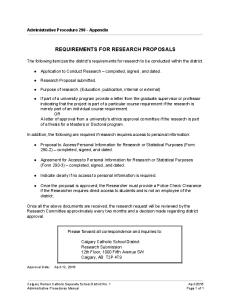
FIGURE 6. Lack of correlation between TNF-␣ and TGF- production in reprogramming. Macrophages were incubated with increasing numbers of apoptotic human neutrophils (0.5– 4 ⫻ 106 apoptotic cells/well of 0.5 ⫻ 106 macrophages). After 2 h, uningested apoptotic cells were removed by gentle washing with PBS, and macrophages were stimulated with LPS. Culture supernatants were harvested 24 h after treatment, and TNF-␣ and TGF-1 levels were determined by ELISA. Data shown are means ⫾ SEM for three wells of macrophages. ⴱ, Significant difference between LPSstimulated and LPS ⫹ apoptotic human neutrophil (ac)-stimulated macrophages; p ⬍ 0.002, Student’s t test.
Failure of correlation of TGF- production, TNF-␣ inhibition, and phagocytosis The observations above suggested that maximal inhibition of LPSstimulated macrophage cytokine responses by apoptotic cells requires both paracrine effects of TGF- and contact of all cytokineproducing macrophages with apoptotic cells. Furthermore, the data suggest that these two mechanisms must cooperate for TNF-␣ inhibition. Therefore, to further determine the contributions of TGF- and contact with apoptotic cells, we measured TGF- production in cultures of macrophages with increasing numbers of apoptotic cells (Fig. 6). The production of TGF- did not correlate directly with the inhibition of TNF-␣ production; thus, although the levels of TGF- produced reached a plateau of 90 pg/ml when the lowest ratio of apoptotic cells to macrophages was used, the production of TNF-␣ was not significantly inhibited until higher numbers of apoptotic cells were added to macrophage cultures (2– 4 ⫻ 106 cells/well). These data confirm that macrophage-produced TGF- does not account for all of the inhibitory effects of apoptotic cell phagocytosis and that additional signals from contact with apoptotic cells must synergize with TGF- for efficient inhibition of cytokine production.
Discussion Apoptotic cells can be efficiently removed in large numbers both constitutively and following inflammation. The potential for the release of cell contents or for presentation of self Ags makes it clear that this process must be tightly regulated. In this study, we show that suppression of LPS-driven macrophage inflammatory responses by apoptotic cells requires two signals, the first being contact with the dying cell and the second being production and response to TGF-. We would suggest a model for response analogous to the “tether and tickle” model for phagocytosis (25, 35), in which it is proposed that for engulfment by macrophages to proceed apoptotic cells must first bind multiple macrophage tether receptors, with phagocytosis then being stimulated by engagement of PS receptors and other macrophage tickle receptors. Thus, we propose that for response, a similar two-step process is required;
initial signaling events that occur at the point of macrophage contact with the apoptotic cell (kiss) are required for subsequent responsiveness to TGF-. Apoptotic cell binding to tell receptors then stimulates release of TGF- and completes the antiinflammatory signal. The molecules that stimulate TGF- release or responsiveness may of course include the proposed tickle receptors, several of which have been suggested to stimulate TGF- release from macrophages. To study this further, we have used cells from mice deficient in receptors implicated in both apoptotic cell clearance and TGF- activation, namely CD36, ␣v3, ␣v5, or TSP-1. The data presented demonstrate that although CD36 and ␣v3 integrins contribute to phagocytosis, these receptors are redundant at the level of response. Thus, tether/kiss molecules may be partially redundant in vivo, as seen for scavenger receptor A (36) or opsonins such as C1q and other collectins (19, 37), but the processes of engulfment and response may be nonredundant with profound consequences in their absence (17, 38, 39). Numerous reports have demonstrated that internalization of apoptotic cells by macrophages inhibits later responses to proinflammatory factors such as LPS, switching off production of TNF-␣, and stimulating production of TGF- (3, 30, 40). In previous studies using this cell system, we have confirmed these reports of a requirement for TGF- signaling in anti-inflammatory responses (8, 30). However, our observation that simple addition of TGF- to macrophages was significantly less effective at switching off macrophage response suggested that additional signals were required. Additionally, macrophages added back to apoptotic cellprogrammed macrophages continue to respond to LPS by production of TNF-␣, demonstrating conclusively that there is no paracrine effect on bystander macrophages. Our data suggest instead that macrophages respond in a cell-autonomous manner, and that an essential signal allowing response to TGF- originates upon apoptotic cell binding to the surface of the phagocyte. TGF- requires activation before it can signal, a process that involves proteolysis, dissociation of associated proteins, and conformational changes. In vivo these processes are catalyzed by proteases such as plasmin, and importantly, occur on the cell surface where TGF--binding molecules associate with proteases. Therefore, to further understand the relation between TGF- and the requirement for cell contact, we focused on events that we believe must originate at the cell surface. Three lines of evidence suggested a possible role for ␣v3/5, CD36, and TSP in such cell surface events: first, they have been implicated in both dendritic cell and macrophage phagocytosis (41– 45); second, production of TGF- and inhibition of macrophage activation have been recapitulated by ligation of these receptors (6, 34); and third, ␣v integrins and TSP have been shown to bind to and/or activate TGF- (46 – 49). Interestingly, in using these knockout cells, we have shown that these receptors are redundant for response in this in vitro system, despite involvement in phagocytosis. It is tempting to suggest that the ability to recruit or activate TGF- is also redundant such that a deficiency in any single apoptotic cell receptor is not sufficient to result in an absence of response to TGF-. If this latter statement is true, it will require generation of compound knockouts to impair the anti-inflammatory response and/or phagocytosis of apoptotic cells. Apoptosis is often associated with inflammation and infection (50 –52), and the avoidance of unnecessary inflammatory responses to dying cells must be weighed against the need to maintain an appropriate response to pathogens. However, the reported strong suppressive effects of apoptotic cells on macrophage inflammatory cytokine production raise the question of how such a balance is maintained. Our previous work has demonstrated that
The Journal of Immunology macrophages that internalize apoptotic cells continue to release macrophage chemokines MIP-1␣ and MIP-2, suggesting that phagocyte clearance of apoptotic cells need not inhibit recruitment of phagocytes to areas of high cell death (30). Our current observations add a further mechanism for preservation of beneficial inflammatory responses at infected sites, in that macrophages engaging apoptotic cells can suppress their own inflammatory responses, but will nevertheless allow newly recruited macrophages to function autonomously, responding normally to pathogens or proinflammatory stimuli. Thus, macrophages that have internalized apoptotic cells can coexist alongside macrophages that are still able to respond to LPS and invading pathogens. In summary, our experiments demonstrate that antiinflammatory responses of mouse macrophages to apoptotic cells occur independently of the phagocytic mediators ␣v3, CD36, and TSP-1. We confirm that apoptotic cell contact, but not phagocytosis, is required for these responses, and identify a further requirement for apoptotic cell contact in response to secreted TGF-. We propose that apoptotic cell responses are triggered by receptors that are distinct from those used for phagocytosis and occur through a two-step process that limits the extent of their action, effectively reducing immune response to dying self while maintaining host defense.
Acknowledgments We thank Michael Clay for expert technical assistance, and Jack Lawler, Dean Sheppard, and Richard Hynes for provision of knockout mice.
Disclosures The authors have no financial conflict of interest.
References 1. Nathan, C. 2002. Points of control in inflammation. Nature 420: 846 – 852. 2. Gordon, S. 2003. Alternative activation of macrophages. Nat. Rev. Immunol. 3: 23–35. 3. Fadok, V. A., D. L. Bratton, A. Konowal, P. W. Freed, J. Y. Westcott, and P. M. Henson. 1998. Macrophages that have ingested apoptotic cells in vitro inhibit proinflammatory cytokine production through autocrine/paracrine mechanisms involving TGF-, PGE2, and PAF. J. Clin. Invest. 101: 890 – 898. 4. Huynh, M.-L. N., V. A. Fadok, and P. M. Henson. 2002. Phosphatidylserinedependent ingestion of apoptotic cells promotes TGF-1 secretion and the resolution of inflammation. J. Clin. Invest. 109: 41–50. 5. Savill, J., I. Dransfield, C. Gregory, and C. Haslett. 2002. A blast from the past: clearance of apoptotic cells regulates immune responses. Nat. Rev. Immunol. 2: 965–975. 6. Voll, R. E., M. Herrmann, E. A. Roth, C. Stach, J. R. Kalden, and I. Girkontaite. 1997. Immunosuppressive effects of apoptotic cells. Nature 390: 350 –351. 7. Byrne, A., and D. J. Reen. 2002. Lipopolysaccharide induces rapid production of IL-10 by monocytes in the presence of apoptotic neutrophils. J. Immunol. 168: 1968 –1977. 8. Stuart, L. M., M. Lucas, C. Simpson, J. Lamb, J. Savill, and A. Lacy-Hulbert. 2002. Inhibitory effects of apoptotic cell ingestion upon endotoxin-driven myeloid dendritic cell maturation. J. Immunol. 168: 1627–1635. 9. Cvetanovic, M., and D. S. Ucker. 2004. Innate immune discrimination of apoptotic cells: repression of proinflammatory macrophage transcription is coupled directly to specific recognition. J. Immunol. 172: 880 – 889. 10. Gardai, S. J., D. L. Bratton, C. A. Ogden, and P. M. Henson. 2006. Recognition ligands on apoptotic cells: a perspective. J. Leukocyte Biol. 79: 896 –903. 11. Wu, Y., N. Tibrewal, and R. B. Birge. 2006. Phosphatidylserine recognition by phagocytes: a view to a kill. Trends Cell Biol. 16: 189 –197. 12. Asano, K., M. Miwa, K. Miwa, R. Hanayama, H. Nagase, S. Nagata, and M. Tanaka. 2004. Masking of phosphatidylserine inhibits apoptotic cell engulfment and induces autoantibody production in mice. J. Exp. Med. 200: 459 – 467. 13. Bondanza, A., V. S. Zimmermann, P. Rovere-Querini, J. Turnay, I. E. Dumitriu, C. M. Stach, R. E. Voll, U. S. Gaipl, W. Bertling, E. Poschl, et al. 2004. Inhibition of phosphatidylserine recognition heightens the immunogenicity of irradiated lymphoma cells in vivo. J. Exp. Med. 200: 1157–1165. 14. Hanayama, R., M. Tanaka, K. Miwa, A. Shinohara, A. Iwamatsu, and S. Nagata. 2002. Identification of a factor that links apoptotic cells to phagocytes. Nature 417: 182–187. 15. Hanayama, R., M. Tanaka, K. Miwa, and S. Nagata. 2004. Expression of developmental endothelial locus-1 in a subset of macrophages for engulfment of apoptotic cells. J. Immunol. 172: 3876 –3882. 16. Nakano, T., Y. Ishimoto, J. Kishino, M. Umeda, K. Inoue, K. Nagata, K. Ohashi, K. Mizuno, and H. Arita. 1997. Cell adhesion to phosphatidylserine mediated by a product of growth arrest-specific gene 6. J. Biol. Chem. 272: 29411–29414.
4053 17. Scott, R. S., E. J. McMahon, S. M. Pop, E. A. Reap, R. Caricchio, P. L. Cohen, H. S. Earp, and G. K. Matsushima. 2001. Phagocytosis and clearance of apoptotic cells is mediated by MER. Nature 411: 207–211. 18. Botto, M., C. Dell’Agnola, A. E. Bygrave, E. M. Thompson, H. T. Cook, F. Petry, M. Loos, P. P. Pandolfi, and M. J. Walport. 1998. Homozygous C1q deficiency causes glomerulonephritis associated with multiple apoptotic bodies. Nat. Genet. 19: 56 –59. 19. Taylor, P. R., A. Carugati, V. A. Fadok, H. T. Cook, M. Andrews, M. C. Carroll, J. S. Savill, P. M. Henson, M. Botto, and M. J. Walport. 2000. A hierarchical role for classical pathway complement proteins in the clearance of apoptotic cells in vivo. J. Exp. Med. 192: 359 –366. 20. Bottcher, A., U. S. Gaipl, B. G. Furnrohr, M. Herrmann, I. Girkontaite, J. R. Kalden, and R. E. Voll. 2006. Involvement of phosphatidylserine, ␣v3, CD14, CD36, and complement C1q in the phagocytosis of primary necrotic lymphocytes by macrophages. Arthritis Rheum. 54: 927–938. 21. Patel, V. A., A. Longacre, K. Hsiao, H. Fan, F. Meng, J. E. Mitchell, J. Rauch, D. S. Ucker, and J. S. Levine. 2006. Apoptotic cells, at all stages of the death process, trigger characteristic signaling events that are divergent from and dominant over those triggered by necrotic cells: implications for the delayed clearance model of autoimmunity. J. Biol. Chem. 281: 4663– 4670. 22. Stuart, L. M., K. Takahashi, L. Shi, J. Savill, and R. A. Ezekowitz. 2005. Mannose-binding lectin-deficient mice display defective apoptotic cell clearance but no autoimmune phenotype. J. Immunol. 174: 3220 –3226. 23. Devitt, A., K. G. Parker, C. A. Ogden, C. Oldreive, M. F. Clay, L. A. Melville, C. O. Bellamy, A. Lacy-Hulbert, S. C. Gangloff, S. M. Goyert, and C. D. Gregory. 2004. Persistence of apoptotic cells without autoimmune disease or inflammation in CD14⫺/⫺ mice. J. Cell Biol. 167: 1161–1170. 24. Ogden, C. A., A. deCathelineau, P. R. Hoffmann, D. Bratton, B. Ghebrehiwet, V. A. Fadok, and P. M. Henson. 2001. C1q and mannose binding lectin engagement of cell surface calreticulin and cd91 initiates macropinocytosis and uptake of apoptotic cells. J. Exp. Med. 194: 781–796. 25. Hoffmann, P. R., A. M. deCathelineau, C. A. Ogden, Y. Leverrier, D. L. Bratton, D. L. Daleke, A. J. Ridley, V. A. Fadok, and P. M. Henson. 2001. Phosphatidylserine (PS) induces PS receptor-mediated macropinocytosis and promotes clearance of apoptotic cells. J. Cell Biol. 155: 649 – 660. 26. Huang, X., M. Griffiths, J. Wu, R. V. Farese, Jr., and D. Sheppard. 2000. Normal development, wound healing, and adenovirus susceptibility in 5-deficient mice. Mol. Cell. Biol. 20: 755–759. 27. Hodivala-Dilke, K. M., K. P. McHugh, D. A. Tsakiris, H. Rayburn, D. Crowley, M. Ullman-Cullere, F. P. Ross, B. S. Coller, S. Teitelbaum, and R. O. Hynes. 1999. 3-integrin-deficient mice are a model for Glanzmann thrombasthenia showing placental defects and reduced survival. J. Clin. Invest. 103: 229 –238. 28. Febbraio, M., N. A. Abumrad, D. P. Hajjar, K. Sharma, W. Cheng, S. F. Pearce, and R. L. Silverstein. 1999. A null mutation in murine CD36 reveals an important role in fatty acid and lipoprotein metabolism. J. Biol. Chem. 274: 19055–19062. 29. Lawler, J., M. Sunday, V. Thibert, M. Duquette, E. L. George, H. Rayburn, and R. O. Hynes. 1998. Thrombospondin-1 is required for normal murine pulmonary homeostasis and its absence causes pneumonia. J. Clin. Invest. 101: 982–992. 30. Lucas, M., L. M. Stuart, J. Savill, and A. Lacy-Hulbert. 2003. Apoptotic cells and innate immune stimuli combine to regulate macrophage cytokine secretion. J. Immunol. 171: 2610 –2615. 31. Ren, Y., L. Stuart, F. P. Lindberg, A. R. Rosenkranz, Y. Chen, T. N. Mayadas, and J. Savill. 2001. Nonphlogistic clearance of late apoptotic neutrophils by macrophages: efficient phagocytosis independent of 2 integrins. J. Immunol. 166: 4743– 4750. 32. Johnson, C. R., D. Kitz, and J. R. Little. 1983. A method for the derivation and continuous propagation of cloned murine bone marrow macrophages. J. Immunol. Methods 65: 319 –332. 33. Savill, J. S., A. H. Wyllie, J. E. Henson, M. J. Walport, P. M. Henson, and C. Haslett. 1989. Macrophage phagocytosis of aging neutrophils in inflammation: programmed cell death in the neutrophil leads to its recognition by macrophages. J. Clin. Invest. 83: 865– 875. 34. Freire-de-Lima, C. G., D. O. Nascimento, M. B. Soares, P. T. Bozza, H. C. Castro-Faria-Neto, F. G. de Mello, G. A. DosReis, and M. F. Lopes. 2000. Uptake of apoptotic cells drives the growth of a pathogenic trypanosome in macrophages. Nature 403: 199 –203. 35. Henson, P. M., D. L. Bratton, and V. A. Fadok. 2001. The phosphatidylserine receptor: a crucial molecular switch? Nat. Rev. Mol. Cell Biol. 2: 627– 633. 36. Platt, N., H. Suzuki, T. Kodama, and S. Gordon. 2000. Apoptotic thymocyte clearance in scavenger receptor class A-deficient mice is apparently normal. J. Immunol. 164: 4861– 4867. 37. Vandivier, R. W., C. A. Ogden, V. A. Fadok, P. R. Hoffmann, K. K. Brown, M. Botto, M. J. Walport, J. H. Fisher, P. M. Henson, and K. E. Greene. 2002. Role of surfactant proteins A, D, and C1q in the clearance of apoptotic cells in vivo and in vitro: calreticulin and CD91 as a common collectin receptor complex. J. Immunol. 169: 3978 –3986. 38. Cohen, P. L., R. Caricchio, V. Abraham, T. D. Camenisch, J. C. Jennette, R. A. Roubey, H. S. Earp, G. Matsushima, and E. A. Reap. 2002. Delayed apoptotic cell clearance and lupus-like autoimmunity in mice lacking the c-mer membrane tyrosine kinase. J. Exp. Med. 196: 135–140. 39. Teder, P., R. W. Vandivier, D. Jiang, J. Liang, L. Cohn, E. Pure, P. M. Henson, and P. W. Noble. 2002. Resolution of lung inflammation by CD44. Science 296: 155–158. 40. McDonald, P. P., V. A. Fadok, D. Bratton, and P. M. Henson. 1999. Transcriptional and translational regulation of inflammatory mediator production by endogenous TGF- in macrophages that have ingested apoptotic cells. J. Immunol. 163: 6164 – 6172.
4054
APOPTOTIC CELL CONTACT AND MACROPHAGE RESPONSES
41. Savill, J., I. Dransfield, N. Hogg, and C. Haslett. 1990. Vitronectin receptormediated phagocytosis of cells undergoing apoptosis. Nature 343: 170 –173. 42. Savill, J., N. Hogg, Y. Ren, and C. Haslett. 1992. Thrombospondin cooperates with CD36 and the vitronectin receptor in macrophage recognition of neutrophils undergoing apoptosis. J. Clin. Invest. 90: 1513–1522. 43. Albert, M. L., B. Sauter, and N. Bhardwaj. 1998. Dendritic cells acquire antigen from apoptotic cells and induce class I-restricted CTLs. Nature 392: 86 – 89. 44. Rubartelli, A., A. Poggi, and M. R. Zocchi. 1997. The selective engulfment of apoptotic bodies by dendritic cells is mediated by the ␣v3 integrin and requires intracellular and extracellular calcium. Eur. J. Immunol. 27: 1893–1900. 45. Urban, B. C., N. Willcox, and D. J. Roberts. 2001. A role for CD36 in the regulation of dendritic cell function. Proc. Natl. Acad. Sci. USA 98: 8750 – 8755. 46. Ludbrook, S. B., S. T. Barry, C. J. Delves, and C. M. Horgan. 2003. The integrin ␣v3 is a receptor for the latency-associated peptides of transforming growth factors 1 and 3. Biochem. J. 369: 311–318.
47. Munger, J. S., X. Huang, H. Kawakatsu, M. J. Griffiths, S. L. Dalton, J. Wu, J. F. Pittet, N. Kaminski, C. Garat, M. A. Matthay, et al. 1999. The integrin ␣v6 binds and activates latent TGF 1: a mechanism for regulating pulmonary inflammation and fibrosis. Cell 96: 319 –328. 48. Murphy-Ullrich, J. E., S. Schultz-Cherry, and M. Hook. 1992. Transforming growth factor- complexes with thrombospondin. Mol. Biol. Cell 3: 181–188. 49. Schultz-Cherry, S., and J. E. Murphy-Ullrich. 1993. Thrombospondin causes activation of latent transforming growth factor- secreted by endothelial cells by a novel mechanism. J. Cell Biol. 122: 923–932. 50. Gavrilescu, L. C., and E. Y. Denkers. 2003. Apoptosis and the balance of homeostatic and pathologic responses to protozoan infection. Infect. Immun. 71: 6109 – 6115. 51. Behnia, M., K. A. Robertson, and W. J. Martin II. 2000. Lung infections: role of apoptosis in host defense and pathogenesis of disease. Chest 117: 1771–1777. 52. Zheng, L., M. He, M. Long, R. Blomgran, and O. Stendahl. 2004. Pathogeninduced apoptotic neutrophils express heat shock proteins and elicit activation of human macrophages. J. Immunol. 173: 6319 – 6326.