Brain Imaging and Behavior (2008) 2:105–116 DOI 10.1007/s11682-008-9020-9
Reduced Hippocampal Activation During Recall is Associated with Elevated FMR1 mRNA and Psychiatric Symptoms in Men with the Fragile X Premutation Kami Koldewyn & David Hessl & John Adams & Flora Tassone & Paul J. Hagerman & Randi J. Hagerman & Susan M. Rivera
Received: 15 October 2007 / Accepted: 4 January 2008 / Published online: 18 January 2008 # Springer Science + Business Media, LLC 2008
Abstract Recent studies reveal that young carriers of the fragile X premutation are at increased risk for psychiatric conditions, memory problems and executive deficits. Post mortem and structural MRI studies suggest the hippocampus is preferentially affected by the premutation. The
K. Koldewyn : D. Hessl : J. Adams : F. Tassone : P. J. Hagerman : R. J. Hagerman : S. M. Rivera Medical Investigation of Neurodevelopmental Disorders (M.I.N.D.) Institute, University of California-Davis, Medical Center, Sacramento, USA K. Koldewyn : S. M. Rivera (*) Center for Mind and Brain, University of California-Davis, 202 Cousteau Place, Davis, CA 95618, USA e-mail:
[email protected] S. M. Rivera Department of Psychology, University of California-Davis, Davis, CA, USA D. Hessl Department of Psychiatry and Behavioral Sciences, University of California-Davis, Medical Center, Sacramento, USA F. Tassone : P. J. Hagerman Department of Biochemistry and Molecular Medicine, University of California-Davis, School of Medicine, Sacramento, USA R. J. Hagerman Department of Pediatrics, University of California-Davis, Medical Center, Sacramento, USA
current study utilized magnetic resonance imaging (MRI) to explore the relationship between hippocampal structure and function as well as molecular/genetic and psychiatric measures in men with the fragile X premutation. Although the groups did not differ in hippocampal volume, the premutation group showed reduced left hippocampal activation and increased right parietal activation during a recall task relative to controls. These results suggest that brain function underlying memory recall is affected by premutation status. Left hippocampal activation was negatively correlated with both FMR1 mRNA level and psychiatric symptomology in the premutation group. These associations support the theory that increased levels of FMR1 mRNA affect brain function and contribute to psychiatric symptoms. Keywords Fragile X premutation . FMR1 mRNA . Hippocampus . fMRI . Recall . Memory
Introduction Carriers of premutation expansions (55 to 200 CGG repeats) of the fragile X mental retardation 1 (FMR1) gene are at increased risk for social, emotional, and cognitive problems and of developing a late-onset neurodegenerative disorder, fragile X-associated tremor/ataxia syndrome (FXTAS; Dorn et al. 1994; Franke et al. 1998; Tassone et al. 2000a, b, c; Johnston et al. 2001; Hagerman and Hagerman 2002; Borghgraef et al. 2004; Moore et al. 2004a, b; Cornish et al. 2005; Hessl et al. 2005; Farzin et al. 2006). FXTAS involvement in premutation carriers, when it occurs, typically manifests itself in carriers over the age of 50, though in rare cases it has been reported earlier.
106
Recently, data from our laboratory has suggested that brain function is also affected by premutation status in relatively young premutation carriers without FXTAS who demonstrate no overt neurological symptoms. Compared with controls on an fMRI task, men with the premutation showed diminished brain activation in the amygdala and several brain areas that mediate social cognition while viewing fearful faces (Hessl et al. 2007). The reduced amygdala activation in this group was also significantly associated with self-report of psychiatric symptoms on the Symptom Checklist-90-Revised (SCL-90-R). Additionally, these men displayed a lack of startle potentiation while viewing fearful faces and showed reduced skin conductance response when greeting an unfamiliar experimenter in comparison with the control group. Several studies have suggested that brain structure itself is also affected by premutation status and report the hippocampus to be significantly affected. In a structural brain MRI study, Jäkälä and colleagues (Jäkälä et al. 1997) showed that, compared to controls, males and females with the premutation had significantly reduced hippocampal volumes and associated memory deficits. In a more recent study of 20 male premutation carriers and 20 age and IQ matched controls, Moore et al. (2004a, b) demonstrated significantly reduced grey matter density in several brain regions in the premutation group, including the amygdala– hippocampal complex. Within this group, increased age, increased CGG repeat size and decreases in the percentage of blood lymphocytes expressing fragile X mental retardation protein (FMRP) were associated with decreased grey matter density in the amygdala–hippocampal complex. Although these studies did not control for the possibility of FXTAS involvement, they show significant differences between groups even at younger ages, when the presence of FXTAS is unlikely. Repeat lengths in the premutation range result in elevated FMR1 mRNA levels (Tassone et al. 2000a, b, c), and mild reductions in FMRP production in some carriers with CGG repeat expansions in the upper premutation range (Tassone et al. 2000a, b, c; Kenneson et al. 2001). Results from several studies suggest that the hippocampus may be especially vulnerable to these molecular effects of the premutation. During normal fetal development, the hippocampus is one of the areas in which FMR1 transcription is the highest (Abitbol et al. 1993) and also demonstrates one of the highest expression rates of FMR1 mRNA in the human brain in adults (Tassone et al. 2004). In a study of the knock-in mouse model of the premutation (Entezam et al. 2007), FMRP expression was significantly reduced in several brain regions, including the hippocampus. In those brain areas sampled in post mortem studies of brain tissue from older premutation males with FXTAS, the hippocampus shows the largest percentage of cells with intranuclear
Brain Imaging and Behavior (2008) 2:105–116
inclusions, again suggesting that this brain region may be particularly affected in FXTAS (Greco et al. 2002, 2006). Our current working hypothesis for psychiatric and cognitive involvement among carriers of premutation alleles posits that clinical features arise through a combination of RNA toxicity and mild reductions of FMRP. If carrying the premutation allele has neural consequences, and if the hippocampus is particularly vulnerable, it is in this area that we might expect to see the clearest CNS manifestations through various imaging approaches. Based on the previous molecular, genetic, and clinical findings illustrating the effects of the premutation on the hippocampus, we conducted a magnetic resonance brain imaging study to determine whether men with the FMR1 premutation show functional changes in this brain region. Additionally, we sought to explore whether altered hippocampal function in this group is related to FMR1 genetic measures, memory task performance, or psychiatric symptoms. The current study was restricted to males to avoid the confounding effect of X-chromosomal activation ratio in females.
Methods Participants Participants included 11 men with a confirmed premutation FMR1 allele and a comparison group of 11 men without the premutation. All subjects whose data is reported here also participated in a study of amygdala function whose results are reported in the Hessl et al. (2007) paper referenced above. The two groups did not differ in age, Full Scale IQ (117.9, 113.4; t=0.54, p=0.60), level of education and overall psychiatric symptomology as measured by the SCL90-R Global Severity Index (see Table 1). All participants except one control were right-handed. Four individuals were Latino (two in control group, two in premutation group), one East Indian (control group), and the remaining participants were Caucasian (self-report). Males with the premutation were recruited through screening of fragile X pedigrees of probands with fragile X syndrome. Controls were non-carrier males in families affected by fragile X. No participants were ascertained due to clinical symptoms or referred to a clinic after participation in this study. Neurological examinations on all participants were normal, including absence of tremor and ataxia. Psychological assessment Intelligence Cognitive ability was based on Full Scale IQ using the Wechsler Adult Intelligence Scale, Third Edition (WAIS-III; (Wechsler 1997).
Brain Imaging and Behavior (2008) 2:105–116
107
Table 1 Participant descriptive statistics and FMR1 measures Brain area
Age WAIS-III full scale IQ Education level (years) SCL-90-R GSI FMR1 CGG repeat size FMR1 mRNA level
Control (n=11)
Premutation (n=11)
Mean
SD
Range
Mean
SD
Range
40.1 113.4 15.0 59.8 27.4 1.2
7.7 18.5 2.5 12.0 5.5 0.2
26–55 84–148 12–18 41–81 17–32 0.90–1.5
42 117.9 15.5 54.5 94.3 3.2
7.4 19.7 3.4 10.9 31.18 1.0
28–56 83–152 10–20 42–81 59–150 1.2–5.1
Psychiatric symptoms We assessed psychiatric symptoms using the SCL-90-R (Derogatis 1994), a standardized selfreport inventory of current psychiatric symptoms. Although not standard for thorough diagnostic assessment, the SCL90-R has been extensively used in research paradigms to assess current psychiatric symptoms. Ninety items, each rated on a five-point scale of distress, are clustered into the following symptom dimensions: Somatization, Obsessive– Compulsive, Interpersonal Sensitivity, Depression, Anxiety, Hostility, Phobic Anxiety, Paranoid Ideation, and Psychoticism. The Global Severity Index (GSI) is an indicator of overall level of psychiatric disturbance within the past week.
t
p-value
−0.59 −0.54 −0.43 1.10 −7.01 −6.19
0.56 0.60 0.67 0.29 <0.0001 <0.0001
GE whole head coil. FMRI was performed using a singleshot gradient recalled echo–echo planar imaging sequence with TR 2,000 ms, TE 32 ms, Flip angle 90°, FOV 22 cm, 4 mm slice thickness, 1 mm slice gap, 64×64 matrix, 27 slices, 194 NEX, and 62.5 KHz bandwidth and coronal orientation. A T1 weighted spoiled grass gradient recalled (SPGR) 3D MRI sequence with 1.3 mm3 resolution, 256× 256 matrix, Flip angle=15° and FOV 22 cm was acquired in the same scan session to aid in localization of functional data. The functional tasks were programmed using Presentation™ software and presented visually using a head-coil mounted mirror and projection to a screen at the participant’s feet. Initiation of scan and task were synchronized using a TTL pulse delivered to the scanner
Molecular genetic measures CGG repeat size Genomic DNA was isolated from peripheral blood lymphocytes (5 ml of whole blood using standard methods (Puregene Kit; Gentra Inc.). For Southern blot analysis, 5–10 μg of isolated DNA was digested with EcoRI and NruI. Hybridization was performed using the FMR1 genomic dig-labeled StB12.3 probe. Genomic DNA was also amplified by PCR using primers c and f (Fu et al. 1991). Hybridization was performed with a dig-end-labeled oligonucleotide probe (CGG)10. Analysis and calculation of the repeat size for both Southern blot and PCR analysis were carried out using an Alpha Innotech FluorChem 8800 Image Detection System. FMR1 mRNA Total cellular RNA was purified from 3–5 ml of peripheral blood using standard methods (Purescript kits; Gentra Inc.; Trizol; BRL). All quantifications of FMR1 mRNA were performed using a 7700 Sequence detector (PE Biosystems) as previously described (Tassone et al. 2000a, b, c).
Brain volume and function Brain image acquisition Images were acquired on a 1.5T GE Signa scanner with Echospeed gradients and a standard
Image preprocessing Images were reconstructed, by inverse Fourier transform, for each of the time points into 64×64× 18 image matrices (voxel size: 3.75×3.75×7 mm) utilizing SPM 99 (Friston et al. 1995). Images were corrected for movement using least square minimization without higherorder corrections for spin history, and normalized to stereotaxic MNI (Montreal Neurological Institute) coordinates. Images were then resampled every 2 mm using sinc interpolation and smoothed with a 4 mm Gaussian kernel to decrease spatial noise. Total brain volume Non-brain elements were manually removed from structural images by operator-guided tracing using a custom-written computer program operating on a UNIX, Solaris platform (Quanta 6.1) These images were automatically segmented into cerebrospinal fluid and brain matter components according to previously published methods in order to obtain a measure of total brain volume (DeCarli et al. 1992, 1995, 1996). Hippocampal volume Quantification of hippocampal volume was performed on coronal 3D SPGR images that were reoriented perpendicular to the long axis of the hippocampus. The sampled hippocampal volume included the CA1– CA4 fields, dentate gyrus, and the subicular complex, and were quantified by operator-guided tracing as described
108
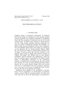
previously (Wu et al. 2002). All hippocampal volumes were adjusted for total brain volume. Intrarater reliability for these methods was good, with intraclass correlation coefficients of 0.96 for the left hippocampus and 0.97 for the right hippocampus. A single rater performed all of the analysis and was blind to participant’s experimental condition and demographic information. FMRI recall task Associative memory recall tasks done during functional MRI scanning have repeatedly been shown to result in robust activation of the hippocampal formation (e.g., Killgore 2000; Sperling et al. 2001; Stark and Squire 2001; Yonelinas et al. 2001; Duzel et al. 2003; Fig. 1 Schematic of task design during the encoding task on day 1 and the in-scanner recall task on day 2
Brain Imaging and Behavior (2008) 2:105–116
Sperling et al. 2003; Giovanello et al. 2004). These studies, and others, document both the role of the hippocampus in recall tasks and the ability of fMRI to measure the functioning of the hippocampus during such tasks. The use of fMRI also allows the study of other brain regions that are active during recall – regions that may be able to compensate for deficits in hippocampal function. To evaluate the activity of the hippocampus during a recall task, we followed a design similar to that used by Yonelinas et al. (2001). Twenty-four hours before the MRI session, participants were presented with 244 line drawings from the Snodgrass set of pictures (Snodgrass and Vanderwart 1980). These pictures were either presented in green or in red and participants were instructed to
Brain Imaging and Behavior (2008) 2:105–116
109
remember the pictures by creating a short, one-sentence explanation of why the object was that particular color. They were given six seconds to memorize each picture before the presentation of the next picture. Immediately after the memorization phase, participants were given a recall test where all pictures presented during the memorization phase were presented again, this time as black line drawings on a white background. Participants responded with the color each picture had been when originally presented and feedback was given. The next day, during the MRI session, subjects were again presented with the black and white drawings, this time for only 2 s, and asked to indicate by a button press whether the picture had initially been presented in red or green. A control task was also presented, during which participants were asked to indicate which simple shape on the screen was larger (right or left). Experimental and control pictures were presented in alternating 24 s blocks, with each block consisting of 12 pictures. A 24 s block of simple fixation occurred at both the beginning and the end of the scanning run. (See Fig. 1.) FMRI analysis Statistical analysis was performed on both individual and group data using the modified General Linear Model and the theory of Gaussian random fields as implemented in SPM99 (Friston et al. 1995). For both within-group and between-group comparisons, significant voxels were defined as those that exceeded a threshold value x equivalent to a one-tailed t-test p<0.05 (Bonferroni corrected for multiple comparisons at the cluster level). Once subjected to threshold analysis, the activation was superimposed on the normalized high-resolution SPGR and localized manually using atlases of the human brain (Talairach and Tournoux 1998; Duvernoy and Bourgouin 1999). Group analyses were overlaid on images created by averaging all individuals’ normalized SPGR images.
utilized in performing group analyses. This model estimates the error variance for each condition of interest across participants rather than across scans (Holmes and Friston 1998). The contrast images for each participant for each effect of interest were generated first, as described above. These contrast images were then analyzed using a general linear model to determine voxel-wise t statistics and generating one contrast image per participant, per effect of interest. Within-group analyses of each contrast were performed to identify voxels/brain regions showing similar response modulation across participants in each group for a given contrast (e.g., recall-control). In addition, between-group analyses were performed to determine how the two groups differed in their average activation in response to each contrast of interest (i.e., which regions were more active in those with the premutation than in controls, and vice versa). Region of interest (ROI) analyses were carried out using Marsbar (Brett et al. 2002), a MATLAB toolbox written to be implemented within SPM. Contrasts were first defined as described above. Each contrast of interest was then analyzed only in voxels that fell either within the MNI (Montreal Neurological Institute Atlas) template of the area of interest (e.g. the hippocampus) provided within Marsbar or in a functionally or statistically defined region of interest (e.g. a region defined functionally by the group which is then assessed within individuals). A t-statistic termed “contrast value” was then calculated as the average of the contrast values of the voxels falling within the defined ROI. The contrast value in these analyses is comparable to the Z score reported in the whole-brain analyses tables.
Results Total brain and hippocampal volume
All effects of interest were modeled using a standard within-subjects procedure for each participant by contrasting experimental and control blocks (e.g. blocks of object recall—blocks of shape comparison). Models for individuals were identical across participants. A random-effects model incorporating a two-stage hierarchical procedure was
Independent sample t-tests revealed no differences between groups in total brain volume or in right, left or total hippocampal volumes (see Table 2) even when hippocampal volumes were adjusted for total brain size (right: t=-0.55, p=0.59, left: t=0.23, p=0.83, total: t=-0.18, p=0.86).
Table 2 Total brain and hippocampal volumes for men with the FMR1 premutation and matched controls Brain area
Total brain (cc3) Hippocampus (cc3) Left Right Total
Control (n=11)
Premutation (n=11)
p-value
Mean
SD
Mean
SD
1,503.8
110.4
1,480.2
110.0
0.60
1.95 2.02 3.99
0.24 0.22 0.43
1.89 2.01 3.90
0.23 0.20 0.42
0.39 0.96 0.64
110
Neither CGG repeat size nor blood levels of FMR1 mRNA was significantly correlated with total brain volume or adjusted hippocampal volumes (Pearson’s r<0.40, p>0.30). Behavioral memory performance For the immediate recall test on day1, a significant difference was found between the accuracy for the control group (M, 82.7; SD, 8.02;) and the premutation group (M, 74.6; SD, 9.65; range, 60.1–90.1; t=2.13, p= 0.045). Behavioral data from the fMRI paradigm on day 2 (Fig. 2) showed no significant differences between groups on accuracy on the recall task (control: M, 71.34; SD, 7.34; premutation: M, 67.97; SD, 9.09; t=0.937, p=0.36), control task (control: M, 93.94; SD, 5.49; premutation: M, 95.76; SD, 3.17; t=−0.919, p=0.37), reaction time (in ms) during recall (control: M, 1017; SD, 86.4; premutation: M, 1224; SD, 523.1; t=−1.30, p=0.21) or reaction time during the control task (control: M, 656; SD, 49.1; premutation: M, 668; SD, 103.6; t=−0.32, p=0.75).
Brain Imaging and Behavior (2008) 2:105–116
and parahippocampal regions. The premutation group showed significant activation in right parietal areas (p< 0.01) while the control group did not. The control group showed significant activation in the left hippocampus and left parahippocampal region (p<0.01) while the premutation group did not (Fig. 4a). Neither group showed significant activation in the right hippocampus or right parahippocampal region. Between-groups fMRI analysis As would be predicted by the within-groups analysis, the control group showed greater activation than the premutation group in the left parahippocampal and hippocampal regions (Fig. 4b), as well as areas in the right cuneus, right lingual gyrus and right caudate nucleus (p<0.01). The premutation group showed more activation than controls in right intraparietal sulcus, supramarginal gyrus and angular gyrus as well as the left caudate nucleus (p<0.01; see Table 4).
Within group fMRI analysis
Correlation analyses
During the associative memory probe, when compared with the control task, premutation carriers showed overall brain activation patterns that were quite similar to those evidenced by the control group (see Fig. 3 and Table 3). These areas included anterior cingulate cortex, bilateral dorsolateral prefrontal areas, left parietal cortex and left fusiform gyrus. Despite these overall similarities, the two groups showed different activation patterns in two areas: within the right parietal cortex and in the left hippocampal
As we expected that abnormal hippocampal activation might be associated with FMR1 measures and/or reflected in psychiatric symptoms, we also investigated hippocampal activity as a function of FMR1 mRNA expression in blood, CGG repeat size and SCL-90-R scores. In conducting these analyses, we entered these variables as covariates of interest in analyzing activation elicited during the recall-control contrast. In those clusters that showed a significant relationship in this group analysis, we then performed a
Fig. 2 Percent correct response on an immediate recall test (day 1) and in-scanner recall and control tasks (day 2)
Brain Imaging and Behavior (2008) 2:105–116
111
Fig. 3 Regions of activation rendered on the surface of a MNI-normalized template image for control (left) and premutation (right) groups during the associative memory recall task. Images are subject to threshold at p<0.01 and corrected for multiple comparisons at the cluster level
ROI analysis to look at the correlation between the mean contrast value for voxels in that cluster for each individual and their molecular measures or SCL-90-R score. We explored the relationship between brain activation and
molecular measures only in the premutation group, as CGG repeat number and FMR1 mRNA levels in the control group lacked sufficient variance to conduct meaningful correlation analyses. No significant correlation between
Table 3 Stereotaxic locations and z-scores of activation peaks in the within-group maps Memory–control contrast Group
Area
Number of voxels in cluster
Z max
Peak coordinates
Control
R superior prefrontal gyrus, cingulate sulcus R lateral fissure L fusiform gyrus L superior, middle and inferior frontal gyrus L intraparietal sulcus, angular gyrus R inferior occipital gyrus R caudate R cerebellum (V) L hippocampus L cingulated gyrus R cingulated gyrus R middle occipital gyrus L inferior frontal gyrus/sulcus, middle frontal gyrus L superior frontal gyrus, cingulate sulcus R inferior occipital gyrus L fusiform gyrus L angular gyrus, intraparietal sulcus R lateral fissure, posterior orbital gyrus R caudate nucleus L caudate nucleus R angular gyrus, intraparietal sulcus R middle frontal gyrus, inferior frontal sulcus
584 667 1,981 3,026 964 305 735 1,724 183 148 128 2,190 1,932 719 375 1,163 642 436 227 441 238 349
4.96 4.94 4.88 4.88 4.76 4.67 4.31 4.27 4.22 3.86 3.78 4.93 4.79 4.7 4.58 4.5 4.32 4.27 4.26 4.04 3.81 3.77
2 34 −46 −50 −34 30 14 32 −28 10 50 36 −48 −2 −32 −34 −32 34 14 −18 38 42
Premutation
All clusters significant at p<0.01, corrected for multiple comparisons.
16 28 −56 12 −62 −94 0 −36 30 20 30 −88 30 26 −96 −36 −80 28 10 −2 −68 32
54 8 −20 38 38 −12 10 −28 0 28 38 −14 30 50 −12 −24 38 −8 10 24 40 26
112
Brain Imaging and Behavior (2008) 2:105–116
Fig. 4 Within-group (a) and between-group (b) activation in the left hippocampus during the recall task. Image threshold is set at p<0.05 and cluster size >10 voxels and images are masked to show only hippocampal activation
CGG repeat number and hippocampal or parietal activation was revealed in the group data. A significant negative association between left hippocampal activation and increased blood levels of FMR1 mRNA was evident in the premutation group (74 voxels, rho=−0.791, p=0.004). This finding must be treated with some caution, however, as the group included a single participant whose blood mRNA level was quite high (5.1-fold above normal) and a statistical
outlier. We primarily addressed this concern by using a nonparametric test to look at the correlation between mRNA and activation, but also looked at the correlation when his data were removed from the analysis. Without his data, the correlation was still strong (rho=−0.721) but reduced in significance (p=0.02). A relationship between greater rightparietal activation (angular gyrus, IPS and supramarginal gyrus) and increased mRNA was also found within the
Table 4 Stereotaxic locations and z-scores of activation peaks in the between-group maps Memory–control contrast Comparison
Area
Number of voxels in cluster
Z Max
Peak coordinates
Control > premutation Premutation > Control
R cuneus, lingual gyrus, caudate L parahippocampal gyrus, hippocampus, lateral fissure R angular gyrus, intraparietal sulcus, superior parietal gyrus, supramarginal gyrus L caudate nucleus
1,042 593 848
3.21 3.06 4.38
20 −10 38
−82 −52 −68
10 8 40
713
3.19
−8
10
12
All clusters significant at p<0.05 corrected for multiple comparisons.
Brain Imaging and Behavior (2008) 2:105–116
premutation group (140 voxels, rho=0.955 p=0.004). Unlike the correlation between mRNA and left hippocampal activation, the strength of this relationship was unaffected by the removal of the subject with the highest mRNA level from the data set. A significant negative correlation between SCL90-R GSI score and left hippocampal activation was also evident in a whole-brain covariate of interest analysis (threshold p<0.05) in the premutation group (see Fig. 5). Despite a similar range of SCL-90-R GSI scores, this association was not evident in the control group. In the premutation group (331 voxels, rho=−0.645, p=0.032), but not in the control group (331 voxels, rho=0.43, p=0.46), severity of psychiatric symptoms as measured by the SCL90-R GSI was negatively correlated with left hippocampal activation. We also investigated correlations between activation in response to the memory-control contrast and accuracy scores from the in-scanner recall task. To assess the strength of these correlations, we performed an ROI analysis looking at the contrast value in response to the memorycontrol contrast for each individual in those voxels in parietal cortex that showed this association in the group data. Contrast values for individuals were then correlated with individual accuracy scores. Neither group showed a significant correlation between hippocampal activation and accuracy. The strongest association between activation and accuracy, in both groups, was in clusters within bilateral parietal cortex. The association was more extensive on the right in the premutation group (291 voxels, rho=0.890, p= 0.001) while the control group showed more voxels correlated with accuracy on the left than the premutation group (137 voxels, rho=0.840, p=0.001). While the higher parietal recruitment in premutation carriers is suggestive of compensation, we were not able to document a negative correlation between hippocampal activation and right
Fig. 5 Bright clusters represent voxels in the left hippocampus in which activation is negatively correlated with psychiatric symptoms as measured by the SCL-90-Revised. Images threshold is set at p<0.05 and a cluster size of >10 voxels and are masked to show this relationship only in the hippocampus
113
parietal activation in the premutation group (rho=−0.182, p=0.503).
Discussion The present study provides evidence that men with the fragile X premutation have a reduced ability to recruit the left hippocampus during recall. Relative to well-matched controls, men with the premutation were significantly worse on an immediate recall test but were not significantly worse on the in-scanner recall task 24 h later. Reduced hippocampal activation in the premutation group was accompanied by increased activation in both frontal and parietal areas, particularly right parietal areas, perhaps allowing them to compensate for decreased hippocampal involvement by compensatory recruitment in these areas. Both the decrease in left hippocampal activation and the increase in right parietal activation were correlated with increased FMR1 mRNA levels in the premutation carriers. Additionally, the clinical relevance of these findings is suggested by the fact that hippocampal activity was negatively correlated with psychiatric symptomology in the men with the premutation. It is of particular interest that this association was absent in controls, who exhibited similar levels of psychiatric symptoms. This difference suggests that carrying the premutation allele causes brain changes that affect both hippocampal activity and psychiatric symptomology, while the etiology of psychiatric symptoms in controls varies and is unrelated to brain activity during recall. That mRNA levels were significantly correlated with brain activation measures while correlations with CGG repeat number did not reach significance may be an important finding but must be treated with caution given our small sample size. Previous data from our group has
114
shown that mRNA levels may be more important to psychiatric symptomology than CGG repeat number in premutation carriers (Hessl et al. 2005), perhaps due to intrinsic variation in mRNA level among individuals with similar CGG repeat expansions. Our data suggests that mRNA levels may also be a stronger factor in brain activation changes specific to premutation carriers. However, these distinctions are difficult to establish unambiguously, as CGG repeat number and FMR1 mRNA levels are strongly correlated. The correlation between CGG repeat number and blood levels of FMR1 mRNA in the current sample, for example, was quite strong (rho=0.764, p= 0.006). Additional factors (other than CGG repeat number) that modulate mRNA levels in premutation carriers have not been identified. Further examination of the factors contributing to increased FMR1 mRNA levels and the relationship between mRNA levels, FMRP levels and brain function will be necessary to untangle how each factor contributes to the neural phenotype in premutation carriers. We chose to investigate hippocampal function through a recall paradigm, expecting that the premutation group would be somewhat impaired on this task. Instead, the premutation group performed as well as the control group during the in-scanner recall task while unexpectedly showing a significant deficit during the immediate recall test. That the two groups performed comparably on the inscanner task simplifies the interpretation of our functional results, giving us confidence that differences between the groups were not primarily a function of differences in their ability to perform the task. The immediate recall deficit demonstrated by the premutation group suggests that there is a significant difference between groups in their ability to encode information into memory and that there is some disparity in the rate of forgetting between the groups. From the data collected we cannot resolve whether hippocampal activation differences between groups are primarily due to differences in encoding ability (which is itself highly dependent on hippocampal function) or is truly reflective of hippocampal activity during recall. In order to address these questions, we are currently investigating hippocampal function during encoding of complex scenes utilizing an event-related design that will allow us to pull apart brain activation during correctly encoded scenes from those scenes that are later forgotten in a post-scan immediate recall test. The reduced activity in the left hippocampus seen in our results could be reflective of premutation-specific developmental changes. There is some indication in the recent literature that boys with the premutation are at increased risk for neurodevelopmental disorders, including autism spectrum disorders and Attention Deficit Hyperactivity Disorder (ADHD; Aziz et al. 2003; Goodlin-Jones et al. 2004; Farzin et al. 2006). Although it is not immediately
Brain Imaging and Behavior (2008) 2:105–116
clear how this connects to possible dysfunction of the hippocampus in premutation carriers, it is an indication that even young boys show some effect of their premutation status. Such developmental consequences could be caused either by elevated FMR1 mRNA (Tassone et al. 2000a, b, c; Jacquemont et al. 2003; Oostra and Willemsen 2003; Allen et al. 2004; Hagerman and Hagerman 2004) or a mild reduction in FMRP as is known to occur in individuals with the premutation, especially with high CGG repeat numbers (Tassone et al. 2000a,b,c; Kenneson et al. 2001; Entezam et al. 2007). We would expect lower FMRP to result in abnormally reduced pruning during development (Irwin et al. 2000; Bagni and Greenough 2005; McKinney et al. 2005; Grossman et al. 2006) which would in theory result in larger and potentially more active hippocampi. It is possible that lowered FMRP and increased mRNA, which can co-occur in carriers with the premutation, are affecting hippocampal structure and function in different ways, complicating the picture and making molecular/fMRI associations difficult to clearly discern, especially in so small a sample. We were unable to measure FMRP levels in the current study but gathering such data in combination with measures of FMR1 mRNA levels, CGG repeat lengths and functional data will be important in future investigations to pinpoint the molecular mechanism at work. Alternatively, or in addition to a developmental effect, our findings could represent early pre-symptomatic brain changes that precede, but which are ultimately associated with, FXTAS. The intranuclear inclusions found in post mortem brain tissue of those who died with FXTAS could be accumulating throughout the lifespan of susceptible premutation carriers. However, it is not known whether inclusion formation, per se, adversely affects brain function. Indeed, although mice develop far greater inclusion loads in neurons than do humans with severe FXTAS, the mice develop only mild neurological features and relatively less neurodegeneration. (Willemsen et al. 2003). As this study was designed and conducted as a preliminary examination of the effect of the fragile X premutation on hippocampal function, it had some limitations. The primary limitation was the small sample sizes of our groups. This limited our power to detect true volumetric differences in the hippocampus that might be present as well as limiting our power in investigating interaction effects between molecular, behavioral and brain imaging data. Our sample size also limited the variance in the molecular measures we collected and our ability to thoroughly investigate relationships between those measures and brain activation in response to our task. Because we expect those with greater levels of FMR1 mRNA to be more affected, our study would have been strengthened by inclusion of more subjects with very high mRNA levels. We did have one individual with the premutation whose
Brain Imaging and Behavior (2008) 2:105–116
blood FMR1 mRNA level was 5 times above normal. This same individual also exhibited the least hippocampal activation, the poorest memory performance, as well as the most severe psychiatric symptoms. Thus, rather than being an “outlier” in our sample, this individual may represent the upper end of a continuum of severity of dysfunction in this population. Indeed, while blood FMR1 mRNA levels as high as tenfold above normal have been reported (Tassone et al. 2000a, b, c), we do not yet have brain MRI data available from individuals in this range. The reduction of hippocampal activity observed in the premutation group was not accompanied by any measurable reduction of hippocampal volume. Although this study is primarily designed as an fMRI study of the hippocampus, our volumetric data strengthens our functional imaging results by showing that the hippocampal activation differences we measured are independent of gross morphological changes in the hippocampus in this particular sample. The sample sizes, however, preclude an adequately powered test of hippocampal volume differences between premutation carriers and typical controls in the wider population. The lack of hippocampal volume effects associated with the premutation in the present study is in contrast both to Jäkälä et al. (1997), who found reduced volumes, and to Loesch et al. (2005) who found increased volumes in this region in male premutation carriers. The lack of consistent findings between these two studies may be due to specific cohort effects, especially if these cohorts included participants with and without FXTAS. The relative effects of decreased FMRP and/or elevated FMR1 mRNA, which may have differential effects on brain development and structure across study participants, may also contribute to these inconsistencies. It is also important to note that CGG repeat size and mRNA measures were ascertained from blood samples. As such, they may not accurately reflect levels in the brain. While a post mortem study carried out on a single premutation carrier and one control reported CGG-repeat length stability between tissue types, FMR1 mRNA expression appears to vary across tissue type and brain region (Tassone et al. 2004). In general, however, the relative brain FMR1 mRNA levels were found to be substantially higher than in peripheral blood leucocytes for both the premutation carrier and control, reflecting the important role this gene plays in brain tissue. FMR1 mRNA levels were substantially higher in brain samples for the premutation compared to the control but not to as great an extent as in blood. From these data it appears likely that increased mRNA levels measured in the blood would be echoed by increased mRNA levels in the brain. Thus, while correlations with brain function measures must be treated with caution, mRNA levels measured in blood are our best current proxy for mRNA levels in the brain.
115
In conclusion, this study demonstrates reduced hippocampal activation during memory recall associated with parietal over-activation, psychiatric problems and abnormal elevation of FMR1 mRNA in men with the fragile X premutation. These findings may be a result of RNA toxicity that in some may reflect developmental changes and in others may develop into a neurodegenerative disease, FXTAS, in later life. Acknowledgements We would like to thank the research participants and their families; Marilyn Juarez for data analysis help, Lisa Cordeiro and Jennifer Cogswell for assistance in scheduling and testing; Louise Gane for assistance with recruitment; Kylee Cook for help with data entry and management and Charles DeCarli for the use of his hippocampal tracing protocol. Funding from the National Institutes of health Grants HD02274 and HD36071 (R.J.H.) and MH77554 (D.H.) and MH078041 (D.H. and S.M.R.) supported this work.
References Abitbol, M., Menini, C., et al. (1993). Nucleus basalis magnocellularis and hippocampus are the major sites of FMR-1 expression in the human fetal brain. Nature Genetics, 4, 147–153. Allen, E. G., He, W., et al. (2004). A study of the distributional characteristics of FMR1 transcript levels in 238 individuals. Human Genetics, 114(5), 439–447. Aziz, M., Stathopulu, E., et al. (2003). Clinical features of boys with fragile X premutations and intermediate alleles. American Journal of Medical Genetics, 121B(1), 119–127. Bagni, C., & Greenough, W. T. (2005). From mRNP trafficking to spine dysmorphogenesis: The roots of fragile X syndrome. Nature Reviews Neuroscience, 6(5), 376–387. Borghgraef, M., Steyaert, J., et al. (2004). Preliminary findings in boys with fragile X premutation: Is there a distinct behavioral phenotype?. Washington, D.C.: International Fragile X Conference. Brett, M., Anton, J. L., et al. (2002). Region of interest analysis using an spm toolbox. Neuroimage, 16(2), S497. Cornish, K., Kogan, C., et al. (2005). The emerging fragile X premutation phenotype: Evidence from the domain of social cognition. Brain and Cognition, 57(1), 53–60. DeCarli, C., Maisog, J., et al. (1992). Method for quantification of brain, ventricular, and subarachnoid CSF volumes from MR images. Journal of Computer Assisted Tomography, 16(2), 274– 284. DeCarli, C., Murphy, D. G., et al. (1995). The effect of white matter hyperintensity volume on brain structure, cognitive performance, and cerebral metabolism of glucose in 51 healthy adults. Neurology, 45(11), 2077–2084. DeCarli, C., Murphy, D. G., et al. (1996). Local histogram correction of MRI spatially dependent image pixel intensity nonuniformity. Journal of Magnetic Resonance Imaging, 6(3), 519–528. Derogatis, L. R. (1994). Symptom Checklist-90-R: Administration, Scoring, and Procedures Manual - Third Edition. Minneapolis: National Computer Systems, Inc. Dorn, M. B., Mazzocco, M. M., et al. (1994). Behavioral and psychiatric disorders in adult male carriers of fragile X. Journal of the American Academy of Child and Adolescent Psychiatry, 33 (2), 256–264. Duvernoy, H. M., & Bourgouin, P. (1999). The Human Brain: Surface, Three-Dimensional sectional Anatomy with MRI, and Blood Supply. New York: Springer.
116 Duzel, E., Habib, R., et al. (2003). Human hippocampal and parahippocampal activity during visual associative recognition memory for spatial and nonspatial stimulus configurations. Journal of Neuroscience, 23(28), 9439–9444. Entezam, A., Biacsi, R., et al. (2007). Regional FMRP deficits and large repeat expansions into the full mutation range in a new Fragile X premutation mouse model. Gene, 395(1–2), 125–134. Farzin, F., Perry, H., et al. (2006). Autism spectrum disorders and attention-deficit/hyperactivity disorder in boys with the fragile X premutation. Journal of Developmental and Behavioral Pediatrics, 27(2 Suppl 2), S137–S144. Franke, P., Leboyer, M., et al. (1998). Genotype–phenotype relationship in female carriers of the premutation and full mutation of FMR-1. Psychiatry Research, 80(2), 113–127. Friston, K. J., Holmes, A. P., et al. (1995). Statistical parametric maps in functional imaging: A general linear approach. Human Brain Mapping, 2, 189–210. Fu, Y. H., Kuhl, D. P., et al. (1991). Variation of the CGG repeat at the fragile X site results in genetic instability: Resolution of the Sherman paradox. Cell, 67(6), 1047–1058. Giovanello, K. S., Schnyer, D. M., et al. (2004). A critical role for the anterior hippocampus in relational memory: Evidence from an fMRI study comparing associative and item recognition. Hippocampus, 14(1), 5–8. Goodlin-Jones, B. L., Tassone, F., et al. (2004). Autistic spectrum disorder and the fragile X premutation. Journal of Developmental and Behavioral Pediatrics, 25(6), 392–398. Greco, C. M., Berman, R. F., et al. (2006). Neuropathology of fragile X-associated tremor/ataxia syndrome (FXTAS). Brain, 129(Pt 1), 243–255. Greco, C. M., Hagerman, R. J., et al. (2002). Neuronal intranuclear inclusions in a new cerebellar tremor/ataxia syndrome among fragile X carriers. Brain, 125(Pt 8), 1760–1771. Grossman, A. W., Elisseou, N. M., et al. (2006). Hippocampal pyramidal cells in adult Fmr1 knockout mice exhibit an immature-appearing profile of dendritic spines. Brain Res, 1084 (1), 158–164. Hagerman, P. J., & Hagerman, R. J. (2004). The fragile-X premutation: A maturing perspective. American Journal of Human Genetics, 74(5), 805–816. Hagerman, R. J., & Hagerman, P. J. (2002). The fragile X premutation: Into the phenotypic fold. Current Opinion in Genetics & Development, 12(3), 278–283. Hessl, D., Rivera, S., et al. (2007). Amygdala dysfunction in men with the fragile X premutation. Brain, 130(Pt 2), 404–416. Hessl, D., Tassone, F., et al. (2005). Abnormal elevation of FMR1 mRNA is associated with psychological symptoms in individuals with the fragile X premutation. American Journal of Medical Genetics B Neuropsychiatria Genetics, 139(1), 115–121. Holmes, A. P., & Friston, K. J. (1998). Generalisability, random effects, and population inference. Human Brain Mapping, 7(4), S754. Irwin, S. A., Galvez, R., et al. (2000). Dendritic spine structural anomalies in fragile-X mental retardation syndrome [In Process Citation]. Cereb Cortex, 10(10), 1038–1044. Jacquemont, S., Hagerman, R. J., et al. (2003). Fragile X premutation tremor/ataxia syndrome: Molecular, clinical, and neuroimaging correlates. American Journal of Human Genetics, 72(4), 869–878. Jäkälä, P., Hanninen, T., et al. (1997). Fragile-X: Neuropsychological test performance, CGG triplet repeat lengths, and hippocampal volumes. Journal of Clinical Investigation, 100(2), 331–338. Johnston, C., Eliez, S., et al. (2001). Neurobehavioral phenotype in carriers of the fragile X premutation. American Journal of Medical Genetics, 103(4), 314–319. Kenneson, A., Zhang, F., et al. (2001). Reduced FMRP and increased FMR1 transcription is proportionally associated with CGG repeat
Brain Imaging and Behavior (2008) 2:105–116 number in intermediate-length and premutation carriers. Human Molecular Genetics, 10(14), 1449–1454. Killgore, W. D., Casasanto, D. J., et al. (2000). Functional activation of the left amygdala and hippocampus during associative encoding. Neuroreport, 11(10), 2259–2263. Loesch, D. Z., Litewka, L., et al. (2005). Magnetic resonance imaging study in older fragile X premutation male carriers. Annals of Neurology, 58(2), 326–330. McKinney, B. C., Grossman, A. W., et al. (2005). Dendritic spine abnormalities in the occipital cortex of C57BL/6 Fmr1 knockout mice. American Journal of Medical Genetics B Neuropsychiatria Genetics, 136(1), 98–102. Moore, C. J., Daly, E. M., et al. (2004a). A neuropsychological investigation of male premutation carriers of fragile X syndrome. Neuropsychologia, 42(14), 1934–1947. Moore, C. J., Daly, E. M., et al. (2004b). The effect of pre-mutation of X chromosome CGG trinucleotide repeats on brain anatomy. Brain, 127(Pt 12), 2672–2681. Oostra, B. A., & Willemsen, R. (2003). A fragile balance: FMR1 expression levels. Human Molecular Genetics, 12(Spec No 2), R249–R257. Snodgrass, J. G., & Vanderwart, M. (1980). A standardized set of 260 pictures: Norms for name agreement, image agreement, familiarity, and visual complexity. Journal of experimental psychology [Hum Learn], 6(2), 174–215. Sperling, R. A., Bates, J. F., et al. (2001). Encoding novel face-name associations: A functional MRI study. Human Brain Mapping, 14 (3), 129–139. Sperling, R., Chua, E., et al. (2003). Putting names to faces: Successful encoding of associative memories activates the anterior hippocampal formation. Neuroimage, 20(2), 1400–1410. Stark, C. E., & Squire, L. R. (2001). Simple and associative recognition memory in the hippocampal region. Learning and Memory, 8(4), 190–197. Talairach, J., & Tournoux, P. (1998). Co-Planar Stereotaxic Atlas of the Human Brain: A 3-dimensional Proportional System, an Approach to Cerebral Imaing. New York: Thieme Medical Publishers. Tassone, F., Hagerman, R. J., et al. (2000a). Clinical involvement and protein expression in individuals with the FMR1 premutation. American Journal of Medical Genetics, 91(2), 144–152. Tassone, F., Hagerman, R. J., et al. (2000b). Elevated levels of FMR1 mRNA in carrier males: A new mechanism of involvement in the fragile-X syndrome. American Journal of Human Genetics, 66 (1), 6–15. Tassone, F., Hagerman, R. J., et al. (2000c). Fragile X males with unmethylated, full mutation trinucleotide repeat expansions have elevated levels of FMR1 messenger RNA. American Journal of Medical Genetics, 94(3), 232–236. Tassone, F., Hagerman, R. J., et al. (2004). Intranuclear inclusions in neural cells with premutation alleles in fragile X associated tremor/ataxia syndrome. J Med Genet, 41(4), e43. Wechsler, D. (1997). Wechsler Adult Intelligence Scale-Third Edition. Manual. San Antonio: The Psychological Corporation. Willemsen, R., Hoogeveen-Westerveld, M., et al. (2003). The FMR1 CGG repeat mouse displays ubiquitin-positive intranuclear neuronal inclusions; implications for the cerebellar tremor/ataxia syndrome. Human Molecular Genetics, 12(9), 949–959. Wu, C. C., Mungas, D., et al. (2002). Brain structure and cognition in a community sample of elderly Latinos. Neurology, 59(3), 383– 391. Yonelinas, A. P., Hopfinger, J. B., et al. (2001). Hippocampal, parahippocampal and occipital-temporal contributions to associative and item recognition memory: An fMRI study. Neuroreport, 12(2), 359–363.