The Journal of Neuroscience, September 15, 1997, 17(18):6947–6951
Protein Phosphorylation and Taurine Biosynthesis In Vivo and In Vitro Xiao Wen Tang,1 Che-Chang Hsu,1 John V. Schloss,2 Morris D. Faiman,3 Elliott Wu,4 Chao-Yuh Yang,4 and Jang-Yen Wu1 Departments of 1Physiology and Cell Biology, 2Medicinal Chemistry, and 3Pharmacology and Toxicology, University of Kansas, Lawrence, Kansas 66045-2106, and 4Department of Medicine, Baylor College of Medicine, Houston, Texas 77030
Taurine is known to be involved in many important physiological functions. Here we report that both in vivo and in vitro the taurine-synthesizing enzyme in the brain, namely cysteine sulfinic acid decarboxylase (CSAD), is activated when phosphorylated and inhibited when dephosphorylated. Furthermore, protein kinase C and protein phosphatase 2C have been identified as the enzymes responsible for phosphorylation and dephosphorylation of CSAD, respectively. In addition, the effect of
neuronal depolarization on CSAD activity and 32P incorporation into CSAD in neuronal cultures is also included. A model to link neuronal excitation and CSAD activation by a Ca 21-dependent protein kinase is proposed.
Taurine, 2-aminoethanesulfonic acid, is one of the most abundant amino acids in the brain (Jacobsen and Smith, 1968). The physiological role of taurine has received much attention because of the reports that cats fed a taurine-deficient diet developed central retinal degeneration (Hayes et al., 1975), cardiomyopathy (Pion et al., 1987), and delay in neural development (Sturman, 1993). So far, taurine has been shown to be involved in various important physiological f unctions; e.g., serving as a trophic factor in the development of the C NS (Sturman, 1993), maintaining the structural integrity of the membrane (Pasantes-Morales and Cruz, 1985), regulating calcium homeostasis (Lazarewicz et al., 1985), modulating protein phosphorylation (L ombardini, 1992), and acting as an osmoregulator (Solis et al., 1988), a neuromodulator (Kuriyama, 1980), and a neurotransmitter (Okamoto et al., 1983; Taber et al., 1986). Despite its importance, little is known about the mechanism of regulation of taurine biosynthesis in the brain. There has been controversy in the past regarding whether GABA and taurine are synthesized in the brain by the same enzyme (Blinderman et al., 1978). It is now well established that two distinctly different enzymes, namely, L-glutamate decarboxylase (GAD) and cysteine sulfinic acid decarboxylase (C SAD) are responsible for GABA and taurine synthesis in the brain, respectively (Wu, 1982). Recently, we have demonstrated that GAD activity is increased when GAD is dephosphorylated and inhibited when it is phosphorylated (Bao et al., 1994, 1995). Furthermore, protein kinase A (PK A) and calcineurin have been identified as the protein kinase and protein phosphatase (PrP) responsible for GAD phos-
phorylation and dephosphorylation, respectively (Bao et al., 1994). In this communication, we report that unlike GAD, CSAD activity is enhanced under conditions favoring protein phosphorylation and is inhibited or inactivated when it is dephosphorylated. Furthermore, direct phosphorylation and concomitant increase of CSAD activity have been demonstrated in three different conditions: namely, in synaptosomal preparations, in purified CSAD, and in cultured neuronal system. In addition, PKC and PrP-2C have been identified as the enzymes involved in phosphorylation and dephosphorylation of CSAD, respectively. A model to link neuronal excitation to activation of CSAD by a Ca 21-dependent protein kinase is also included.
Received May 1, 1997; revised June 24, 1997; accepted July 9, 1997. This work was supported by the National Science Foundation (IBN-9723079), the Office of Naval Research (N00014-94-1-0457), the University of Kansas General Research Fund, and National Institutes of Health (NS20978). We thank Drs. Erik Floor and James Orr for critical review of this manuscript. The expert typing of the manuscript by Sharon Lee Hopkins is greatly appreciated. Correspondence should be addressed to Dr. Jang-Yen Wu, Department of Physiology and C ell Biology, University of Kansas, Lawrence, KS 66045-2106. Copyright © 1997 Society for Neuroscience 0270-6474/97/176947-05$05.00/0
Key words: taurine; cysteine sulfinic acid decarboxylase; protein kinase C; protein phosphatase 2C; protein phosphorylation; protein dephosphorylation
MATERIALS AND METHODS Materials. Fresh porcine brains were obtained from a local slaughter house. Heat-inactivated fetal calf serum, poly-L-lysine, Triton X-100, 1.2-diolein, L-a-phosphatidyl-L-serine, cAM P-dependent protein kinase (PK A), PK A inhibitory peptide, and PK A catalytic subunit were from Sigma (St. L ouis, MO). PKC and PKC inhibitory peptide were from Upstate Biotechnology (Lake Placid, N Y). Okadaic acid was from Alexis (Laufelfingen, Switzerland). [I-14C]C SA was purchased through Research Products International (Santa Cruz, CA). All other radioisotopes were purchased from DuPont N EN (Boston, M A). Nitrocellulose membranes (0.45 mM) were from Bio-Rad (Melville, N Y). T ween-20 was from Fisher (Pittsburgh, PA). Goat anti-rabbit IgG conjugated with alkaline phosphatase and bromochloroindolyl phosphate/nitro blue tetrazolium (BCI P/ N BT) color development substrate were from Promega (Madison, W I). Sepharose protein A resin and cyanogen bromide (C N Br)-activated Sepharose 4B resin were from Pharmacia (Piscataway, NJ). Complete Freund’s adjuvant, incomplete Freund’s adjuvant, Basal Medium Eagle, and glutamine were obtained from Life Technologies (Grand Island, N Y). Preparation of synaptosome. Preparation of crude synaptosomal fractions was conducted as described previously. Briefly, fresh porcine brains were homogenized in 0.32 M sucrose (w/ v 5 15 gm:100 ml) using a glass homogenizer. The homogenate was centrif uged at 1000 3 g for 10 min, and the supernatant solution obtained was f urther centrif uged at 100,000 3 g for 30 min. The resulting pellet was the crude synaptosomal preparation. The pellet was resuspended in Kreb’s–Ringer’s phosphate buffer, pH 7.2, containing 123 mM NaC l, 3 mM KC l, 0.4 mM MgC l2 , 0.5
6948 J. Neurosci., September 15, 1997, 17(18):6947–6951
mM NaH2PO4 , 0.25 mM Na2HPO4 , and 1 mg /ml glucose, and divided into aliquots for f urther studies. E xtraction of CSA D f rom synaptosomal f ractions in the presence of phosphatase or k inase inhibitors. E xtraction of C SAD from synaptosomal fractions in the presence of PrP inhibitors was conducted as described previously for GAD (Bao et al., 1994, 1995). Briefly, fresh porcine brains were homogenized in 0.32 M sucrose, and synaptosomal fractions were prepared as described above. Aliquots of the synaptosomal fractions were centrif uged, and the pellets were resuspended in standard C SAD buffers [50 mM potassium phosphate, pH 7.2, 1 mM reduced glutathione (GSH), 2 mM 2-aminoethylisothiouronium bromide (AET), and 0.4 mM pyridoxal-59-phosphate (PL P)] containing either phosphatase inhibitors or kinase inhibitors as indicated. The synaptosomes were then ruptured by sonication (3 3 1 sec). The suspensions obtained were kept at room temperature for 45 min with constant shaking. C SAD activity was then determined by the C SAD activity assay as described (Wu, 1982), except that a final concentration of 10 mM glutamate was included in the assay to block any C SAD activity attributable to GAD. Phosphor ylation of CSA D in synaptosomal f ractions. Phosphorylation of C SAD by endogenous kinase in the presence of [g-32P]ATP was performed as described previously (Bao et al., 1994, 1995). Synaptosomal fractions were lysed, and a phosphorylation reaction was performed under the following conditions: 50 mM Tris/citrate, pH 7.3, 1 mM AET, 2 mM GSH, 0.10 mM [g-32P]-ATP (200 mC i /ml), and protein phosphatase inhibitors or protein kinase inhibitors as indicated. The reaction mixture was incubated at 37°C for 60 min and then centrif uged for 10 min at 14,000 rpm. The supernatant solution obtained was applied to an antiC SAD IgG immunoaffinity column and eluted as described (Bao et al., 1994; Tang et al., 1996). For calf intestine phosphatase (CI P) treatment, 200 U CI P was added to the affinity column as described previously (Bao et al., 1994, 1995). The eluates were analyzed on a 10% SDS-PAGE. The gel was first stained for protein by the silver staining method, followed by autoradiographic visualization as described (Bao et al., 1994). Phosphor ylation and dephosphor ylation of CSA D in purified preparations. Aliquots of purified C SAD were dialyzed at 4°C in 50 mM Tris/ citrate buffer, pH 7.2, containing 1 mM GSH, 1 mM AET, and 0.2 mM PL P for 18 hr, with three changes. The C SAD samples were treated under the following conditions: (1) PKC buffer alone (containing 1 mM C aC l2 , 5 mM MgC l2 , 0.3 mg /ml L-a-phosphatidyl-L-serine, 0.06 mg /ml diolein, 0.03% Triton X-100, 0.1 mM ATP, and 100 mC i [g-32P]-ATP); (2) PKC buffer plus 200 ng /ml PKC; (3) the same as (2), to be used later for CI P treatment; (4) the same as (2) plus 100 nM staurosporine; (5) the same as (2) plus 200 ng /ml PKC inhibitory peptide; (6) PK A buffer alone (containing 5 mM MgC l2 , 0.1 mM cAM P, 0.1 mM ATP, and 100 mC i [g-32P]-ATP); and (7) PK A buffer plus 150 U PK A catalytic subunit. The suspensions were incubated at 37°C for 45 min. The reactions were stopped by adding 5 3 SDS sample loading buffer except for group (3), which was f urther incubated with 100 U CI P-agarose resin in the presence of 100 nM staurosporine for another 45 min at 37°C before SDS treatment. The samples were then subjected to SDS-PAGE, followed by autoradiography. To determine the effect of kinase and phosphatase on C SAD activity, purified C SAD samples were treated under the same conditions as those described above, except that [g-32P] ATP was omitted. At the end of treatment, the incubation mixture was transferred immediately for measurement of C SAD activity using the standard C SAD assay as described (Wu, 1982). In the case of the CI P groups, CI P-agarose resin or Sephadex G-25 resin was removed by brief centrif ugation before assaying for C SAD activity. C SAD activity in each control group was used as the reference, 100%. Phosphor ylation of CSA D in cultured neurons. Whole-brain primary neuronal cultures were prepared from fetal rats (17–18 d gestation), using modifications of a previously published method (Lee et al., 1994). Briefly, culture medium was removed, and the cultures were washed and incubated with 1 ml of Earle’s balanced salt solutions (EBSS) (116.4 mM NaC l; 5.4 mM KC l; 0.8 mM MgSO4 ; 1.0 mM NaH2PO4 ; 26.2 mM NaHC O3 ; 1.8 mM C aC l2 ; 5.6 mM D-glucose, pH 7.4). [ 32P]-phosphate (85 mC i) was added to each dish and incubated for 1 hr in the incubator. The culture was treated for an additional 15 min by adding 1 ml of EBSS containing the following different substances: (1) EBSS alone; (2) EBSS plus 2 mM glutamate; (3) EBSS plus 2 mM glutamate and 50 mM taurine; or (4) high K 1 EBSS (20.6 mM NaC l; 100.7 mM KC l; 0.8 mM MgSO4 ; 1.0 mM NaH2PO4 ; 26.2 mM NaHC O3 ; 1.8 mM C aC l2 ; 5.6 mM D-glucose, pH 7.4). The cells were harvested in C SAD buffer containing phosphatase inhibitor (0.2 mM vanadate), protease inhibitors (1 mM phenylmethylsul-
Tang et al. • Protein Phosphorylation and Taurine Biosynthesis
Figure 1. Relative CSAD activity in synaptosomal extracts in the presence of phosphatase inhibitors. CSAD was extracted from synaptosomal preparations in the presence of different protein phosphatase inhibitors. 1, Control; 2, 2 mM EDTA and 2 mM EGTA; 3, 0.2 mM sodium orthovanadate; 4, 2 mM sodium fluoride; 5, 0.2 mM sodium pyrophosphate; 6, mixture of 2-5; 7, 7.5 mM okadaic acid. Each value is the mean 6 SE (n 5 4). fonyl fluoride, 5 mM benzamidine), and 100 mM amino-oxyacetic acid (AOAA). The suspensions were then sonicated and centrif uged. The supernatants thus obtained were kept on ice for 1 hr to allow the removal of PL P by AOAA. Each supernatant fraction was then passed twice through an anti-C SAD affinity column and analyzed by immunoblotting and autoradiography. For C SAD activity determination, a parallel experiment was conducted as described above except that no [ 32P]-phosphate was included. Cultures were harvested, sonicated in C SAD buffer containing protease and phosphatase inhibitors, and assayed for C SAD activity as described previously (Wu, 1982).
RESULTS Effects of protein kinase and phosphatase inhibitors on CSAD activity in synaptosomal fractions When CSAD was extracted under conditions favoring protein phosphorylation, e.g., in the presence of PrP inhibitors, CSAD activity was markedly increased (Fig. 1). When CSAD was extracted from synaptosomal fractions in a mixture of general PrP inhibitors, CSAD activity increased to 245 6 20% of the control level. Vanadate alone increased CSAD activity to an extent of 259 6 29% and hence can account for all the activation produced by the mixture of general PrP inhibitors mentioned above; however, okadaic acid, a potent PrP inhibitor, had no effect on CSAD activity even at 7.5 mM. Because PrP-2C is sensitive to vanadate but not to okadaic acid, whereas the other three types of PrPs, namely PrP-1, PrP-2A, and PrP-2B, are known to be highly sensitive to okadaic acid at the concentration used (7.5 mM), the results suggest that PrP-2C is probably involved in dephosphorylation of CSAD. On the other hand, CSAD activity decreased when it was extracted in the presence of protein kinase inhibitors. Among the kinase inhibitors used, only those that inhibited PKC activity decreased CSAD activity. PKC inhibitory peptide (100 ng/ml), staurosporine (1 mM), H-8 (10 mM), and chelerythrine (50 mM) decreased CSAD activity to 48 6 9, 48 6 2, 54 6 6, and 57 6 14% of the control level, respectively. No significant effect on CSAD activity was observed with PKA inhibitory peptide, indicating that PKC but not PKA is involved in the regulation of CSAD activity. In addition, when 5 mM ATP was included in the extraction, it increased CSAD activity to 172 6 19% of the control level. In short, CSAD activity increased under conditions that favored protein phosphorylation, whereas CSAD activity
Tang et al. • Protein Phosphorylation and Taurine Biosynthesis
Figure 2. Autoradiograph of C SAD in synaptosomal preparations in the presence of protein kinase inhibitors, phosphatase inhibitors, and C a 21chelators. Incorporation of 32P was performed in the lysed synaptosomal preparations under the condition indicated below. Lane 1: C SAD buffer 1 PKC activators (1 mM C aC l2 , 5 mM MgC l2 , 0.31 mg /ml, L-a-phosphatidyl-L-serine; 0.06 mg /ml 1.2-diolein); lane 2: C SAD buffer only; lane 3: CSAD buffer 1 PKC activators 1 0.2 mM vanadate; lane 4: C SAD buffer 1 PKC activators 1 PKC inhibitory peptide (100 ng /ml); lane 5: CSAD buffer 1 PKC activators 1 0.1 mM staurosporine; lane 6: C SAD buffer 1 PKC activators 1 2 mM EGTA; lane 7: C SAD buffer 1 PKC activators 1 2 mM EDTA; lane 8: C SAD buffer 1 PKC activators 1 200 U CIP; lane 9: silver staining of immunoaffinity-purified C SAD. Arrow indicates the subunit of C SAD, a 43 kDa protein (Tang et al., 1996). The standard molecular weight markers (in kilodaltons) are also indicated.
decreased under the conditions that favored dephosphorylation of proteins.
Effect of protein kinase and phosphatase activators and inhibitors on phosphorylation of CSAD in synaptosomal fractions In addition to activity measurements, direct phosphorylation of CSAD by an endogenous kinase in the presence of [g-32P]-ATP was demonstrated in lysed synaptosomal preparations. CSAD phosphorylation was enhanced in the presence of PKC activators (Fig. 2, lane 1) compared with the control (lane 2). PKC activators plus vanadate greatly increased C SAD phosphorylation (lane 3). Furthermore, C SAD phosphorylation was greatly reduced by PKC inhibitors, e.g., 100 ng /ml PKC inhibitory peptide (lane 4 ), 10 mM staurosporine (lane 5), 2 mM EGTA (lane 6 ), or 2 mM EDTA (lane 7 ). Incorporation of 32P into C SAD was also abolished by treatment with CI P (lane 8).
Effect of PKA and PKC on phosphorylation and activity of CSAD in purified preparations To ascertain that the above observations were not caused by interference from substances present in the crude extract, similar studies were conducted using a highly purified C SAD preparation. It was found that purified C SAD was phosphorylated by PKC (Fig. 3, lane 2) but not PK A (lane 6 ). Incorporation of 32P into C SAD was greatly reduced in the presence of PKC inhibitors, e.g., staurosporine (lane 4 ) or PKC inhibitory
J. Neurosci., September 15, 1997, 17(18):6947–6951 6949
Figure 3. Autoradiograph of C SAD in highly purified preparations. Purified C SAD obtained as described (Tang et al., 1996) was phosphorylated using conditions similar to those described (Bao et al., 1994, 1995), except that exogenous kinase and CI P were included as indicated. Lane 1: control in PKC buffer; lane 2: PKC treatment; lane 3: PKC 1 CIP; lane 4: staurosporine 1 PKC; lane 5: PKC inhibitory peptide 1 PKC; lane 6: PK A; lane 7: control in PK A buffer; lane 8: Coomassie blue staining of the purified C SAD used for the above phosphorylation experiments. Table 1. Effect of kinase and phosphatase on CSAD activity in the purified preparation
Treatment
CSAD activity (% of control)
PKC control PKC treatment PKC 1 staurosporine PKC 1 CI P CI P control CI P treatment PK A control PK A treatment
100 6 5 352 6 14 107 6 6 102 6 4 100 6 5 662 100 6 5 85 6 6
Purified CSAD was treated under the following conditions. For the PK A control, the same incubation conditions were used as in the group treated with PK A, except that ATP and the catalytic subunit of PKA were omitted. For the phosphatase (CI P) control, the conditions were the same as those for the group treated with CI P, except that no CIP was included. CSAD activity was determined by the radiometric method, as described (Wu, 1982). Each value is the mean 6 SE (n 5 4).
peptide (lane 5). Furthermore, phosphorylated C SAD could be dephosphorylated by CI P (lane 3). In addition to [ 32P] incorporation experiments, a parallel group was treated under the same conditions as described above, except that [g-32P]ATP was omitted and the preparation was assayed for C SAD activity. The activity of C SAD was found to correlate at least qualitatively with its state of phosphorylation as shown in Figure 3 and Table 1. Activation of C SAD activity was observed by treatment with PKC but not PK A (Table 1), similar to the phosphorylation pattern of C SAD (Fig. 3). Furthermore, PKC -mediated activation of C SAD activity was abol-
6950 J. Neurosci., September 15, 1997, 17(18):6947–6951
Tang et al. • Protein Phosphorylation and Taurine Biosynthesis
with glutamate and high K 1 was blocked by taurine as indicated by the decrease of CSAD activity to 70 and 83% of the control value, respectively.
DISCUSSION
Figure 4. Autoradiograph of C SAD in cultured neurons. Lane 1: control; lane 2: CIP treatment after stimulation; lane 3: 1 mM glutamate treatment; lane 4: 1 mM glutamate and 25 mM taurine; lane 5: 1 mM H-8; lane 6: high K 1 treatment; lane 7: high K 1 and 25 mM taurine; lane 8: 1 mM calphostin C; lane 9: immunoblotting test of cultured neuronal preparation with anti-CSAD serum.
ished by CI P treatment (Table 1), corresponding to complete dephosphorylation of C SAD (Fig. 3, lane 3).
Effect of neuronal stimulation on phosphorylation of CSAD in cultured neurons To demonstrate that C SAD activity is regulated through phosphorylation in vivo, and also to determine its physiological significance, neuronal primary cultures were used in these studies. Cultured neurons were first incubated with [ 32P]-phosphate, followed by treatment under various conditions as indicated: (1) control, medium alone, (2) medium plus 2 mM glutamate, (3) medium plus 2 mM glutamate and 25 mM taurine, and (4) medium containing high K 1. As shown in Figure 4, direct phosphorylation of C SAD by endogenous kinase was demonstrated in primary neuronal cultures (lane 1), and the degree of 32P- incorporation into C SAD was greatly reduced either by CI P treatment (lane 2) or in the presence of PKC inhibitors, e.g., H-8 (lane 5) and calphostin C (lane 8), suggesting that PKC is likely to be responsible for C SAD phosphorylation in vivo. Stimulation by either glutamate (lane 3) or high K 1 (lane 6 ) enhanced 32P incorporation into C SAD. Furthermore, glutamate- or high K 1-induced increase of C SAD phosphorylation was greatly inhibited by taurine (lanes 4, 7 ).
Effect of neuronal stimulation on CSAD activity in cultured neurons In a parallel experiment, C SAD activity was measured under the same conditions as described above for C SAD phosphorylation except that no [ 32P]-phosphate was included. It was found that CSAD activity was increased to 187 and 150% by stimulation with glutamate and high K 1, respectively. The control group, which had a specific activity of 7.14 nmol /mg protein/hour, was used as 100%. The potentiation of C SAD activity by stimulations
Although CSAD was established as the taurine-synthesizing enzyme in the brain more than a decade ago (Wu, 1982), little progress has been made regarding its regulation. In the peripheral tissues, it has been reported that hepatic CSAD activity was decreased in female rats, adrenalectomy rats, and rats fed with L-methionine, and the decrease of CSAD activity was attributable to changes in CSAD protein (Jerkins and Steele, 1992). The same authors reported further that CSAD activity in liver of rats fed sulfur amino acids, e.g., cystine-, homocystine-, methionine-, or ethionine-supplemented diets, was reduced to 60, 40, 40 and 8%, respectively (Jerkins and Steele, 1991a), suggesting that CSAD activity may be specifically regulated by sulfur amino acids through the S-adenosylmethionine-dependent pathway of methionine metabolism. Recently, it has been shown that in hyperthyroidism the decrease in liver CSAD activity was caused by a decrease in CSAD mRNA (Jerkins and Steele, 1991b; Kaisakia et al., 1995). In this communication, we report that CSAD activity is activated when it is phosphorylated and inhibited when it is dephosphorylated. Furthermore, we suggest that PKC and PrP-2C are responsible for phosphorylation and dephosphorylation of CSAD, respectively. This conclusion is based on the following observations. (1) In crude synaptosomal preparations, CSAD activity is greatly enhanced when it is extracted in the presence of PrP inhibitors. Furthermore, only vanadate can account for all the activation of CSAD by PrP inhibitors. Okadaic acid seems to have no effect, even at high concentration. These results suggest that PrP-2C is probably responsible for CSAD dephosphorylation because PrP-2C is the only PrP that is sensitive to vanadate but not to okadaic acid. (2) Incorporation of [ 32P] into CSAD is enhanced by PKC activator and reduced by PKC inhibitors in crude synaptosomal preparations. In addition, there is a good correlation between the degree of [ 32P] incorporation into CSAD and CSAD activity. (3) Incorporation of [ 32P] into CSAD is also demonstrated by PKC but not PKA in purified CSAD preparations. Again CSAD activity is greatly enhanced under phosphorylation conditions mediated by PKC but not by PKA. All of these observations are compatible with the notion that PKC, but not PKA, is likely to be responsible for CSAD phosphorylation. It should be pointed out, however, that our results cannot rule out the possibility that other Ca 21-dependent protein kinases, e.g., calcium/calmodulin protein kinase II, may also be involved in phosphorylation and activation of CSAD. In addition to the mechanisms discussed above, CSAD activity seems to be regulated by taurine itself. This is supported by the observation that the enhanced [ 32P] incorporation and activation of CSAD activity by neuronal stimulation, e.g., L-glutamate or high K 1, are inhibited in the presence of taurine. Although the concentration of taurine (25 mM) used in these studies seems to be high, it is still within physiological range of taurine in the brain. The concentration of taurine in the synaptosomes of the developing rat brain is reported to be ;25–35 mM and is gradually decreased to ;5–10 mM in adult rat brain (Lleu et al., 1992). It is conceivable that taurine is taken up into the cell where it regulates intracellular Ca 21, [Ca 21]i , by inhibiting the reverse mode of Na 1/Ca 21 exchanger. This will lead to a decrease in [Ca 21]i , resulting in the inhibition of Ca 21-dependent protein kinases such as PKC and the subsequent phosphorylation and activation
Tang et al. • Protein Phosphorylation and Taurine Biosynthesis
J. Neurosci., September 15, 1997, 17(18):6947–6951 6951
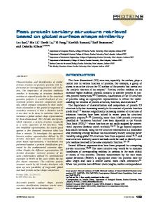
taurine has accumulated in the cells, it then inhibits the activation of PKC directly or indirectly (possibly through regulating Ca 21 availability), thus shutting down activation of CSAD through inhibition of CSAD phosphorylation by PKC. The activated CSAD soon returns to its inactive state through the action of a protein phosphatase, most likely PrP-2C (step 7).
REFERENCES
Figure 5. A proposed model on the role of protein phosphorylation in the regulation of taurine biosynthesis in the brain. The sequence of events leading from neuronal stimulation to increased synthesis of taurine is as follows: ( 1) arrival of action potential; ( 2) opening of voltage-dependent Ca 21 channels; ( 3) release of taurine; ( 4) activation of PKC by elevated [Ca 21]i ; ( 5) activation of C SAD by PKC -mediated protein phosphorylation; ( 6) increase of taurine biosynthesis by activated C SAD; and ( 7) termination of taurine biosynthesis by inactivation of C SAD through PrP-2C-mediated dephosphorylation.
of C SAD. Indeed, taurine has been shown to reduce Ca 21 influx into cells by inhibiting the Na 1/C a 21 exchanger (Matsuda et al., 1995). Furthermore, it has been reported that taurine inhibits phosphorylation of certain proteins in the retina or in the heart (Lombardini, 1992). In summary, it seems that there is a good correlation between changes of C SAD activity and the extent of C SAD phosphorylation in both in vitro (purified preparation) and in vivo (cultured neurons) systems. For instance, an increase of C SAD activity under depolarization conditions is accompanied by an increase in 32 P incorporation into C SAD. Similarly, when C SAD activity is inhibited, such as in the presence of taurine or PKC inhibitors, the extent of 32P incorporation into C SAD is also reduced. On the basis of the above results, the following model is proposed, as shown in Figure 5. When neurons are stimulated, the arrival of action potential (step 1) will open the voltage-dependent Ca 21channel (step 2), resulting in an increase of intracellular free Ca 21, [C a 21]i. Elevation of [C a 21]i will trigger release of taurine (step 3) as well as activation of PKC (step 4), which in turn activates C SAD through protein phosphorylation (step 5). The activated C SAD then synthesizes more taurine (step 6) to replenish that lost because of release by stimulation. When enough
Bao J, Nathan B, Wu JY (1994) Role of protein phosphorylation in the regulation of brain L-glutamate decarboxylase activity. J Biomed Sci 1:237–244. Bao J, Cheung W Y, Wu JY (1995) Brain L-glutamate decarboxylase: inhibition by phosphorylation and activation by dephosphorylation. J Biol Chem 270:6464 – 6467. Blinderman JM, Maitre M, Ossola L, Mandel P (1978) Purification and some properties of L-glutamate decarboxylase from human brain. Eur J Biochem 86:143–152. Hayes KC, C arey RE, Schmidt SY (1975) Retinal degeneration associated with taurine deficiency in the cat. Science 188:949 –951. Jacobsen JG, Smith L H (1968) Biochemistry and physiology of taurine and taurine derivatives. Physiol Rev 48:424 –511. Jerkins AA, Steele RD (1991a) Dietary sulf ur amino acid modulation of cysteine sulfinic acid decarboxylase. Am J Physiol 261:551–555. Jerkins AA, Steele RD (1991b) C ysteine sulfinic acid decarboxylase activity in response to thyroid hormone administration in rats. Arch Biochem Biophys 286:428 – 432. Jerkins AA, Steele RD (1992) Quantification of cysteine sulfinic acid decarboxylase in male and female rats: effect of adrenalectomy and methionine. Arch Biochem Biophys 294:534 –538. Kaisakia PJ, Jerkins AA, Goodspeed DC, Steele RD (1995) Cloning and characterization of rat cysteine sulfinic acid decarboxylase. Biochim Biophys Acta 1262:79 – 82. Kuriyama K (1980) Taurine as a neuromodulator. Fed Proc 39:2680 –2684. Lazarewicz JW, Noremberg K , Lehmann A, Hamberger A (1985) Effects of taurine on calcium binding and accumulation in rabbit hippocampal and cortical synaptosomes. Neurochem Int 7:421– 428. Lee YH, Deupree DL, Chen SC, Kao L S, Wu JY (1994) Role of Ca 21 in AM PA-mediated poly phosphoinositides turnover in primary neuronal cultures. J Neurochem 62:2325–2332. L leu PL, Croswell S, Huxtable RJ (1992) Phospholipids, phospholipid methylation and taurine content in synaptosomes of developing rat brain. Adv E xp Med Biol 315:221–228. L ombardini JB (1992) Taurine: nutritional value and mechanisms of action. Adv E xp Med Biol 315:309 –318. Matsuda T, Takuma K , Azuma J, Baba A (1995) Protective effect of taurine against calcium paradox-induced injury in cultured rat astrocytes. Abstract presented at the International Taurine Symposium ’95, Osaka, Japan, June 27–July 1, 1995, p. 80. Okamoto K, Kimura H, Sakai Y (1983) Evidence for taurine as an inhibitory neurotransmitter in cerebellar stellate interneurons: selective antagonism by TAG (6-aminomethyl-3-methyl-4H, 1, 2, 4-benzothiadiazine-1, 1-dioxide). Brain Res 265:163–168. Pasantes-Morales H, Cruz C (1985) Taurine and hypotaurine inhibit light-induced lipid peroxidation and protect rod outer segment structures. Brain Res 330:154 –157. Pion PD, K ittleson MD, Rogers QR, Morris JG (1987) Myocardial failure in cats associated with low plasma taurine: a reversible cardiomyopathy. Science 237:764 –768. Solis JM, Herranz AS, Herreras J, Lerma J, Del Rio RM (1988) Does taurine act as an osmoregulatory substance in the rat brain? Neurosci Lett 91:53–58. Sturman JA (1993) Taurine in development. Physiol Rev 73:119 –147. Taber TC, Lin C T, Song GX, Thalman RH, Wu JY (1986) Taurine in the rat hippocampus: localization and postsynaptic action. Brain Res 386:113–121. Tang X W, Hsu CC, Sun Y, Wu E, Yang C Y, Wu JY (1996) Multiplicity of brain cysteine sulfinic acid decarboxylase-purification, characterization and subunit structures. J Biomed Sci 3:442– 453. Wu JY (1982) Purification and characterization of cysteic/cysteine sulfinic acids decarboxylase and L-glutamate decarboxylase in bovine brain. Proc Natl Acad Sci USA 79:4270 – 4274.