Developmental Cell, Vol. 7, 107–116, July, 2004, Copyright 2004 by Cell Press
PlexinD1 and Semaphorin Signaling Are Required in Endothelial Cells for Cardiovascular Development Aaron D. Gitler,1,2 Min Min Lu,1 and Jonathan A. Epstein1,2,* 1 Cardiovascular Division Department of Medicine 2 Department of Cell and Developmental Biology University of Pennsylvania Health System 954 BRB II/III 421 Curie Boulevard Philadelphia, Pennsylvania 19104
Summary The identification of new signaling pathways critical for cardiac morphogenesis will contribute to our understanding of congenital heart disease (CHD), which remains a leading cause of mortality in newborn children worldwide. Signals mediated by semaphorin ligands and plexin receptors contribute to the intricate patterning of axons in the central nervous system. Here, we describe a related signaling pathway involving secreted class 3 semaphorins, neuropilins, and a plexin receptor, PlexinD1, expressed by endothelial cells. Interruption of this pathway in mice results in CHD and vascular patterning defects. The type of CHD caused by inactivation of PlexinD1 has previously been attributed to abnormalities of neural crest. Here, we show that this form of CHD can be caused by cellautonomous endothelial defects. Thus, molecular programs that mediate axon guidance in the central nervous system also function in endothelial cells to orchestrate critical aspects of cardiac morphogenesis.
Introduction Since the time of Vesalius, an association between the complex patterning of the vasculature and that of the peripheral nervous system has been apparent to anatomists. Nerves and blood vessels often course together, and it has been proposed that axons provide guidance cues that pattern the vasculature (Mukouyama et al., 2002). Alternatively, it is possible that blood vessels utilize parallel signaling pathways that result in predictable relationships between vascular and neural networks. In the nervous system, axonal pathways are shaped by repulsive cues provided by ligands of the semaphorin family that are sensed by migrating neuronal growth cones through neuropilin and plexin receptors (Raper, 2000). Semaphorins represent a family of membrane bound and secreted factors characterized by a conserved semaphorin domain (Kolodkin et al., 1993; Luo et al., 1993). In vertebrates, there are 19 semaphorins divided into five subclasses based on structural features and whether they are membrane bound or secreted (Good*Correspondence:
[email protected]
man et al., 1999). Semaphorins are expressed in the central nervous system, where function has been extensively analyzed (Luo et al., 1995). However, semaphorins also have been shown to be important regulators of diverse biological processes. The transmembrane semaphorins Sema4A and Sema4D function in specific immune responses (Hall et al., 1996; Kumanogoh et al., 2002). Sema7A is expressed by lymphocytes and activates monocytes (Holmes et al., 2002). Sema3A is expressed in the developing lung where it inhibits branching morphogenesis (Ito et al., 2000). Class 3 semaphorins (SEMA3 proteins) are secreted glycoproteins whose functions to guide axonal growth cones have been well characterized. Sema3A functions as a potent axonal repellant and can repel axonal projections from neurons of dorsal root ganglia (DRG) in vitro (Luo et al., 1993; Messersmith et al., 1995; Puschel et al., 1996; Varela-Echavarria et al., 1997). Mice with a targeted disruption of sema3A have severe axon guidance defects during early embryonic development (Behar et al., 1996; Taniguchi et al., 1997). Additional SEMA3 proteins cause neuronal growth cone collapse in similar in vitro assays. Sema3F has recently been shown to play a critical role in regulating peripheral nervous system connectivity (Sahay et al., 2003). Recently, receptors for some semaphorin ligands have been identified. In Drosophila, semaphorins directly bind the transmembrane receptor plexin (Winberg et al., 1998). Plexins are a large family of receptors that are also conserved in vertebrates, and some vertebrate semaphorin/plexin pairs have been identified. However, SEMA3 proteins do not appear to bind plexin receptors directly. Instead, they bind to receptors that include members of the neuropilin family (Kolodkin et al., 1997). Furthermore, the neuropilin receptor complex contains a plexin subunit, which is thought to transduce intracellular signals (He and Tessier-Lavigne, 1997; Rohm et al., 2000; Takahashi et al., 1999). In vertebrates, there are two neuropilins and at least ten plexins. Hence, diverse cellular responses may be achieved by the large variety of potential ligand-receptor combinations (Raper, 2000; Takahashi et al., 1999; Tamagnone et al., 1999). In order to understand this diversity, it will be important to characterize additional semaphorin-plexin and semaphorinneuropilin/plexin interactions. In the cardiovascular system, the analysis of neuropilins as semaphorin receptors is complicated by the fact that they also function as receptors for vascular endothelial growth factor (VEGF) (Soker et al., 1998). Neuropilin-1 (Npn-1) and Neuropilin-2 (Npn-2), in addition to their ability to heterodimerize with plexins, can interact with the VEGF receptor KDR (kinase-insert domain-containing receptor) on endothelial cells (ECs) and can bind VEGF. Mouse knockouts of neuropilins have revealed essential roles for these receptors in vascular development (Takashima et al., 2002). Several lines of evidence suggest an important role for semaphorins in the cardiovascular system. First, in addition to severe nervous system defects, Sema3A-
Developmental Cell 108
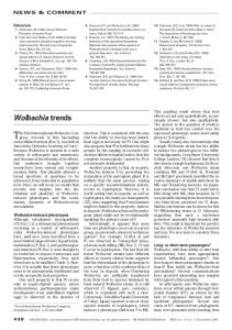
Figure 1. Endothelial-Specific Expression of plexinD1 (A) Whole-mount in situ hybridization of an E9.5 mouse embryo shows plexinD1 expression in the intersomitic blood vessels (arrow). (B–H) Radioactive in situ hybridizations of sections of E9.5–E11.5 mouse embryos. (B–D) At E9.5, strong plexinD1 expression is seen in intersomitic vessels (arrows, [B]), pharyngeal arch arteries (arrows, [C]), and outflow tract endothelium (arrows, [D]). At E10.5, plexinD1 expression is seen in endothelium lining the atria (at, [E]) and the dorsal aorta (ao, [F]). Notably, expression is seen in blood vessels surrounding the dorsal root ganglia (drg, [F]) but not within the drg. (G) At E11.5, expression is seen in endothelium of the branchial arches (ba), heart (ht), lining the dorsal aorta (ao), and other organs, as well as in a punctate pattern throughout the embryo where blood vessels are located. (H) Endothelium lining the outflow tract (ot) of the heart expresses plexinD1, but not the mesenchyme of the endocardial cushions, populated at this time point by neural crest cells. (I and J) PlexinD1 protein is expressed in endothelial cells in a pattern similar to the pan-endothelial marker PECAM. (K) Northern blot showing plexinD1 expression in cultured murine endothelial cell lines (1, Eoma; 2, MS-1; 3, MS-1/VEGF). Ethidium bromide stained agarose gel shows 28S rRNA as loading control. (L–R) Immunostaining confirms endothelial-specific PlexinD1 expression (L and N) that overlaps with that of von Willebrand factor (vWF, [M and O]). PlexinD1 expression is non-overlapping with that of neurofilament indicated by staining with 2H3 antibody (P–R) even within the region of the dorsal root ganglia (drg, high power shown in [R]).
PlexinD1 and Cardiovascular Development 109
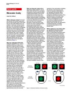
Figure 2. Targeted Inactivation of plexinD1 (A) A targeting vector was constructed to replace the first coding exon of plexinD1, containing the sema domain, with a neomycin resistance cassette. (B) Southern blot of BamH1-digested genomic DNA isolated from ES cell clones, probed with a 5⬘ probe demonstrates correct targeting of the plexinD1 locus. (C) PCR was used to genotype offspring from plexinD1⫹/⫺ intercrosses. (D and E) plexinD1 transcripts are detected in wild-type (D) but not knockout (E) hearts of E11.5 embryos. (F and G) PlexinD1 protein is also present in wild-type (F) but not knockout (G) E11.5 hearts. (H–K) Cardiac expression of SEMA3 signaling components. (H) plexinD1 is expressed specifically in endothelium (white arrows), whereas Npn-1 (I) and Npn-2 (J) are expressed in both myocardium (yellow arrows) and endothelium (white arrows). (K) sema3C expression is robust in OT myocardium (yellow arrows).
deficient mice exhibit cardiovascular defects on some genetic backgrounds (Behar et al., 1996). The cardiac defects are characterized by right ventricular hypertrophy and a dilated right atrium. sema3A⫺/⫺ embryos also exhibit vascular defects (Serini et al., 2003). Second, Sema3C-deficient mice die in the perinatal period with severe congenital cardiovascular defects (Feiner et al., 2001). These defects are reminiscent of those resulting from ablation of cardiac neural crest cells in the chick. Third, an elegant mouse model by Gu and colleagues, in which the ability of Npn-1 to transduce semaphorin signals is disrupted, suggests that neuropilin-mediated semaphorin signaling is required for cardiovascular patterning (Gu et al., 2003). Thus, semaphorins and neuropilins function outside of the nervous system to regulate cardiovascular development. Since neuropilins are expressed in both endothelium and neural crest, it is not clear in which cell type neuropilin-mediated semaphorin signaling functions. Furthermore, plexin coreceptors required for cardiovascular
development have not been identified, though recent evidence suggests that PlexinA1 may mediate signaling by Sema6D during cardiac development (Toyofuku et al., 2004). Here, we identify a plexin receptor that is essential for cardiac morphogenesis and determine the cell type in which semaphorin signaling is required during heart development. Our results suggest that ECs play an important role in orchestrating outflow tract septation and morphogenesis, processes previously attributed primarily to neural crest. Finally, we propose a model that integrates VEGF and semaphorin signaling pathways in cardiovascular development. Results and Discussion Identification of plexinD1 and Generation of plexinD1⫺/⫺ Mice Using a bioinformatics approach, we identified a mouseexpressed sequence tag (EST #AA138454) with homology to members of the plexin family, to a human cDNA
Developmental Cell 110
Table 1. Genotypes of Offspring from plexinD1⫹/⫺ Intercrosses Age
⫹/⫹
⫹/⫺
⫺/⫺
# of Litters
E10.5 E11.5 E12.5 E13.5-E16.5 P0 3 weeks
14 4 9 14 36 32
31 10 21 30 77 66
13 4 8 22a 26b 0
6 2 4 8 17 14
a b
Three embryos were partially resorbed. All became cyanotic and died within 24 hr.
(KIAA0620) and to a predicted human gene designated PLEXIND1 (Tamagnone et al., 1999). Therefore, we designated the mouse cDNA plexinD1. In situ hybridization analysis indicates that plexinD1 is expressed by embryonic vascular ECs. Between E9.5 and E11.5 (Figures 1A–1H), expression is robust in most, if not all, vascular endothelium including endocardium, intersomitic vessels, pulmonary vasculature, aorta, and pharyngeal arch arteries. Notably, plexinD1 is not expressed in neural crest-derived structures including the dorsal root ganglia at these early time points (Figure 1F). Expression in vascular endothelium persists throughout embryogenesis (data not shown). In the adult, plexinD1 is expressed in heart, brain, lung, kidney, and testis (data not shown; van der Zwaag et al., 2002), consistent with broad expression in the vasculature. Primary and immortalized EC lines including human umbilical vein endothelial cells (HUVECs) also express plexinD1 (Figure 1K and data not shown). To detect protein expression, we generated a polyclonal antibody against a portion of the extracellular domain of PlexinD1. PlexinD1 is expressed in a pattern similar to the pan-endothelial cell marker PECAM (platelet and endothelial cell adhesion molecule) (Figures 1I and 1J). Finally, we performed double and tripleimmunostaining to verify endothelial-specific expression of PlexinD1 (Figures 1L–1R). We generated mice with targeted inactivation of plexinD1 by homologous recombination in embryonic stem (ES) cells (Figures 2A–2C). Inactivation of plexinD1 was confirmed by both in situ hybridization (Figures 2D and 2E) and immunohistochemistry (Figures 2F and 2G). We were unable to detect either plexinD1 mRNA or PlexinD1 protein in homozygous knockout embryos, suggesting that we have generated a null allele.
Congenital Heart Disease in plexinD1⫺/⫺ Mice To determine if plexinD1 is required for survival, we analyzed offspring from intercrosses of heterozygous mice (Table 1). Most of the expected homozygous knockout mice are born; however, all mutant pups become cyanotic shortly after birth and succumb within 24 hr. All newborn homozygotes that we examined (n ⫽ 26) have structural cardiovascular defects involving the outflow tract (OT) of the heart and derivatives of the aortic arch arteries. In each case, persistent truncus arteriosus (PTA) is present (Figures 3A–3C). The atria are enlarged and thin-walled, and there is a uniform abnormality characterized by an ectopic origin of a coronary artery which arises from the mid portion of the
ascending aorta instead of from the coronary sinus (arrows, Figures 3C–3E). Increased vascularity is apparent on the surface of the ventricular chambers (Figure 3B). Some null pups (5/26) display a right-sided aortic arch (Figure 3C). To better visualize vascular patterning, we performed polymer corrosion casts at E18.5 (Figures 3D and 3E). In wild-type embryos, the pulmonary artery and aorta are clearly distinct vessels, and the ductus arteriosus is patent. In plexinD1 knockout embryos, the right and left pulmonary arteries arise from the truncus arteriosus, indicating a failure of OT septation. In some cases (Figure 3E) the right subclavian artery arises from a more cranial (distal) location than in wild-type, and the left carotid artery is missing or atretic. The ductus arteriosus is absent, and the abnormal origin of the coronary artery is apparent (arrow, Figure 3E). These cardiovascular defects are sufficient to account for perinatal lethality of plexinD1-deficient mice. Thus, the cardiovascular phenotype of plexinD1-deficient mice reveals an unexpected role for endothelium in OT morphogenesis. Cardiac Neural Crest Migration Is Unaffected in plexinD1⫺/⫺ Mice Failure of OT septation has been previously attributed to defects in cardiac neural crest (Creazzo et al., 1998; Kirby et al., 1983). To determine if loss of PlexinD1 affected neural crest migration, we analyzed a set of molecular markers including endothelin receptor A (ednrA; Figures 4A and 4B), FoxC1 (Figures 4C and 4D), and plexinA2 (Figures 4E and 4F). Neural crest cell migration appears normal in E12.5 plexinD1⫺/⫺ embryos. Neural crest cells migrate properly to and surround the pharyngeal arches and populate the endocardial cushions of the OT. In addition, patterning of the peripheral nervous system appeared unaffected in E10.5 mutant embryos, as assessed by anti-neurofilament antibody staining (Figures 4G and 4H and data not shown). Pharyngeal Arch Patterning Defects and Failure to Recruit Smooth Muscle in plexinD1⫺/⫺ Mice Defects in cardiovascular development are evident as early as E10.5. Normally, the 3rd, 4th, and 6th aortic arch arteries can be easily identified within the segmented pharyngeal arches in frontal sections at this time point (Figure 3F). In plexinD1 mutants, however, the 4th aortic arch arteries are diminutive and the 6th aortic arch arteries are extremely small or absent (Figure 3G). ECs are present in the 3rd and 4th arch arteries of plexinD1-deficient embryos as determined by expression of PECAM (Figures 3H and 3I). Intriguingly, there is an excessive number of ECs in the vicinity of the 6th arch artery that fail to coalesce into a vascular tube (Figures 3H and 3I). The defective 6th arch artery explains the loss of ductus arteriosus in the newborn mutant mice, and the abnormal 4th arch artery is consistent with the incidence of right-sided aortic arch that we observe. Deficient aortic arch artery development could reflect a primary defect in endothelium that results in secondary failure to recruit vascular smooth muscle, a process that is required for maintenance of aortic arch artery integrity (Kochilas et al., 2002; Lindsay and Baldini, 2001). Consistent with this hypothesis, there is a striking deficiency of differentiated
PlexinD1 and Cardiovascular Development 111
Figure 3. Congenital Heart Disease and Vascular Abnormalities in plexinD1⫺/⫺ Mice (A) Two great vessels, aorta (Ao) and pulmonary artery (PA), arise from the wild-type P0 mouse heart. (B) Only one great vessel, truncus arteriosus (TA), arises from plexinD1⫺/⫺ hearts at P0, and there is an abnormal origin of the coronary artery (arrow). (C) Some of the mutants also have a right-sided aortic arch and retro-esophageal right subclavian artery. (D and E) Acrylic corrosion casts of P0 wild-type (D) and mutant (E) demonstrate a coronary artery anomaly in the mutants (arrows) and truncus arteriosus. (F and G) Frontal sections through the pharyngeal arch arteries (numbered) at E10.5 demonstrate that plexinD1⫺/⫺ embryos (G) have diminutive 4th pharyngeal arch arteries and are missing the 6th arch arteries compared to wild-type (F). (H and I) PECAM staining confirms diminutive 4th arch arteries in mutant embryos (I) and excessive numbers of PECAM-expressing endothelial cells in the vicinity of missing 6th arch arteries. (J and K) Smooth muscle differentiation is defective in the 4th arch arteries of the mutants (K), compared to wild-type (J), as visualized by ␣SMA staining. (L and M) By E12.5, the wild-type outflow tract (L) has septated into aorta (Ao) and pulmonary artery (PA), but septation does not occur in the mutants (M), resulting in a persistent truncus arteriosus (TA), and the atria are enlarged. (N and O) The myocardium is thinned in E12.5 mutants. There are also ectopic coronary vessels (arrows, [O]) on the epicardial surface of mutant hearts. (P and Q) Higher magnification of boxed regions shown in (N) and (O). Arrowheads (Q) show blood-filled vascular structures. (R and S) PECAM whole-mount immunostaining labels well-organized intersomitic blood vessels of an E10.5 wild-type embryo (R), whereas the intersomitic vessels of the mutant are disorganized (S). (T and U) Intercostal blood vessels of plexinD1 mutant newborns (U) are also disorganized compared to wild-type (T).
Developmental Cell 112
Figure 4. Preserved Cardiac Neural Crest Migration and Neuronal Patterning in plexinD1 Mutant Embryos (A–F) Radioactive in situ hybridization of E12.5 wild-type (A, C, and E) or mutant (B, D, and F) embryos shows preserved expression of cardiac neural crest markers ednrA (A and B), FoxC1 (C and D), and plexinA2 (E and F). Note comparable amounts of neural crest cells populating the endocardial cushions of the outflow tract (arrows). (G and H) Normal neuronal patterning, as visualized by immunostaining frontal sections of E10.5 wild-type (G) or mutant (H) embryos with monoclonal antibody 2H3 directed against neurofilament.
smooth muscle cells expressing ␣ smooth muscle actin (␣SMA) surrounding the 4th and 6th arch arteries of mutant embryos compared to wild-type (Figures 3J and 3K). Mutations in the endothelin pathway, by targeted disruption of endothelin-1 (ET-1), endothelin converting enzyme-1, or endothelin receptor A (ednrA), result in neural crest-related defects involving the pharyngeal arch arteries (Kurihara et al., 1995; Yanagisawa et al., 1998). This pathway functions in pharyngeal arch endothelium and provides signals to surrounding mesenchyme, resulting in proper remodeling of the great vessels. We did not observe differences in ET-1 or ednrA expression in plexinD1 mutant embryos (Supplemental Data [http:// www.developmentalcell.com/cgi/content/7/1/107/ DC1] and Figures 4E and 4F). However, subtle defects in endothelin expression or protein processing remain possible and future work will be required to address these possibilities and to examine potential genetic interactions between these signaling pathways. We examined the expression of a series of genes previously implicated in endothelial cell signaling and aortic arch artery patterning (Abramsson et al., 2003; Hellstrom et al., 1999; Kuo et al., 1997; Shin et al., 2001; Yanagisawa et al., 1998). endothelin-1, ephrinB2, PDGF-b, PDGFR-, and LKLF were expressed in the OT and pharyngeal arch artery endothelium at comparable levels in both wild-type and mutant embryos (data not shown and Supplemental Figure S1). Coronary and Intersomitic Blood Vessel Defects in plexinD1⫺/⫺ Mice At E12.5, cardiac development is notably abnormal in knockout embryos. The atria are enlarged and thinwalled (Figures 3L and 3M). The OT of wild-type embryos
is septated by E12.5 (Figure 3L), while there is no evidence of conotruncal septation in null embryos (Figure 3M). On the ventricular surface of mutant hearts, there are an excessive number of small vessels (Figures 3O and 3Q) compared to wild-type (Figures 3N and 3P). These vascular structures contain blood and express PECAM, indicating that they are lined by endothelium (data not shown). Loss-of-function mutations in the zebrafish plexinD1 ortholog result in patterning defects of intersomitic blood vessels (Torres-Vazquez et al., 2004). In the mouse, plexinD1 is strongly expressed by intersomitic vessels at E9.5 (Figure 1A). By E10.5, patterning of intersomitic vessels is clearly abnormal in plexinD1 knockout embryos, as evidenced by whole-mount PECAM immunostaining (Figures 3R and 3S). The vessels of mutant embryos are disorganized and are not confined to regions between somites. Analogous defects in the intercostal blood vessels of newborn plexinD1 knockout mice are also present (Figures 3T and 3U). These defects are less severe than those seen in embryos, suggesting a partial recovery of intersomitic vessel development at later stages. Furthermore, we do not observe changes in cell death or proliferation in mutant embryos, as determined by TUNEL and phospho-histone H3 staining, respectively (data not shown). These results, coupled with those in zebrafish, suggest that PlexinD1 signaling may mediate guidance cues responsible for some aspects of vascular patterning. PlexinD1 Associates with Neuropilin to Form a Receptor for Class 3 Semaphorins The cardiac OT defects of plexinD1 knockout mice are remarkably similar to sema3C knockouts (Feiner et
PlexinD1 and Cardiovascular Development 113
to Npn-1 is enhanced in the presence of PlexinA1 (Takahashi et al., 1999). However, no plexin previously has been described to enhance binding of Sema3C to neuropilins. Our data indicate that binding of both Sema3A and Sema3C to COS cells expressing Npn-1 is greatly enhanced by cotransfection of PlexinD1 (Figures 5B– 5D). PlexinD1 also enhanced binding of Sema3C to Npn-2 (Figure 5E). Thus, we propose that PlexinD1 heterodimerizes with Npn-1 or Npn-2 to form a receptor capable of binding SEMA3 proteins.
Figure 5. PlexinD1 Interacts with Npn-1, and This Association Enhances Binding to Sema3C (A) Coimmunoprecipitation experiments show that full-length PlexinD1 (PlexinD1-V5) and a cytoplasmic domain truncation (PlexinD1⌬cyt-V5) can form a complex with Npn-1 and that this interaction requires the sema domain (PlexinD1⌬sema-V5). (B–D) AP-Sema3C binding to Cos-7 cells transfected with Npn-1 alone (B), Npn-1 and PlexinA1 (C), or Npn-1 and PlexinD1 (D) was measured colorimetrically. Weak binding of Sema3C to Npn-1 or to Npn-1/PlexinA1 was detected after 20 min incubation in alkaline phosphatase staining solution (B and C), whereas strong binding was detected to Npn-1/PlexinD1 transfected cells (D). (E) Summary of AP-Sema binding experiments. Shown is one of five experiments with similar results. Data are displayed as mean pixel intensity (arbitrary units) after subtraction of background (mean ⫾ SEM, n ⫽ 5). *p ⬍ 0.01, **p ⬍ 0.05.
al., 2001), endothelial-restricted neuropilin-1 (Npn-1) knockouts (Gu et al., 2003), and Npn-1[-sema];Npn-2 knockouts (Gu et al., 2003). Furthermore, plexinD1, Npn-1, and Npn-2 are coexpressed in endothelium and sema3C is expressed by outflow tract myocardium (Figures 2H– 2K). Therefore, we sought to determine if PlexinD1 could heterodimerize with Npn-1 to form a functional receptor for Sema3C. Coimmunoprecipitation experiments indicate that PlexinD1 and Npn-1 can associate, and this interaction requires the sema domain of PlexinD1 (Figure 5A). Different SEMA3 proteins bind to Npn-1 or Npn-2 with varying affinities. Sema3A binds to Npn-1 but not Npn-2, while Sema3C can bind to both neuropilins (Chen et al., 1997; He and Tessier-Lavigne, 1997; Kolodkin et al., 1997; Takahashi et al., 1998). Plexins can enhance the binding of SEMA3 proteins to neuropilins (He and Tessier-Lavigne, 1997; Kolodkin et al., 1997; Takahashi et al., 1999; Tamagnone et al., 1999). Sema3A binding
A Model for the Integration of VEGF and Semaphorin Signaling in Cardiovascular Development The closely related phenotypes involving OT and aortic arch defects of sema3C, plexinD1, and Npn-1 knockout mice (summarized in Table 2), coupled with our biochemical data, strongly suggest that semaphorin signaling mediated by ECs expressing PlexinD1 and neuropilins composes a receptor-ligand paracrine signaling pathway that orchestrates septation of the OT and development of aortic arch artery derivatives. Congenital heart defects in humans frequently involve OT and aortic arch defects, and SEMA3C, PLEXIND1, and NPN-1 are candidate genes for congenital heart disease. The ability of neuropilin to bind distinct ligands (VEGF165 and SEMA3 proteins), coupled with recent elegant genetic studies (Feiner et al., 2001; Gu et al., 2003; Soker et al., 1998; Stalmans et al., 2003), has previously suggested that neuropilin facilitates VEGF signaling in endothelium and semaphorin signaling in non-ECs including cardiac neural crest (Gu et al., 2003). Endothelial-specific loss of Npn-1 has been interpreted in terms of loss of VEGF165 signaling in endothelium (Gu et al., 2003). However, our biochemical and genetic data identify a direct role for neuropilin-mediated semaphorin signaling in endothelium. This result demands reinterpretation of existing data concerning the role of neuropilin, VEGF, and semaphorin signaling in cardiovascular development and leads us to propose a unifying model (Figure 6). We suggest that neuropilin, in ECs, functions in both VEGF and semaphorin signaling and that both pathways are required for proper cardiac OT development. Either Npn-1 or Npn-2 is able to cooperate with PlexinD1 to bind Sema3C. This explains why specific inhibition of Npn-1-dependent semaphorin signaling does not result in OT defects unless the redundant Npn-2 is also inactivated (Gu et al., 2003), whereas inactivating either the ligand Sema3C (Feiner et al., 2001) or the coreceptor PlexinD1 is sufficient to produce OT defects. This model also explains why loss of the VEGF signaling component, by virtue of either an isoform specific VEGF165 knockout (Stalmans et al., 2003) or endothelial restricted Npn-1 knockout (Gu et al., 2003) leads to OT defects. Finally, our model explains defects in atrial development associated with disruptions in semaphorin signaling in ECs. We propose that Sema3A provides this signal to PlexinD1 and Npn-1. Accordingly, sema3A, endothelial-restricted Npn-1, and plexinD1 knockout mice present with similar atrial defects (Behar et al., 1996; Gu et al., 2003), although these studies do not address the question of whether atrial defects are primary or secondary to distinct cardiovascular abnormalities. Additional SEMA3 genes are expressed during cardiac development in regionalized patterns (A.D.G. and
Developmental Cell 114
Table 2. Cardiac Phenotypes in Semaphorin and VEGF Signaling Mutants Genotype
PlexinD1⫺/⫺ Sema3C⫺/⫺ Sema3A⫺/⫺ Npn1ecKO Npn1[Sema-] Npn1[Sema-];Npn2⫺/⫺ VEGF165⫺/⫺
PTA
⫹ ⫹ ⫺ ⫹ ⫺ ⫹ ⫹
VSD
⫹ ⫹ ⫺ ⫹ ⫺ ⫹ ⫹
Cardiac Phenotypes
Reference
Coronary Artery Anomaly
Aortic Arch Defects
Atrial Defects
⫹ ⫺ ⫺ ⫹ ⫺ ⫹ ⫺
⫹a ⫹b ⫺ ⫺ ⫺ ⫺ ⫹c
⫹ ⫺ ⫹ ⫹ ⫹ ⫹ ⫺
Feiner et al., 2001 Behar et al., 1996 Gu et al., 2003 Gu et al., 2003 Gu et al., 2003 Stalmans et al., 2003
Comparison of frequency of specific cardiac embryonic phenotypes between Semaphorin and VEGF signaling pathway mutants. Endothelialrestricted Neuropilin-1 knockout (Npn1ecKO); persistent truncus arteriosus (PTA); ventricular septal defect (VSD). a, subset of mutants have right-sided aortic arch. b, interrupted aortic arch. c, mutants can present with either right-sided arch or interrupted aortic arch. (“⫺” indicates not present or not reported).
J.A.E., unpublished data). It is likely that PlexinD1 and neuropilin function together in ECs to transduce multiple semaphorin signals required for various aspects of cardiovascular development. In zebrafish, PlexinD1 functions to mediate repulsive signals during sprouting and growth of the intersomitic vessels (Torres-Vazquez et al., 2004) while cardiac morphogenesis appears normal. However, fish do not have a septated outflow tract. The observation of vascular and cardiac outflow tract defects in mice lacking PlexinD1 suggests that this endothelial receptor has acquired new functions during evolution coincident with increased complexity of cardiovascular patterning. Recent data indicate that SEMA3 proteins play a role in
vascular development and angiogenesis via autocrine signals in ECs, resulting in alterations in integrin activation and adhesiveness to extracellular matrix (Serini et al., 2003). Interestingly, SEMA3C expression was not detected in vitro in human ECs (Serini et al., 2003). This is consistent with our data, which suggest sema3C, expressed by OT myocardium, is involved in paracrine signaling to ECs expressing the receptors Npn-1, Npn-2, and PlexinD1. Taken together, these results demonstrate unexpected similarities between neural patterning, vascular patterning, and endothelial function during cardiovascular morphogenesis. Experimental Procedures In Situ Hybridization Whole-mount and in situ hybridization protocols are available at http://www.uphs.upenn.edu/mcrc/histology/histologyhome.html. The plexinD1 antisense riboprobe derived from EST #AA138454 corresponding to bp 5901–6955 of mouse plexinD1 mRNA (Accession # XM_149784). 35S-labeled antisense riboprobes were synthesized by in vitro run-off transcription of linearized plasmids, using SP6, T7 or T3 RNA polymerase, and 35S-UTP. Probes for FoxC1 (Kume et al., 2001), ednrA (Yanagisawa et al., 1998), plexinA2 (Brown et al., 2001), Npn-1 (Brown et al., 2001), Npn-2 (Brown et al., 2001), and ET-1 (Yanagisawa et al., 1998) have been described previously. The sema3C probe was prepared from a 3.2 kb mouse cDNA cloned into pBSII/KS (provided by Jonathan Raper). The plasmid was linearized with Not 1 and transcribed with T3 RNA polymerase. The PDGF-b and PDGFR- probes derived from EST #BE554525 and #BF120498, respectively, by linearization with EcoR1 and transcription with T7 RNA polymerase. The EphrinB2 and LKLF templates were kindly provided by John Lee and Mark Kahn, and we used T7 polymerase to synthesize antisense riboprobes.
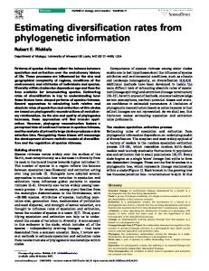
Figure 6. A Model for the Integration of VEGF and Semaphorin Signaling in Cardiovascular Development Endothelial cells express neuropilin receptors capable of heterodimerization with PlexinD1 or the VEGF receptor KDR. Both VEGF signaling (mediated by VEGF165 and KDR) and semaphorin signaling (mediated by Sema3C and PlexinD1) utilize neuropilin receptor complexes and are required for outflow tract development. Disruptions in Sema3A-dependent semaphorin signaling cause atrial defects.
Immunohistochemistry Immunohistochemistry protocols are available at http://www.uphs. upenn.edu/mcrc/histology/histologyhome.html. To detect PlexinD1 protein, we used rabbit polyclonal antisera raised against a 60 amino acid peptide (corresponding to aa 186–237) of mouse PlexinD1 (Accession # XP_149784) at a dilution of 1:500. To detect PECAM, we used CD31 antiserum at a dilution of 1:500. To detect ␣ smooth muscle actin, we used A2547 (Sigma) at a dilution of 1:1000. To detect vWF, we used Von Willebrand Factor antiserum (Sigma) at a dilution of 1:20 with the Zenon labeling kit (Molecular Probes). To detect neurofilament, we used monoclonal antibody 2H3 (Developmental Studies Hybridoma Bank) at a dilution of 1:10. Northern Blotting Total RNA was isolated from endothelial cell lines using Trizol (Invitrogen). 20 g of total RNA was used for Northern blots hybridized
PlexinD1 and Cardiovascular Development 115
with a 32P-labeled probe generated from EST #AA138454, corresponding to bp 5901–6955 of mouse plexinD1 (Accession # XP_149784). Generating plexinD1 Knockout Mice We constructed a targeting vector based on pPNT-loxP (Tybulewicz et al., 1991). PCR was used to amplify 6 kb and 2 kb long and short homology arms from R1 ES cell genomic DNA. We screened 600 drug-resistant R1 ES clones by Southern blotting and identified five correctly targeted clones. Two clones were injected into C57Bl/6 blastocysts to generate chimeric mice that were crossed to C57Bl/6 to generate heterozygous plexinD1 knockout mice. (Primer sequences for PCR genotyping are available upon request.) Coimmunoprecipitation We transfected 1 ⫻ 106 HEK293T cells with 5 g myc-Npn-1 and 5 g of pcDNA3.1, pcDNA3.1-plexinD1-V5, pcDNA3.1-plexinD1⌬Cyt-V5, pcDNA3.1-plexinD1⌬Sema-V5, or pcDNA3.1-plexinA2⌬Cyt-V5. These constructs were derived from cDNA KIAA0620 (Accession # AB014520). 48 hr after transfection, cells were lysed in NP-40 lysis buffer (150 mM NaCl, 50 mM Tris [pH 8.0], 1% NP-40) and immunoprecipitated with a monoclonal V5 antibody (Invitrogen) for 2 hr at 4⬚C. Complexes were incubated with protein A/G sepharose (Santa Cruz), separated by SDS-PAGE and transferred to PVDF membranes. We used a monoclonal myc antibody (9E10) to detect Npn-1 by Western blotting. AP-Sema Binding Experiments HEK293T cells were transfected with pAG-AP-Sema3C. 48 hr after transfection, conditioned medium was diluted 1:10 in DMEM with 10% FBS. Separately, we transfected 2 ⫻ 105 COS-7 cells with 0.4 g Npn-1 and 2 g pcDNA3.1, PlexinA1, or PlexinD1. Transfected COS-7 cells were incubated with diluted AP-Sema3C conditioned media for 90 min at room temperature, washed with PBS, and fixed with 4% paraformaldehyde. Endogenous alkaline phosphatases were inactivated by heating at 65⬚C for 3 hr. AP-Sema binding was detected by incubating cells in AP buffer (100 mM Tris [pH 9.5], 100 mM NaCl, 50 mM MgCl2–0.1% Tween 20, 0.33 mg/ml nitroblue tetrazolium, and 0.05 mg/ml BCIP) for 20–30 min at room temperature. Stained cells were digitally photographed and staining intensity was quantified using ImageJ software (NIH). Acknowledgments We are grateful to Brant Weinstein, Jesus Torres-Vazquez, Sarah Childs, and members of the Epstein lab for helpful discussions. We thank the Kazusa DNA Research Institute, Japan, for providing the KIAA0620 cDNA clone. We also thank Li Jia and Jonathan Raper for reagents and advice. This work was supported by grants from the National Institutes of Health and the American Heart Association to J.A.E. A.D.G. is supported by the Department of Cell and Developmental Biology predoctoral training grant from the National Institutes of Health. Received: February 18, 2004 Revised: May 5, 2004 Accepted: May 5, 2004 Published online: June 10, 2004 References Abramsson, A., Lindblom, P., and Betsholtz, C. (2003). Endothelial and nonendothelial sources of PDGF-B regulate pericyte recruitment and influence vascular pattern formation in tumors. J. Clin. Invest. 112, 1142–1151. Behar, O., Golden, J.A., Mashimo, H., Schoen, F.J., and Fishman, M.C. (1996). Semaphorin III is needed for normal patterning and growth of nerves, bones and heart. Nature 383, 525–528. Brown, C.B., Feiner, L., Lu, M.M., Li, J., Ma, X., Webber, A.L., Jia, L., Raper, J.A., and Epstein, J.A. (2001). PlexinA2 and semaphorin signaling during cardiac neural crest development. Development 128, 3071–3080. Chen, H., Chedotal, A., He, Z., Goodman, C.S., and Tessier-Lavigne,
M. (1997). Neuropilin-2, a novel member of the neuropilin family, is a high affinity receptor for the semaphorins Sema E and Sema IV but not Sema III. Neuron 19, 547–559. Creazzo, T.L., Godt, R.E., Leatherbury, L., Conway, S.J., and Kirby, M.L. (1998). Role of cardiac neural crest cells in cardiovascular development. Annu. Rev. Physiol. 60, 267–286. Feiner, L., Webber, A.L., Brown, C.B., Lu, M.M., Jia, L., Feinstein, P., Mombaerts, P., Epstein, J.A., and Raper, J.A. (2001). Targeted disruption of semaphorin 3C leads to persistent truncus arteriosus and aortic arch interruption. Development 128, 3061–3070. Goodman, C.S., Kolodkin, A.L., Luo, Y., Puschel, A.W., and Raper, J.A. (1999). Unified nomenclature for the semaphorins/collapsins. Semaphorin Nomenclature Committee. Cell 97, 551–552. Gu, C., Rodriguez, E.R., Reimert, D.V., Shu, T., Fritzsch, B., Richards, L.J., Kolodkin, A.L., and Ginty, D.D. (2003). Neuropilin-1 conveys semaphorin and VEGF signaling during neural and cardiovascular development. Dev. Cell 5, 45–57. Hall, K.T., Boumsell, L., Schultze, J.L., Boussiotis, V.A., Dorfman, D.M., Cardoso, A.A., Bensussan, A., Nadler, L.M., and Freeman, G.J. (1996). Human CD100, a novel leukocyte semaphorin that promotes B-cell aggregation and differentiation. Proc. Natl. Acad. Sci. USA 93, 11780–11785. He, Z., and Tessier-Lavigne, M. (1997). Neuropilin is a receptor for the axonal chemorepellent Semaphorin III. Cell 90, 739–751. Hellstrom, M., Kalen, M., Lindahl, P., Abramsson, A., and Betsholtz, C. (1999). Role of PDGF-B and PDGFR-beta in recruitment of vascular smooth muscle cells and pericytes during embryonic blood vessel formation in the mouse. Development 126, 3047–3055. Holmes, S., Downs, A.M., Fosberry, A., Hayes, P.D., Michalovich, D., Murdoch, P., Moores, K., Fox, J., Deen, K., Pettman, G., et al. (2002). Sema7A is a potent monocyte stimulator. Scand. J. Immunol. 56, 270–275. Ito, T., Kagoshima, M., Sasaki, Y., Li, C., Udaka, N., Kitsukawa, T., Fujisawa, H., Taniguchi, M., Yagi, T., Kitamura, H., and Goshima, Y. (2000). Repulsive axon guidance molecule Sema3A inhibits branching morphogenesis of fetal mouse lung. Mech. Dev. 97, 35–45. Kirby, M.L., Gale, T.F., and Stewart, D.E. (1983). Neural crest cells contribute to normal aorticopulmonary septation. Science 220, 1059–1061. Kochilas, L., Merscher-Gomez, S., Lu, M.M., Potluri, V., Liao, J., Kucherlapati, R., Morrow, B., and Epstein, J.A. (2002). The role of neural crest during cardiac development in a mouse model of DiGeorge syndrome. Dev. Biol. 251, 157–166. Kolodkin, A.L., Matthes, D.J., and Goodman, C.S. (1993). The semaphorin genes encode a family of transmembrane and secreted growth cone guidance molecules. Cell 75, 1389–1399. Kolodkin, A.L., Levengood, D.V., Rowe, E.G., Tai, Y.T., Giger, R.J., and Ginty, D.D. (1997). Neuropilin is a semaphorin III receptor. Cell 90, 753–762. Kumanogoh, A., Marukawa, S., Suzuki, K., Takegahara, N., Watanabe, C., Ch’ng, E., Ishida, I., Fujimura, H., Sakoda, S., Yoshida, K., and Kikutani, H. (2002). Class IV semaphorin Sema4A enhances T-cell activation and interacts with Tim-2. Nature 419, 629–633. Kume, T., Jiang, H., Topczewska, J.M., and Hogan, B.L. (2001). The murine winged helix transcription factors, Foxc1 and Foxc2, are both required for cardiovascular development and somitogenesis. Genes Dev. 15, 2470–2482. Kuo, C.T., Veselits, M.L., Barton, K.P., Lu, M.M., Clendenin, C., and Leiden, J.M. (1997). The LKLF transcription factor is required for normal tunica media formation and blood vessel stabilization during murine embryogenesis. Genes Dev. 11, 2996–3006. Kurihara, Y., Kurihara, H., Oda, H., Maemura, K., Nagai, R., Ishikawa, T., and Yazaki, Y. (1995). Aortic arch malformations and ventricular septal defect in mice deficient in endothelin-1. J. Clin. Invest. 96, 293–300. Lindsay, E.A., and Baldini, A. (2001). Recovery from arterial growth delay reduces penetrance of cardiovascular defects in mice deleted for the DiGeorge syndrome region. Hum. Mol. Genet. 10, 997–1002.
Developmental Cell 116
Luo, Y., Raible, D., and Raper, J.A. (1993). Collapsin: a protein in brain that induces the collapse and paralysis of neuronal growth cones. Cell 75, 217–227. Luo, Y., Shepherd, I., Li, J., Renzi, M.J., Chang, S., and Raper, J.A. (1995). A family of molecules related to collapsin in the embryonic chick nervous system. Neuron 14, 1131–1140. Messersmith, E.K., Leonardo, E.D., Shatz, C.J., Tessier-Lavigne, M., Goodman, C.S., and Kolodkin, A.L. (1995). Semaphorin III can function as a selective chemorepellent to pattern sensory projections in the spinal cord. Neuron 14, 949–959. Mukouyama, Y.S., Shin, D., Britsch, S., Taniguchi, M., and Anderson, D.J. (2002). Sensory nerves determine the pattern of arterial differentiation and blood vessel branching in the skin. Cell 109, 693–705. Puschel, A.W., Adams, R.H., and Betz, H. (1996). The sensory innervation of the mouse spinal cord may be patterned by differential expression of and differential responsiveness to semaphorins. Mol. Cell. Neurosci. 7, 419–431. Raper, J.A. (2000). Semaphorins and their receptors in vertebrates and invertebrates. Curr. Opin. Neurobiol. 10, 88–94. Rohm, B., Ottemeyer, A., Lohrum, M., and Puschel, A.W. (2000). Plexin/neuropilin complexes mediate repulsion by the axonal guidance signal semaphorin 3A. Mech. Dev. 93, 95–104. Sahay, A., Molliver, M.E., Ginty, D.D., and Kolodkin, A.L. (2003). Semaphorin 3F is critical for development of limbic system circuitry and is required in neurons for selective CNS axon guidance events. J. Neurosci. 23, 6671–6680. Serini, G., Valdembri, D., Zanivan, S., Morterra, G., Burkhardt, C., Caccavari, F., Zammataro, L., Primo, L., Tamagnone, L., Logan, M., et al. (2003). Class 3 semaphorins control vascular morphogenesis by inhibiting integrin function. Nature 424, 391–397. Shin, D., Garcia-Cardena, G., Hayashi, S., Gerety, S., Asahara, T., Stavrakis, G., Isner, J., Folkman, J., Gimbrone, M.A., Jr., and Anderson, D.J. (2001). Expression of ephrinB2 identifies a stable genetic difference between arterial and venous vascular smooth muscle as well as endothelial cells, and marks subsets of microvessels at sites of adult neovascularization. Dev. Biol. 230, 139–150. Soker, S., Takashima, S., Miao, H.Q., Neufeld, G., and Klagsbrun, M. (1998). Neuropilin-1 is expressed by endothelial and tumor cells as an isoform-specific receptor for vascular endothelial growth factor. Cell 92, 735–745. Stalmans, I., Lambrechts, D., De Smet, F., Jansen, S., Wang, J., Maity, S., Kneer, P., von der Ohe, M., Swillen, A., Maes, C., et al. (2003). VEGF: a modifier of the del22q11 (DiGeorge) syndrome? Nat. Med. 9, 173–182. Takahashi, T., Nakamura, F., Jin, Z., Kalb, R.G., and Strittmatter, S.M. (1998). Semaphorins A and E act as antagonists of neuropilin-1 and agonists of neuropilin-2 receptors. Nat. Neurosci. 1, 487–493. Takahashi, T., Fournier, A., Nakamura, F., Wang, L.H., Murakami, Y., Kalb, R.G., Fujisawa, H., and Strittmatter, S.M. (1999). Plexinneuropilin-1 complexes form functional semaphorin-3A receptors. Cell 99, 59–69. Takashima, S., Kitakaze, M., Asakura, M., Asanuma, H., Sanada, S., Tashiro, F., Niwa, H., Miyazaki Ji, J., Hirota, S., Kitamura, Y., et al. (2002). Targeting of both mouse neuropilin-1 and neuropilin-2 genes severely impairs developmental yolk sac and embryonic angiogenesis. Proc. Natl. Acad. Sci. USA 99, 3657–3662. Tamagnone, L., Artigiani, S., Chen, H., He, Z., Ming, G.I., Song, H., Chedotal, A., Winberg, M.L., Goodman, C.S., Poo, M., et al. (1999). Plexins are a large family of receptors for transmembrane, secreted, and GPI-anchored semaphorins in vertebrates. Cell 99, 71–80. Taniguchi, M., Yuasa, S., Fujisawa, H., Naruse, I., Saga, S., Mishina, M., and Yagi, T. (1997). Disruption of semaphorin III/D gene causes severe abnormality in peripheral nerve projection. Neuron 19, 519–530. Torres-Vazquez, J., Gitler, A.D., Fraser, S.D., Berk, J.A., Pham, V.N., Fishman, M.C., Childs, S., Epstein, J.A., and Weinstein, B.M. (2004). Semaphorin-Plexin signaling guides patterning of the developing vasculature. Dev. Cell 7, this issue, 117–123. Toyofuku, T., Zhang, H., Kumanogoh, A., Takegahara, N., Suto, F.,
Kamei, J., Aoki, K., Yabuki, M., Hori, M., Fujisawa, H., and Kikutani, H. (2004). Dual roles of Sema6D in cardiac morphogenesis through region-specific association of its receptor, Plexin-A1, with off-track and vascular endothelial growth factor receptor type 2. Genes Dev. 18, 435–447. Tybulewicz, V.L., Crawford, C.E., Jackson, P.K., Bronson, R.T., and Mulligan, R.C. (1991). Neonatal lethality and lymphopenia in mice with a homozygous disruption of the c-abl proto-oncogene. Cell 65, 1153–1163. van der Zwaag, B., Hellemons, A.J., Leenders, W.P., Burbach, J.P., Brunner, H.G., Padberg, G.W., and Van Bokhoven, H. (2002). PLEXIN-D1, a novel plexin family member, is expressed in vascular endothelium and the central nervous system during mouse embryogenesis. Dev. Dyn. 225, 336–343. Varela-Echavarria, A., Tucker, A., Puschel, A.W., and Guthrie, S. (1997). Motor axon subpopulations respond differentially to the chemorepellents netrin-1 and semaphorin D. Neuron 18, 193–207. Winberg, M.L., Noordermeer, J.N., Tamagnone, L., Comoglio, P.M., Spriggs, M.K., Tessier-Lavigne, M., and Goodman, C.S. (1998). Plexin A is a neuronal semaphorin receptor that controls axon guidance. Cell 95, 903–916. Yanagisawa, H., Hammer, R.E., Richardson, J.A., Williams, S.C., Clouthier, D.E., and Yanagisawa, M. (1998). Role of Endothelin-1/ Endothelin-A receptor-mediated signaling pathway in the aortic arch patterning in mice. J. Clin. Invest. 102, 22–33.