CARDIOVASCULAR
Physiological Consequences of Bipolar Radiofrequency Energy on the Atria and Pulmonary Veins: A Chronic Animal Study Sunil M. Prasad, MD, Hersh S. Maniar, MD, Michael D. Diodato, MD, Richard B. Schuessler, PhD, and Ralph J. Damiano, Jr, MD Division of Cardiothoracic Surgery, Washington University School of Medicine, St. Louis, Missouri
Background. Alternative energy sources have been proposed for the transvenous and surgical treatment of atrial fibrillation. This study examined the physiologic consequences of a novel energy source, bipolar radiofrequency energy, in a chronic animal model in order to determine its ability to produce transmural lesions on the beating heart. Methods. Five dogs underwent baseline pacing from the following target areas: right and left atrial appendage, superior and inferior vena cavae, and right and left pulmonary veins. A cuff of atrial myocardium, proximal to the target tissue was clamped and ablated between the arms of the bipolar radiofrequency energy device. Tissue conductance was used as a transmural indicator. After ablation, the pacing protocol was repeated. Baseline and postablation pulmonary vein flows were measured. Animals were survived for 30 days, and permanent electrical isolation was evaluated by pacing, epicardial mapping, and histology.
Results. Mean ablation time was 5.0 ⴞ 1.8 seconds and mean peak tissue temperature was 46.7°C ⴞ 2.8°C. All lesions (30/30) acutely and permanently isolated atrial tissue. There was no change in pulmonary vein flow. Mapping studies with pacing of atrial tissue on both sides of the lesion confirmed isolation. Histology demonstrated that all lesions were linear, continuous, and transmural with no thrombus formation or stenosis. Conclusions. Bipolar radiofrequency energy rapidly produced permanent transmural linear lesions on the beating heart. Measurement of tissue conductance reliably predicted transmural lesions. This new technology may enable the development of a less invasive, surgical approach to atrial fibrillation.
A
Recently, interest has turned toward the development of a less complicated procedure. Ideally this procedure would be technically simpler and require less operative time, and would be performed through a smaller incision on the beating heart. One strategy for developing such a procedure has been to replace the surgical incisions with linear lines of ablation. This eliminates the timeconsuming cut-and-sew method of the traditional CoxMaze procedure. Numerous energy sources have been used to create linear ablation lesions. These have included cryoablation, laser, ultrasound, and microwave [6, 7]. One of the most widely applied energy sources has been radiofrequency [8]. Radiofrequency energy (RF) has been used in surgical operations for nearly a century. McLean [9] reports that Bovie introduced the first electrosurgical unit in 1928, and with technologic advances over the last several decades, applications for RF energy have been developed in virtually all medical and surgical specialties. Recent clinical studies in cardiac surgery have investigated the use of unipolar RF energy as an adjunct to atrial fibrillation (AF)
trial fibrillation is the most common arrhythmia in the United States. Atrial fibrillation effects 0.5% of the population at age 50, which increases to approximately 10% of the population greater than 80 years of age [1]. Medical therapy has consisted of antiarrhythmic medications and anticoagulation, but fails to restore normal sinus rhythm in the majority of patients [2]. The development of the Cox-Maze procedure has offered a cure for atrial fibrillation [3]. This surgical approach has achieved success rates greater than 90% and has been established as the therapeutic gold standard. The CoxMaze procedure is based on the theory that atrial fibrillation is the result of macro-reentrant circuits [4, 5]. By abolishing theses circuits with lines of conduction block created by carefully placed surgical incisions, atrial fibrillation can be eliminated. Despite the clinical success of the Cox-Maze procedure, it has not gained widespread acceptance because of its invasiveness and the length of time required to perform the intricate set of lesions.
Presented at the Thirty-eighth Annual Meeting of The Society of Thoracic Surgeons, Fort Lauderdale, FL, Jan 28 –30, 2002. Address reprint requests to Dr Damiano, Washington University School of Medicine, One Barnes-Jewish Plaza, Queeny Tower Suite 3108, St. Louis, MO 63110; e-mail:
[email protected].
© 2003 by The Society of Thoracic Surgeons Published by Elsevier Inc
(Ann Thorac Surg 2003;76:836 – 42) © 2003 by The Society of Thoracic Surgeons
Drs Schuessler and Damiano disclose that they have a financial relationship with AtriCure, Inc.
0003-4975/03/$30.00 PII S0003-4975(03)00716-1
PRASAD ET AL ISOLATION OF ATRIAL MYOCARDIUM USING BIPOLAR RF ENERGY
surgery with varied success [10]. This may in part be due to the inability to confirm transmural lesions at the time of surgery. This study was designed to evaluate a new technology, bipolar RF energy. With this technology, radiofrequency energy is delivered between two electrodes rather than from a monopolar source. This results in a more focused delivery of energy, which potentially could minimize lesion width and collateral tissue injury. Another advantage of placing the tissue between two electrodes is that it becomes possible to measure realtime tissue conductance and energy delivery. Conductance was hypothesized to represent a way to define precisely when the lesion became transmural. As the tissue is irreversibly desiccated, tissue conductance should fall and reach a stable minimum value. At this point, ablation can be stopped, minimizing unnecessary energy delivery. It was our hypothesis that bipolar RF energy could reproducibly create transmural lesions capable of chronically isolating atrial tissue in a rapid and controlled manner. This study examined the physiologic consequences of bipolar RF energy on the structure and integrity of the atrial myocardium and the pulmonary veins.
Material and Methods Experimental Design Five adult dogs weighing 20 to 30 kg were used in this study. The animals were anesthetized with intravenous profolol (7 mg/kg) bolus, followed by inhaled 1%–5% isoflurane. The femoral arterial pressure and electrocardiogram were monitored continuously. A median sternomtomy was performed. The pericardium was opened and the inferior vena cava and the right and left pulmonary veins were circumferentially dissected. The animal was given intravenous heparin (200 units/kg) and an activated clotting time of greater than 200 seconds was maintained throughout the study.
837
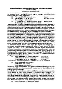
Fig 1. The Atricure ablation device (Atricure, Inc, Cincinnati, OH) consists of two 5-cm long arms, each with a 1-mm wide electrode. The target tissue is clamped between the two arms.
embedded 1-mm electrode. The RF generator was able to independently control both voltage and current delivery to the electrodes. A laptop computer with Labview version 5.1 (National Instruments, Austin, TX) was used to monitor and record temperature, time, current, voltage, impedance, conductance and energy delivered. Temperature was recorded by a themocouple 1 mm from the electrode edge. The computer system displayed these variables in a graphical format in real time. Atrial lesions were created by clamping a cuff of atrial tissue between the two electrode arms. (Fig 2) Radiofrequency energy was delivered at 75 volts and 750 milliamps. It was hypothesized that a steep fall in tissue conductance would be an indicator of transmurality. Therefore, the ablation was continued until the tissue conductance between the two electrodes decreased below 0.0025 Siemens and achieved a steady state for 2 seconds. (Fig 3) After ablation, bipolar pacing was performed from each of the previous sites. Electrical isolation was defined as
Data Acquisition: Acute Study Prior to ablation, the heart was paced from the free wall of the right and left atria and the right and left atrial appendage, and at the junction of the atrium and the right and left pulmonary veins, and the superior and inferior vena cavae using a bipolar epicardial electrode. Pacing thresholds were recorded at all sites at a fixed pulse width of 0.5 ms. Pulmonary vein flows were recorded using a 4-mm ultrasonic flow probe (Transonics, Ithaca, NY) attached to a flow meter ([Model T206]), Transonics, Ithaca, NY). During flow measurements, ventilation was temporarily discontinued and the heart was paced at a constant rate of 100 bpm. Blood pressure was maintained at a mean of 40 mm Hg. Pre-ablation flows were recorded twice for 30 seconds from each of the four main pulmonary veins. After ablation, pulmonary vein flows were similarly recorded from the identical sites. A bipolar RF ablation device was used to create the lesions (AtriCure Inc, Cincinnati, OH) (Fig 1). The bipolar hand piece consisted of two 5-cm long arms, each with an
Fig 2. The lesion set in this study was designed to evaluate circumferential ablations on the beating heart. The lesion sites are shown by the dotted lines. (IVC ⫽ inferior vena cava; SVC ⫽ superior vena cava; RT ⫽ right.)
CARDIOVASCULAR
Ann Thorac Surg 2003;76:836 – 42
CARDIOVASCULAR
838
PRASAD ET AL ISOLATION OF ATRIAL MYOCARDIUM USING BIPOLAR RF ENERGY
Ann Thorac Surg 2003;76:836 – 42
automatically determined by the maximum dV/dT of the unipolar deflection, and were manually edited for verification. Electrophysiologic data were recorded under four experimental conditions: (1) normal sinus rhythm; (2) pacing from the tip of the isolated right atrial appendage; (3) pacing from the tip of the isolated left appendage; and (4) pacing from the free wall of the right atrium.
Histologic Preparation Fig 3. Real-time graphical output from the ablation sensing unit displays conductance in Siemens in the y-axis over time. At 5.5 seconds, conductance falls rapidly. In this study, ablation was discontinued when conductance remained below 0.0025 Siemens for 2 seconds.
the inability to capture the free wall of the atrium by pacing at stimulus strengths up to 20 mA.
Data Acquisition: Chronic Follow-Up The animals were survived for 30 days. They received 325 mg of aspirin each morning after surgery. On day 30, a median sternotomy was performed and the heart was exposed. Pacing at 20 mA from the previously isolated sites (inferior vena cava, right and left atrial appendange, right and left pulmonary veins, superior vena cava) was used to document chronic isolation. A custom 256-channel epicardial atrial mapping system was used to map the atria. The system has been previously described in detail [11]. Custom made silastic plaques were secured to cover the entire atrial epicardial surface. These plaques contained 256 silver unipolar electrodes 1 mm in diameter with an inter-electrode distance of 5 mm. Data were recorded with a gain of 1,000 and a frequency response of 50 to 1,000 Hz. The electrode data were digitized at 1,000 Hz. Activation times were
The animals were sacrificed and the heart and lungs excised en block. The vena cava, pulmonary artery, and aorta were clamped, and the tissue was fixed through the left atrial appendage by infusion of 10% formalin at a pressure of 20 mm Hg for 30 minutes. The heart was placed in formalin for an additional 24 hours. After fixation, the endocardium of the left atrium and each of the pulmonary vein ostia were examined for evidence of thrombus or stricture. The pulmonary veins were grossly examined and classified as free of lumenal narrowing, stenosis, or occlusion. The tissue was then sent to an independent pathology lab (Pathology Associates, Westchester, OH) for analysis of transmural lesions, lesion width, endothelial growth, thrombus, and stricture. To better evaluate the chronic histologic changes in the pulmonary veins, both transverse and longitudinal sections were taken from each of the pulmonary veins. The tissue was examined by microscope for structural changes (ie, transmural, lesion width, and endothelial growth) with Masson’s trichrome and hematoxylin and eosin staining. All animals received humane care in compliance with the “Guide for the Care and Use of Laboratory Animals” published by the National Institutes of Health (NIH Publication No. 85–23, revised 1985).
Statistics The data were expressed as mean ⫾ standard deviation. The comparisons of ablation time, energy, and temperature were performed by the student’s t test.
Results Operative Results None of the animals had any perioperative complications, and there were no operative mortalities. There was no clinical evidence of embolic events or heart failure. A total of 30 circumferential lesions were performed on cuffs of atrial myocardium. The mean ablation time was 5.0 ⫾ 1.6 seconds (range, 2.7–9.3 seconds) with a mean peak temperature of 46.7°C ⫾ 2.8°C (range, 41.8°C– 51.4°C), and an average delivered energy of 92 ⫾ 35 (range, 43–205) Joules (Table 1).
Acute and Chronic Pacing Fig 4. Endocardial view of a lesion surrounding the superior vena cava at 30 days; this representative lesion was linear and continuous. The broken arrow points to the lesion. (RA ⫽ right atrium; SVC ⫽ superior vena cava.)
Before ablation, pacing was performed on atrial tissue 1 cm distal to the planned ablation site (30/30). Pacing thresholds were less than or equal to 1.5 mA at every site. After ablation, pacing stimuli from all sites (30/30) were unable to capture the body of the right atrium at stimulus
PRASAD ET AL ISOLATION OF ATRIAL MYOCARDIUM USING BIPOLAR RF ENERGY
839
Table 1. Ablation Summary
Lesion Site
N
Time (Seconds)
Temperature (C)
Energy (Joules)
Acute Isolation (%)
RAA LAA RPV LPV SVC IVC
5 5 5 5 5 5
4.5 ⫾ 1.9 4.1 ⫾ 0.8 6.6 ⫾ 1.6 6.0 ⫾ 2.2 4.5 ⫾ 0.4 4.3 ⫾ 0.4
46.0 ⫾ 3.4 46.8 ⫾ 4.5 47.5 ⫾ 1.5 46.7 ⫾ 4.1 48.4 ⫾ 0.8 44.8 ⫾ 1.4
63 ⫾ 22 74 ⫾ 26 120 ⫾ 19 132 ⫾ 48 85 ⫾ 5 77 ⫾ 18
100 100 100 100 100 100
Chronic Isolation (%) 100 100 100 100 100 100
Data are represented as mean ⫾ standard deviation. IVC ⫽ inferior vena cava; LAA ⫽ left atrial appendage; pulmonary veins; SVC ⫽ superior vena cava.
LPV ⫽ left pulmonary veins;
strengths up to 20 mA. After 30 days, pacing studies documented continued electrical isolation at all 30 sites.
Pulmonary Vein Flows Pulmonary vein flow was documented from the four pulmonary veins of each animal before and after acute ablation. In animals with more than four total pulmonary veins, the two largest veins from each side were used. The average pre-ablation pulmonary vein flow was 226 ⫾ 55 mL/min. After ablation, the average pulmonary vein flow was 213 ⫾ 48 mL/min. There were no significant differences between acute pre-ablation and postablation flows.
Atrial Mapping Bi-atrial mapping at 30 days demonstrated complete circumferential lines of conduction block by each bipolar lesion. The cuffs of atrial myocardium surrounded by the ablation were completely isolated from the remainder of the heart. By recording data during both normal sinus rhythm and pacing from inside the completely isolated atrial tissue, bi-directional electrical isolation was documented in each animal.
RAA ⫽ right atrial appendage;
RPV ⫽ right
Comment This chronic animal study has demonstrated an efficacious method to create transmural atrial lesions from the epicardium on the beating heart using bipolar radiofrequency energy. Circumferential bipolar RF lesions did not acutely change pulmonary vein flow, and there was no acute or chronic stenosis of the pulmonary veins. Both pacing and epicardial mapping documented bidirectional isolation with every lesion. Chronic histologic examination demonstrated discrete and narrow lesions without evidence of thrombus, stricture, or aneurysm formation. Finally, the real-time measurement of tissue conductance reliably predicted both acute and chronic transmural lesions. Our findings suggest that this technology can be safely and reliably used to replace surgical incisions with ablative lesions on the beating heart.
Histology At 30 days, gross examination of the atria, vena cava, and pulmonary veins revealed no evidence of thrombus or stricture formation (Fig 4). Of note, both the isolated right and left atrial appendages were viable with no evidence of necrosis. Both isolated appendages could be paced and were contractile. By microscopic examination, all lesions (30/30) were transmural, continuous, and discrete. Lesion width varied depending on tissue depth and the time of the lesion. In areas of thin walls (ie, inferior vena cava), the lesion width was fairly constant at 1 mm. On thicker areas of the atrium (ie, the appendage) where the wall thickness was up to 5 mm, the lesion width was greater but was never more than 2.5 mm. There was no evidence of endothelial proliferation or thrombus formation at the site of ablation. The pulmonary vein lesions demonstrated no stricture or luminal narrowing. The lesion widths on the pulmonary vein lesions were never wider than 1.5 mm (Fig 5).
Fig 5. Trichrome staining of a cross-sectional view of an ablation site surrounding the left pulmonary vein demonstrated a linear and transmural lesion with minimal surrounding tissue injury. The pulmonary vein lesions were discrete with no evidence of stenosis in both cross-sectional and longitudinal histology. The red stain represents muscle, cytoplasm, and erythrocytes. The blue stain represents collagen.
CARDIOVASCULAR
Ann Thorac Surg 2003;76:836 – 42
CARDIOVASCULAR
840
PRASAD ET AL ISOLATION OF ATRIAL MYOCARDIUM USING BIPOLAR RF ENERGY
Despite the proven long-term efficacy of the Cox-Maze III procedure, widespread adoption of this procedure has been limited by its length, technical difficulty, and prolonged cardiopulmonary bypass times [5]. The majority of patients in the United States undergoing mitral valve surgery with concomitant AF have been deprived of a curative operative procedure for AF because of the perceived complexity of the Cox-Maze procedure. Ablation technologies have been introduced in an attempt to facilitate the surgical treatment of atrial fibrillation by creating myocardial lesions that replace surgical incisions. By substituting linear lines of ablation for the traditional cut-and-sew approach, it has been hypothesized that the procedure could be performed more simply, potentially decreasing its morbidity, and making it more applicable to the entire spectrum of patients with AF.
Limitations of Unipolar RF Energy None of the currently available unipolar RF devices are able to document real-time transmural lesions. Because of this limitation, it is necessary to use prolonged ablation times and multiple applications of energy to give the surgeon some reassurance of transmural ablation. High temperatures are needed at the tissue-electrode interface to insure penetration of the myocardium. This results in coagulum at the single electrode, which increases the resistance to the flow of energy [12]. This leads to the energy traveling laterally around the developing lesion instead of directly through the tissue. This uncontrolled, multidirectional flow of energy results in wider lesions and collateral tissue injury [13]. Unipolar RF energy has been shown to produce wide lesions ranging up to 1 to 2 cm [13]. There have also been reports of delayed esophageal perforation with the use of the unfocused unipolar RF energy during surgery for AF [14]. High temperatures also lead to protein denaturization in surrounding structures, including red blood cells and matrix proteins. Although this is an advantage for unipolar RF energy in certain organs, such as the liver in the prevention of bleeding, it is a drawback in myocardial tissue. In the heart this can lead to the formation of thrombus and subsequent embolization [15]. Contracture of the myocardial matrix, especially near the pulmonary veins can lead to stenosis. Previous studies have suggested that unipolar radiofrequency energy causes acute pulmonary vein injury leading to luminal narrowing, stenosis, thrombus formation, and even complete occlusion of the pulmonary veins [16, 17]. Admittedly, these complications have been reported during catheter ablation procedures. In contrast, the careful surgical use of unipolar RF energy to dry endocardial fields in the arrested heart has not been associated with a significant number of reported complications. It should be noted that most described surgical techniques have avoided energy application to the actual pulmonary vein orifices. A final drawback of unipolar thermal ablation is the difficulty in obtaining transmural lesions on the beating
Ann Thorac Surg 2003;76:836 – 42
heart. The circulating blood volume on the endocardial surface constitutes virtually infinite heat sink preventing the device from achieving the high ablation temperatures required for permanent tissue injury at the endocardial surface. Our laboratory has shown that unipolar RF is unable to create transmural lesions on the beating heart [18]. This study demonstrated the ability of bipolar radiofrequency energy to produce permanent transmural linear lesions on the beating heart. Moreover, several advantages of bipolar RF energy were clearly defined in our experiments. As opposed to other unipolar energy sources, lesion transmurality can be reliably correlated with an objective real-time endpoint. In this study, a drop of tissue conductance below 0.0025 Siemens for 2 seconds resulted in a 100% correlation with the development of a permanent transmural scar. By using this physiologic parameter of transmural ablation, ablation time can be tailored to the particular tissue characteristics as opposed to using an empirical method to derive ablation time or tissue temperature. The duration of bipolar RF ablation is determined by the thickness and resistivity of the tissue between the electrodes. The ability to determine transmural ablation in realtime leads to several interwined advantages of bipolar RF ablation. First, the tissue receives just enough energy to produce a transmural lesion, shortening ablation time to seconds as opposed to minutes with other energy sources. By shortening ablation time, the duration that surrounding tissue is exposed to excessive resistive heating is reduced, thereby minimizing thermal spread to surrounding tissue and structures. Tissue temperature averaged only 40°C to 50°C 1 mm from the electrode edge as opposed to temperatures of 80°C to 90°C with other thermal ablation devices. By reducing thermal spread, only the tissue immediately adjacent to the electrode is exposed to the elevated temperatures required to produce necrosis and scarring. This limits the contracture of tissue to a few millimeters and removes the danger of stenosis or aneurysm formation. In this study the average lesion width was less than 2 mm as opposed to several centimeters as seen with some of the other devices. Another advantage of bipolar RF energy is that the ablative energy is focused between the two electrodes. The current passes from one electrode to the other transversing through tissue in the shortest, least resistant path, and this results in discrete lesion widths of less than 2.5 mm. Moreover, this makes it difficult to injure surrounding structures (ie, coronary arteries, esophagus). The lower temperatures and more discrete lesions may decrease the risk of both intraoperative and postoperative thromboembolic complications or stricture. No intracavitary thrombi were found in this chronic study. Moreover, there was also no evidence of pulmonary vein stenosis. This complication has been reported to be as high as 39% acutely and 8% chronically after transvenous catheter ablation using unipolar RF energy [16]. A potential benefit of the narrow discrete lesions may be better preservation of atrial transport function (a critical out-
PRASAD ET AL ISOLATION OF ATRIAL MYOCARDIUM USING BIPOLAR RF ENERGY
come measurement of the Cox-Maze procedure). However, this has yet to be proven.
Study Limitations There are several limitations of this device. It is not malleable and it can be difficult to maneuver in the chest. The adaptation of this bipolar RF energy ablator to a totally endoscopic approach will be difficult because of the rigidity of the electrode arms. The length of the instrument (5 cm) is adequate for canine lesions, but may be short for large human atria and pulmonary veins. A theoretical drawback in using bipolar technology is that it is possible to receive a false positive if there is air between the tissue and the electrode. Air will act as an insulator and will feed back to the computer system that the exposed portion of the electrode has a conductance below 0.0025 Siemens. Although it did not happen in this study, care must be taken to firmly clamp the tissue between the jaws of the device to eliminate this possibility. In addition, this study has the drawbacks of all animal studies in that direct extrapolation to the clinical situation must be done with caution. These animals had normal atria with thinner tissue and less fat than diseased human myocardium. Finally, there was no attempt to perform an entire Cox-Maze III procedure with this device. The ability of bipolar radiofrequency energy to fully replicate all the lesions of a Cox-Maze III procedure remains unknown. In summary, the surgical treatment of AF has been established for more than a decade. The complexity of the surgery has limited its widespread application to all patients with atrial fibrillation. New technologies that simplify and shorten the operation by producing transmural lesions to replace the multiple surgical incisions of the Cox-Maze III procedure offer promise. This study documented the creation of permanent lines of electrical isolation in atrial myocardium with bipolar energy. Moreover, this novel technology can produce linear, transmural lesions in the beating heart. The measurement of tissue conductance as a real-time determinant of transmural lesions is one of the unique advantages of bipolar energy. Our results suggest that bipolar RF energy may be an enabling technology to allow surgeons to perform curative surgery for atrial fibrillation on the beating heart.
This study was funded by an unrestricted research grant from Enable, Inc, NIH 5 R01 HL32257 and NIH T32 HL07275. The authors would like to thank Diane Toeniskoetter, Kathy Fore, and Dennis Gordon for their technical assistance.
841
References 1. Cameron A, Schwartz MJ, Kronmal RA, et al. Prevalence and significance of atrial fibrillation in coronary artery disease (CASS registry). Am J Cardiol 1988;61:714 –7. 2. Coplen SE, Antman EM, Berlin JA, Hewitt P, Chalmers TC. Efficacy and safety of quinidine therapy for maintenance of sinus rhythm after cardioversion. A meta analysis of randomized control trials. Circulation 1990;82:1106 –16. 3. Smith PK, Holman WL, Cox JL. Surgical treatment of supraventicular tachyarrhythmias. Surg Clin North Am 1985; 65:553–70. 4. Cox JL, Canavan TE, Schuessler RB, et al. The surgical treatment of atrial fibrillation: II. Intraoperative electrophysiologic mapping and description of the electrophysiologic basis of atrial flutter and atrial fibrillation. J Thorac Cardiovasc Surg 1991;101:406 –26. 5. Cox JL, Ad N, Palazzo T, et al. Current status of the MAZE procedure for the treatment of atrial fibrillation. Semin Thorac Cardiovasc Surg. 1999;118:628 –35. 6. Shah DC, Haissaguerre M, Jais P. Catheter ablation of pulmonary vein foci for atrial fibrillation. Thorac Cardiovasc Surg 1999;47(Suppl):352–6. 7. Natale A, Pisano E, Shewchik, et al. First human experience with pulmonary vein isolation using a through-the-balloon circumferential ultrasound ablation system for recurrent atrial fibrillation. Circulation 2000:102:1879 – 86. 8. Pappone C, Rosanio S, Oreto G, et al. Circumferential radiofrequency ablation of pulmonary vein ostia. Circulation 2000;102:2619 –28. 9. McLean A. The Bovie electrosurgical current generator. Arch Surg 1929;18:1863. 10. Khargi K, Deneke T, Haardt H, et al. Related articles salineirrigated, cooled-tip radiofrequency ablation is an effective technique to perform the maze procedure. Ann Thorac Surg 2001;72(3):S1090 –95. 11. Rodefeld MD, Branham BH, Schuessler RB, et al. Global electrophysiological mapping of the atrium: computerized three-dimensional mapping system. PACE 1997;20(9 Pt 1): 2227–36. 12. McRury ID, Panescu D, Mitchell MA, Haines DE. Nonuniform heating during radiofrequency catheter ablation with long electrodes. Circulation 1997;96:4057–64. 13. Langberg JJ, Lee MA, Chin MC, Rosenquist M. Radiofrequency catheter ablation: the effect of electrode size on lesion volume in vivo. PACE 1990;13:1242–8. 14. Gillinov AM, Pettersson G, Rice TW. Esophageal injury during radiofrequency ablation for atrial fibrillation. J Thorac Cardiovasc Surg 2001;122(6):1239 –40. 15. Epstein MR, Knapp LD, Martindill M, et al. Embolic complications associated with radiofrequency catheter ablation. Am J Cardiol 1996;77:655–8. 16. Yu WC, Hsu TL, Tai CT, et al. Acquired pulmonary vein stenosis after radiofrequency catheter ablation of paroxysmal atrial fibrillation. J Cardiovasc Electrophysiol 2001;12(8): 887–92. 17. Taylor GW, Kay GN, Zheng X, et al. Pathological effects of extensive radiofrequency energy applications in the pulmonary veins in dogs. Circulation 2000;101(14):1736 –42. 18. Hoenicke EM, Strange RG, Patel H, Griffith JW, Damiano RJ. Epicardial radiofrequency ablation on the beating heart ovine heart: moving towards a less invasive MAZE procedure. Surg Form 2000;51:79 – 82.
DISCUSSION DR TAKASHI NITTA (Tokyo, Japan): One reason that you did not see any significant decrease in the pulmonary vein (PV) flow could be that you clamped the posterior left atrium around the
pulmonary veins, and not the pulmonary veins themselves. Should you have clamped and ablated the pulmonary veins directly? What affect would that have had on the PV flow?
CARDIOVASCULAR
Ann Thorac Surg 2003;76:836 – 42
CARDIOVASCULAR
842
PRASAD ET AL ISOLATION OF ATRIAL MYOCARDIUM USING BIPOLAR RF ENERGY
Ann Thorac Surg 2003;76:836 – 42
DR PRASAD: This is actually our fourth animal study with this type of technology. There have been previous chronic studies that did demonstrate that we actually have very similar lesions on the pulmonary veins themselves, which was presented at the American College of Surgeons, but one of the advantages of doing surgical isolation of the pulmonary veins is that we do not have to go on the pulmonary veins, we can actually isolate a cuff of atrial myocardium around the pulmonary veins and thereby isolate any sort of initiation in the pulmonary veins.
that were circumferential and we could pace from tissue within the isolated segment. This has nothing to do with any sort of lesion set to stop atrial fibrillation, although some would argue that PAF may be treated with isolated pulmonary vein isolation.
DR VERDI J. DISESA (West Chester, PA): Does this set of lesions cure atrial fibrillation?
DR PRASAD: Correct. What we have demonstrated in this study is not a curative operation for atrial fibrillation. What we have demonstrated is that we can reliably replace the surgical incisions that are done for the Maze procedure, or any sort of arrhythmia surgery procedure, with ablations, and that we may be able to place these lesions. We do have an ongoing study right now to actually confirm all the Cox-Maze III lesions on the beating heart in dogs. So that would be the final step.
DR PRASAD: No. This set of lesions was specifically designed so that we could maximize the number of lesions we could construct on the heart in areas we could document electrical isolation by pacing. So we used the atrial appendages and the vena cava and the pulmonary veins so that we had six lesion sets
DR DISESA: But is not part of your argument that you can do this off pump with all epicardial lesions, and therefore it is a little disingenuous to put the two together without acknowledging that this is not an operation for atrial fibrillation?