[CANCER RESEARCH 64, 64 –71, January 1, 2004]
Loss of Heterozygosity and Its Correlation with Expression Profiles in Subclasses of Invasive Breast Cancers Zhigang C. Wang,1 Ming Lin,5 Lee-Jen Wei,5 Cheng Li,3,5 Alexander Miron,1 Gabriella Lodeiro,1 Lyndsay Harris,4,7 Sridhar Ramaswamy,6,7 David M. Tanenbaum,7 Matthew Meyerson,7 James D. Iglehart,1,4 and Andrea Richardson2 Departments of 1Surgery and 2Pathology, Brigham and Women’s Hospital, Boston; Departments of 3Biostatistical Science and 4Cancer Biology, Dana-Farber Cancer Institute, Boston; 5Department of Biostatistics, Harvard School of Public Health, Boston; 6Whitehead Institute/Massachusetts Institute of Technology, Center for Genomic Research, Cambridge; and 7Department of Medical Oncology, Dana-Farber Cancer Institute, and Brigham and Women’s Hospital, Boston, Massachusetts
high diversity of LOH patterns in breast cancers (10), and complicates the effort to identify the chromosomal regions truly associated with pathogenesis in these tumors. The high diversity and frequency of LOH in breast cancer has made it difficult to classify this disease solely according to LOH profiles (11). However, recent studies have revealed associations among levels and patterns of LOH, chromosomal instability, and histological grades (10, 11). Nevertheless, breast cancers of similar grade may display diverse clinical and molecular features, and may not represent distinct tumor classes. To elucidate molecular events and pathways in this highly diverse tumor type, recent studies using gene expression profiling identified a number of subclasses with distinct expression profiles and different histopathology (12, 13). Expression array profiling may identify breast cancers sharing genomic alterations in the same molecular pathway(s). Indeed, HER-2 gene amplification underlies the distinct gene expression profile of the HER-2-positive group of tumors (12). Similarly, inactivation of particular tumor suppressor gene(s) by mutation and deletion may affect key functional pathways and result in different gene expression profiles. Supporting evidence comes from gene expression analysis of cancers from BRCA1 or BRCA2 germ-line mutation carriers (with somatic allelic loss), which demonstrated gene expression signatures that distinguish these genetically distinct tumors (14). Similar links between cancer genotypes and expression signatures in sporadic cancers may stimulate exploration of genomic events that alter distinct molecular pathways and give rise to different groups of breast tumors. The present study linked LOH profiles to the subclasses of breast cancer defined by gene expression signatures. Genome-wide LOH patterns were determined by a new high-throughput technique using single nucleotide polymorphism (SNP) arrays (Affymetrix Inc.) to allelotype tumor and autologous normal samples for ⬃1494 SNP alleles distributed on all human chromosomes. Our group and others have validated that SNP analysis is an effective and accurate method to detect tumor LOH across the genome (15, 16). The method has been successfully used for studies of LOH in a number of tumor types (15–19), including validation in a few breast cancers (20). Using this method, our study demonstrates distinct LOH patterns in subclasses of breast cancers defined by gene expression profiles.
ABSTRACT Gene expression array profiles identify subclasses of breast cancers with different clinical outcomes and different molecular features. The present study attempted to correlate genomic alterations (loss of heterozygosity; LOH) with subclasses of breast cancers having distinct gene expression signatures. Hierarchical clustering of expression array data from 89 invasive breast cancers identified four major expression subclasses. Thirty-four of these cases representative of the four subclasses were microdissected and allelotyped using genome-wide single nucleotide polymorphism detection arrays (Affymetrix, Inc.). LOH was determined by comparing tumor and normal single nucleotide polymorphism allelotypes. A newly developed statistical tool was used to determine the chromosomal regions of frequent LOH. We found that breast cancers were highly heterogeneous, with the proportion of LOH ranging widely from 0.3% to >60% of heterozygous markers. The most common sites of LOH were on 17p, 17q, 16q, 11q, and 14q, sites reported in previous LOH studies. Signature LOH events were discovered in certain expression subclasses. Unique regions of LOH on 5q and 4p marked a subclass of breast cancers with “basal-like” expression profiles, distinct from other subclasses. LOH on 1p and 16q occurred preferentially in a subclass of estrogen receptorpositive breast cancers. Finding unique LOH patterns in different groups of breast cancer, in part defined by expression signatures, adds confidence to newer schemes of molecular classification. Furthermore, exclusive association between biological subclasses and restricted LOH events provides rationale to search for targeted genes.
INTRODUCTION Breast cancer is a highly heterogeneous disease, and its initiation and early progression is associated with mutations, chromosomal deletions, and amplifications. Chromosomal amplifications may result in oncogenic gains of function and are best detected by such methods as fluorescence in situ hybridization (1) and comparative genomic hybridization (CGH; Ref. 2). Chromosomal deletions, best detected by methods that identify loss of heterozygosity (LOH; Ref. 3), may occur in combination with germ-line or sporadic mutation of the retained allele and bring about loss of gene function (4, 5). The paradigm of mutation and allelic loss of genetic information on opposite chromosomes is exemplified by p53, the target of mutation on one allele and somatic deletion, detected as LOH, of the remaining normal allele (6, 7). Accordingly, LOH may indicate regions harboring tumor suppressor genes. For example, mutations of tumor suppressor genes p53, BRCA1, and BRCA2 are heralded by LOH on 17p, 17q21, and 13q12–13, respectively (5, 7–9). Alternatively, LOH may reflect random chromosomal instability and may not specifically target pathogenic genes. Such genetic instability contributes to the
MATERIALS AND METHODS Subjects and Tissues. Major tumor subclasses were identified by hierarchical cluster analysis performed on a gene expression array data set which included previously published array data of 89 frozen breast carcinoma samples (21) and array data from 7 additional samples of bulk normal breast tissue. The tumors were scored for modified Bloom-Richardson grade and immunoreactivity for estrogen receptor (ER), HER-2 receptor, and p53. The 7 normal samples were from unaffected areas of the same breast as 7 of the tumor samples and were confirmed normal by histology. RNA extraction, cRNA synthesis, and hybridization to Affymetrix U95A arrays were performed as described previously (21). From the 89 cases, a subset of 34 cases representative of the different gene expression classes were chosen for additional LOH analysis. Formalin-fixed, paraffin-embedded tumor samples of these 34 cases were retrieved from the Department of Pathology, Brigham and Women’s
Received 8/18/03; revised 10/22/03; accepted 11/10/03. Grant support: Breast Cancer Research Foundation (BCRF), by the Dana-Farber/ Harvard Specialized Programs of Research Excellence in Breast Cancer from the National Cancer Institute, and by the Department of Defense Center of Excellence at Harvard. The costs of publication of this article were defrayed in part by the payment of page charges. This article must therefore be hereby marked advertisement in accordance with 18 U.S.C. Section 1734 solely to indicate this fact. Requests for reprints: Andrea Richardson, Department of Pathology, Brigham and Women’s Hospital, 75 Francis Street, Boston, MA 02115. E-mail: arichardson@ partners.org.
64
GENOTYPE OF BREAST CANCER SUBCLASSES
previously for linkage analysis (23) and the Genome Database.10 PCR amplifications with DNA from microdissected tumor cells and autologous normal cells, sequencing gel electrophoresis, and fluorescence analysis were performed as described elsewhere (23). LOH was determined by comparison of heterozygous markers in normal tissue with the same marker in tumor tissue. A ⬎5-fold decrease in the relative signal of one allele was considered LOH. Statistical Analysis. The fractional LOH in each tumor is calculated as the proportion of LOH calls per overall informative calls for that case. The fractional LOH reflects the overall level of allelic loss and may also indicate the degree of chromosomal instability. Because of the low informative rate of SNP markers in the population (⬍30% in our data set), common sites of LOH among a group of tumors cannot be tabulated by observed frequency of LOH for individual SNP markers. To identify common regions of LOH, we used a newly developed method programmed into the dChipSNP software. A complete description of the method and application is reported elsewhere (19, 22). In brief, the method scores the sum proportion of LOH events among all of the informative markers in a local chromosomal region for a selected set of tumors. Scoring the proportion of LOH events per informative loci within a chromosomal region alleviates the influence of different informative marker densities for each patient and at different chromosomal regions. A large summary score indicates a substantial increase in LOH in that chromosomal region in that set of patients. In this study, we set the parameters to determine the summary LOH proportion score for chromosomal blocks of 6 Mb moved along the chromosome by 1-Mb increments, giving a rolling summary LOH score along the chromosome. In addition, the dChipSNP analysis software uses a permutation method to evaluate for measurement errors and to make statistical inference as to the significance of the LOH score (P). The software displays the LOH scores with P curves along the chromosomes to aid with determination of chromosomal regions with statistically significant high rates of shared LOH among a group of tumors. Statistical differences among subclasses were analyzed using Fisher’s exact test with the R statistical software package.11 Significance values were reported as a P. Subclasses differed with respect to the overall frequency of LOH events, possibly indicating a difference in chromosomal stability and the random level of LOH. To correct for this, differences between subclasses in the frequency of LOH in individual chromosomal regions were adjusted for the fractional level of LOH by using the Exact Score test of exact logistic regression (LogXact version 4.1; Cytel Software Co., Cambridge, MA). After adjustment, significance values were reported as a customized P (Pc).
Hospital. Tissue sections were examined for adequate tumor component for microdissection and DNA extraction. Each tumor also had autologous, nonmetastatic axillary lymph node tissue or peripheral blood mononuclear cells available for normal DNA. All of the tissues and blood were collected under an Institutional Review Board-approved protocol. Hierarchical Clustering Analysis. Our previous study reported a gene expression array data set of 89 primary invasive carcinomas generated on Affymetrix U95A gene arrays (21). Hierarchical cluster analysis of the 89 tumors using a subset of 2130 genes selected by a relatively low stringency filter for variation of gene expression highlighted the basal-like subclass of tumors in one cluster, with a robust expression signal relative to the other cases. For this study, we included the normal samples in the cluster analysis. To find structure in the expression array data that may be obscured by the “noise” of less variably expressed genes, we chose to perform the cluster analysis using a smaller set of 672 genes selected using a higher stringency filter for variation of expression. As in our prior study, raw expression values obtained with Affymetrix GENECHIP software were additionally analyzed with DNA-Chip Analyzer (dChip) custom software.8 The set of highly variable genes were selected using filtering criteria requiring coefficient of variation (SD/mean) between 0.8 and 10.0, and a mean intensity value of ⬎100 units in at least 10% of samples. Duplicate probes for the same genes were eliminated leaving 672 unique gene probes. Hierarchical clustering of the 89 tumors and 7 normal samples was carried out with the dChip clustering function. Laser Capture Microdissection and DNA Extraction. Tumor cells were isolated from H&E-stained tissue sections by laser capture microdissection using a PixCell laser capture microscope (Arcturus Engineering, Mountain View, CA). Approximately 10,000 pulses were used to dissect tumor cells for each case. Lobular and tubular cancers were excluded from analysis due to the difficulty of accurately microdissecting these cancer types. Duplicate microdissections were performed on 24 cases. To minimize discrepancies resulting from intratumoral heterogeneity, duplicate microdissections were performed at the same area of two adjacent tumor tissue sections. DNA was extracted by overnight digestion of tumor cells with proteinase K followed by heat inactivation. Genomic DNA from lymph nodes was additionally purified by standard phenol-chloroform extraction and ethanol precipitation protocols. WBC DNA was extracted using the QIAamp DNA kit (Qiagen Inc., Valencia, CA). HuSNP Chip Analysis. Affymetrix HuSNP mapping assay system (Affymetrix Inc., Santa Clara, CA) was used to determine tumor and normal allelotypes. This system uses the HuSNP chip, an array of oligonucleotide probes for 1494 SNP loci distributed on all human chromosomes with an average of 2.57 cM between each SNP marker. Multiplexed PCR amplification of the SNP templates from DNA extracts, labeling, and hybridization of the PCR products to the Affymetrix HuSNP chips, and staining of chips were performed following the instructions provided by the manufacturer as described elsewhere (16). A GeneArray Scanner scanned chips and genotype “calls” (heterozygous or homozygous) were made from the collected hybridization signals using Affymetrix HuSNP 3.1 software. Tumor and normal samples were allelotyped on separate chips. For each patient tumor, each SNP locus was scored as LOH, retention of heterozygosity, uninformative, or uncertain by comparing the genotype calls for tumor and normal (autologous) pairs. The scoring process was conducted by dChipSNP custom software.8 Twenty-nine tumor samples were genotyped in duplicate. In 24 cases, duplicate microdissections were performed. In 5 cases, a single microdissected sample was subjected to duplicate PCR amplification and chip hybridization. The reproducibility of experiments was evaluated by comparing the duplicates of each tumor sample to obtain the error rate of informative genotype calls (discordant informative calls per total informative calls). LOH mapping analysis was performed by dChipSNP software based on updated map locations of SNP loci from University of California Santa Cruz Biotechnology genome assembly (hg15 human genome assembly).9 Common regions of LOH were identified using the dChipSNP permutation method (19, 22). Simple Sequence Length Polymorphism (SSLP) Analysis. The HuSNP method was additionally validated by confirming the LOH results for a representative region in a subset of tumors using standard SSLP analysis. Microsatellite markers on 5q (D5S612, D5S1414, D5S614, D5S644, D5S1215, D5S339, D5S436, D5S422, and D5S400) were selected from the panel used 8 9
RESULTS Classification of Breast Tumors by Gene Expression Profiles. This study explored the correlation of LOH patterns with subclasses identified by gene expression profiles in invasive breast cancers. We first determined the major expression subclasses by hierarchical clustering analysis of expression array data from our previously published set of 89 breast cancers (21) together with 7 additional normal breast tissues samples. Hierarchical clustering was performed in the space of 672 unique gene probes stringently filtered for highest variation of expression across the sample set (Fig. 1). As shown by the dendrogram at the top of the figure, two major expression classes were identified, denoted clusters I and II. Cluster I is characterized by higher-grade tumors, the majority of ER-negative tumors, HER-2positive tumors, and p53 expression-positive tumors. In contrast, cluster II has predominantly ER-positive cases and has the majority of lower-grade tumors. The two main classes further split into two minor subclasses, sublabeled A and B. The subclass IA is dominated by tumors with high HER2 expression and a prominent lymphoid gene expression signature. The high expression of the lymphocyte-associated genes correlates with the histological finding of a significant lymphocytic infiltrate in the stroma of these tumors. The subclass IB is composed of tumors with high grade, and an ER-negative and 10
Wong, W. H. and Li, C. Internet address: http://www.dchip.org. Internet address: http://www.genome.ucsc.edu.
11
65
Internet address: http://www.gdb.org. Internet address: http://www.r-project.org.
GENOTYPE OF BREAST CANCER SUBCLASSES
Fig. 1. Hierarchical clustering of 89 invasive breast cancers and 7 normal samples for 672 probe sets (genes) filtered for high variation in expression across the sample set. Clustering orders the cancers according to greatest similarity of gene expression, shown by the dendrogram at the top, and orders the genes by similarity of expression level among the sample set, shown by the dendrogram along the side. The top (5 shaded rows) displays the clinical and immunophenotypic data for each sample, color-coded as follows: dark rose, ductal histology; light rose, lobular cancers; dark gray, histological grade III; medium gray, grade II; light gray, grade I; dark blue, estrogen receptor (ER) -positive; light blue, ER-negative; dark green, HER2-positive (3⫹); light green, HER2-negative (0 –2⫹); dark yellow, p53 immunohistochemical staining-positive; light yellow, p53 immunohistochemical staining-negative. The 7 normal samples clustered together and are shown by the white area in the clinical information colorgram panel. Relative gene expression levels are represented as follows: mean expression levels are shown by white, expression levels above the mean in progressively darker shades of red, and expression levels below the mean in progressively darker shades of blue. The tumor dendrogram shows the two main clusters, clusters I and II, and second-order clustering is labeled IA, IB, IIA, and IIB.
well represented in the LOH analysis. Tumor and paired normal samples from the 34 cases were genotyped using HuSNP arrays to determine LOH patterns and frequency. The average genotype call rates for tumor and normal samples were 82.2 ⫾ 3.8% and 84.8 ⫾ 2.8%, respectively. On average, 349 of the 1494 SNP loci were informative (both heterozygous in normal samples and callable in the tumor samples), corresponding to 1 marker/8.6 Mb (7.9 cM) on
HER2-negative immunophenotype. The gene expression signature of this small subclass is consistent with that of “basal-like” tumors including expression of basal cytokeratins and high expression of S phase and M phase cell cycle genes (12, 13) indicative of the high proliferation rate in these cancers. Cluster II further split into two subclasses of luminal-type ER-positive and HER2-negative breast cancers. Lower-grade ductal carcinomas and the majority of lobular carcinomas distinguish subclass IIA. The normal breast samples clustered together and were located with the low-grade tumors in subclass IIA. Subclass IIB had more of the higher-grade ER-positive tumors. This division of invasive breast cancers into a basal-like subclass, a HER-2-positive subclass, and two ER-positive luminal-type subclasses is quite similar to the gene expression clusters reported by others (12), supporting the validity of the classification results. LOH Analysis in Breast Tumors Using HuSNP Arrays. Thirtyfour invasive breast carcinomas were selected from the 89 tumors described above for additional LOH analysis. The 34 cases were chosen to represent the four gene expression subclasses. The clinical features of these cases are summarized in Table 1, organized by membership in the different subclasses. Because the low-grade tubular and lobular cancers were difficult to microdissect to adequate tumor purity and amount, such tumors from cluster II subclass IIA were not
Table 1 Summary of pathologic features and loss of heterozygosity (LOH) levels for the 34 tumors used for LOH analysis grouped according to membership in subclasses of a larger cohort of 89 tumors Cluster I Subclasses
IA (n ⫽ 15)
IB (n ⫽ 7)
IIA (n ⫽ 4)
IIB (n ⫽ 8)
Tumor Grade I II III Estrogen receptor ⫹ HER2–3⫹ P53 stain ⫹ Mean fractional LOHb
20a 27 53 53 60 47 14
0 0 100 0 0 57 37
25 25 50 100 0 0 6
25 13 62 100 0 0 14
a b
66
Cluster II
Percentage of tumors. Fraction (%) of LOH per all informative (heterozygous) loci for each tumor.
GENOTYPE OF BREAST CANCER SUBCLASSES
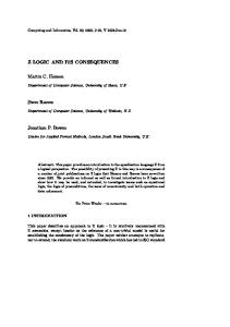
Fig. 2. Identification of regions containing LOH on chromosomes 5 and 16. Each column represents a tumor-normal pair. The colorgram at the top displays the clinical and immunophenotypic data for each case color-coded as follows: dark gray, histological grade III; medium gray, grade II; light gray, grade I; dark blue, estrogen receptor (ER) -positive; light blue, ER-negative; dark green, HER2-positive (3⫹); light green, HER2-negative (0 –2⫹); dark yellow, p53 immunohistochemical staining-positive; light yellow, p53 immunohistochemical staining-negative. The single nucleotide polymorphism (SNP) markers are mapped onto the G-banded chromosomes shown on the left with banding nomenclature, to give approximate locations. The LOH and retention of heterozygosity at the SNP loci for each tumor-normal pair is displayed in the corresponding columns, with blue bars representing the regions of LOH and yellow representing the regions of retention. The white areas represent regions with no genotype call, uninformative markers, or lack of SNP markers. The curve on the right plots the permutation P calculated as a rolling average of overlapping 6 megabase chromosome blocks, and indicates the level of significance of identified LOH events in that region in that tumor set. Peaks indicate the most significant regions of frequent LOH. The red line shows the P ⫽ 0.05 threshold. The top and bottom panels show the data for chromosomes 16 and 5, respectively.
Fig. 2. The plot on the right side of the figure denotes regions of statistically significant LOH. Setting the permutation P ⱕ 0.05 as a cutoff for significance, 46 common LOH regions were identified across the genome (Table 2). By visual inspection of the LOH patterns, we determined that LOH affected each of these regions in at least 18% of the tumors. Overall, the most commonly involved regions were 17p13, 17q11.2–12, 17q21, 11q23–24, 16q23–24, 14q32, 10q23–24, and 13q14, each lost in ⬎40% of tumors. Most of these common sites are consistent with those reported by numerous previous LOH studies (10, 11, 24), lending validity to both the method and results. Signature LOH Profiles Associated with Particular Expression Classes of Breast Cancer. Signature LOH patterns were sought for the tumor classes defined by expression profiling. Comparison of the two major classes and four subclasses revealed differences in the fractional LOH rates (Table 3). The mean fractional LOH rate was significantly higher in cluster I relative to cluster II, and highest (37%) in the basal-like tumors in subclass IB. The frequency of LOH at specific chromosomal locations also differed between the two major classes. In general, tumors in cluster I possessed frequent and widely dispersed LOH events, often on chromosomal regions rarely affected in cluster II cancers. The tumors in cluster II had fewer overall losses, and the sites of LOH were more restricted in distribution. The relative proportion of cases from the two major clusters showing LOH at some of the more frequently involved chromosomal sites is shown in Fig. 3. Most chromosomal sites were affected in a higher proportion of cases from cluster I than cluster II, consistent with the higher overall LOH rate in cluster I. The frequency of loss at some of the more common
average. The LOH calls were highly reproducible in the 29 duplicated comparisons. For the 5 cases with duplicate PCR and chip hybridization from a single microdissected sample, the median error rate of discrepancy for informative calls was 1.3% (range, 0.25–3.0%). In the 24 cases with duplicate microdissections, the median error rate was 1.9% (range, 0.27– 8.7%). Despite performing duplicate microdissections from similar tumor areas to minimize the effect of heterogeneity within tumors, we did see a higher discrepancy rate for the duplicate microdissections compared with the duplicate PCR from a single microdissection. This may indicate some level of intratumoral heterogeneity in some of the samples. A more detailed study is required to evaluate the level and effect of intratumoral heterogeneity on LOH determination in breast tumors. Confirming LOH at a representative chromosomal region using the standard method of SSLP or microsatellite genotyping further validated the HuSNP chip method for LOH analysis. LOH on 5q was mapped in 12 of the tumors by SSLP analysis using a panel of nine microsatellite markers. The two methods of LOH determination were concordant in these 12 tumors on this chromosomal arm, with 4 tumors showing no LOH on 5q, and 8 tumors showing 5q LOH over a similar extent by both methods (data not shown). All 34 of the tumors had at least some allelic loss, but the overall level of allelic loss within tumors, represented by the fractional LOH rate, varied greatly (range, 0.3– 62.7%, median 16.4%; see Table 1). Within the tumor set, LOH was found on all of the chromosomes. The dChipSNP analysis software determined chromosomal regions containing a high frequency of LOH events. An example of the dChipSNP display of LOH patterns is shown for two chromosomes in 67
GENOTYPE OF BREAST CANCER SUBCLASSES
region at 16q was lost in 88% of the tumors comprising subclass IIB, 50% of tumors in IIA, and overall in 33% of tumors in cluster I (Pc ⫽ 0.001; Table 3). The region at 1p34 was lost in 50% of IIB tumors, none of the IIA tumors, and in 13% of tumors in major cluster I. Conversely, subclass IB, the basal-like tumors with the highest overall rate of LOH, had the lowest rate of LOH at 16q23–24 of all of the four subclasses. This finding is consistent with the report of preferential 16q allelic loss by CGH analysis in ER-positive and HER2-negative tumors, but not in tumors with basal-like phenotypes (25). Subclass IB, the basal-like tumors, had a significantly higher fre-
Table 2 Common loss of heterozygosity (LOH) regions in invasive breast cancers identified using single nucleotide polymorphism array analysisa Chromosomal regions
Tumors affected (n ⫽ 34)b %c
Chromosomal regions
Tumors affected (n ⫽ 34) %
17p13 17q11.2–12 17q21 11q23–24 16q23–24 14q32 10q23–24 13q14 17q25 1p36 9p21 11p15.4 15q15 5q14 5q21–32 8p21.2 13q32 1p13 3p22–24 3p12 5q11.2 8p22–23 9p23–24
56 53 53 50 47 44 41 41 38 35 35 32 32 29 29 29 29 26 26 26 26 26 26
10q21–22 10q25.3 12q24 14q21 15q25 22q11.23–13 1p31 2q35–36 7q22 9q22 21q21–22.1 1p34 4p15.3 9q34 12q22–23 13q12 4p14 6q22 6q26 13q22 18q22–23 19q13.4 Xq28
26 26 26 26 26 26 24 24 24 24 24 21 21 21 21 21 18 18 18 18 18 18 18
a Common regions of LOH among the entire tumor set determined by permutation testing for significance with P set at ⱕ0.05. b Total number of tumors. c Percentage of tumors affected determined by visual inspection of LOH patterns as displayed in Fig. 2.
sites such as 17p13, 14q32, and 11q24 was not significantly different between the two major classes or four subclasses (Table 3), perhaps indicating a more general association of these LOH sites in breast cancer. Two chromosomal regions, 1p34 and 16q23–24, were affected more frequently in cluster II, despite a lower average fractional LOH rate in this group of tumors. Chromosomal alterations in these regions may be more specifically associated with the ER-positive cancers of this class (Fig. 3; Table 3). Comparison of the LOH frequencies demonstrated signature genotypes for some of the subclasses as well. Allelic imbalance at 16q23–24 and 1p34 was seen most commonly in the subclass IIB. The
Fig. 3. Loss of heterozygosity (LOH) frequency of common LOH sites in cluster I and II tumors identified by gene expression profiles. The LOH frequencies of 12 more commonly involved sites of LOH are plotted separately for each of the two main gene expression clusters. LOH frequencies of clusters I and II are shown by the black and the gray bars, respectively. The frequency levels are shown on the vertical axis, and the chromosomal regions on the horizontal axis. The chromosomal locations are ordered by ratio of the LOH frequencies in the two clusters, with regions higher in cluster I at the far left, and regions higher in cluster II at the far right. Regions with a significant difference in frequency between the two clusters at P ⬍ 0.05 are indicated with ⴱ.
Table 3 Frequency of loss of heterozygosity (LOH) in subclasses of breast cancersa Clusters Chromosomal regions 1p34 4p14 4p15.3 5q11.2 5q14 5q21–32 10q23–24 11q23–24 13q14 14q21 14q32 16q23–24 17p13 17q11.2–12 17q21 18q22–23 Mean fractional LOH (%)
I (n ⫽ 22) 13 23 32 41 41 41 50 59 50 32 50 32 59 64 68 27 21
d
Subclasses II (n ⫽ 12)
Pb
IA (n ⫽ 15)
IB (n ⫽ 7)
IIA (n ⫽ 4)
IIB (n ⫽ 8)
P
Pcc
30 8 0 0 8 8 25 33 25 17 33 75 50 33 25 0 11
NSe NS 0.035f 0.01 NS NS NS NS NS NS NS 0.029g NS NS 0.029f NS 0.04h
7 7 13 13 20 20 40 60 40 13 33 33 47 53 60 13 14
29 57 71 100 86 86 71 57 71 71 86 29 86 71 86 57 37
0 0 0 0 25 25 0 0 25 25 25 50 25 25 25 0 6
50 13 0 0 0 0 25 50 13 13 38 88 63 38 25 0 14
0.058 0.04 0.004 ⬍0.0001 0.001 0.004 NS NS NS NS NS 0.053 NS NS 0.046 0.032 ⬍0.001h
0.055 NS NS 0.02 NS NS
a
0.001
NS NS
Subclasses defined by expression hierarchical clustering in Fig. 1. P of Fisher’s exact test in comparing LOH frequencies for the individual chromosomal regions between two clusters or among four subclasses. P of exact score test of exact logistic regression adjusting for fractional LOH in comparing LOH frequencies for the individual chromosomal regions among four subclasses. d Percentage of the tumors affected by LOH. e P ⬎ 0.05; NS ⫽ not significant. f P ⬎ 0.05 after adjusting for fractional LOH in exact score test of exact logistic regression. g P ⫽ 0.005 after adjusting for fractional LOH in exact score test of exact logistic regression. h P from Student’s t test. b c
68
GENOTYPE OF BREAST CANCER SUBCLASSES
ation of SNP arrays containing probes for more SNP markers and SNP markers with a higher informative rate. The present study correlated LOH patterns to classes of breast tumors with distinct gene expression profiles. Subclasses identified by hierarchical clustering in this study were consistent with those reported previously (12). The identification of similar subclasses of breast cancer by several investigators using different gene expression array platforms and different patient sample populations lends confidence to the gene expression-based classification. Signature LOH events were found to be associated with certain gene expression subclasses. In particular, LOH on 1p and 16q was associated with one ER-positive and HER2-negative subclass. Such an association for 1p has not been recognized previously, whereas common loss of 16q in low grade and ER-positive tumors has been reported (25, 27). The 1p34 region described in this study has only rarely been noted in breast cancer, although some reports suggested that loss in this region may be associated with poor prognosis (28). Loss of the 1p32-ter region has been reported in hyperplasia of breast and in monozygotic twins concordant for breast cancer suggesting the 1p32-ter region as a possible site of a breast cancer tumor suppressor gene (29, 30). Because the HuSNP array informative marker density is relatively low at this 1p region, the true frequency and extent of LOH at this site will have to be confirmed using a higher density marker set. The 16q23–24 imbalance has been reported in numerous prior studies as a common region of loss in breast cancers. This region harbors a number of cadherin genes and other putative tumor suppressor genes (31). Our results link these two chromosomal regions specifically to a subset of ER-positive breast tumors identified by gene expression profiles. Another intriguing group of cancers was identified as subclass IB (Fig. 1), the basal-like cluster emphasized previously by Perou et al. (12). The basal-like tumors tend to be both ER- and HER2-negative, high grade, and have frequent p53 inactivation. In the basal-like tumors in this study, LOH was frequently observed at 5q, 14q32, 17p13, 17q, 4p, and 18q. The losses on 5q, 4p, and 18q occurred at a significantly higher rate in this particular subclass of tumors relative to other subclasses, especially the locus at 5q11 lost in 100% of these tumors. These results suggest a genomic signature for high-grade ERand HER2-negative breast cancers, which may better define those with a basal-like phenotype. Recent studies using CGH have investigated the loss and gain of chromosomal regions in basal-like breast tumors (25, 32, 22). Korsching et al. (25) clustered primary breast tumors based on phenotypic and CGH profiles, and found a major cluster arm that contained tumors with basal or myoepithelial markers. This study also compared cancers expressing myoepithelial markers to the more common luminal phenotype and found a higher overall number of genetic alterations in the basal-like tumors. However, these studies have not found unique alterations in this basal-like subclass that serve to differentiate them from other breast cancers. The frequent LOH on 4p, 5q, and 18q in basal-like tumors described in the present study were not reported by previous studies using CGH. The reason for this may be different sensitivities of the two methods for detecting allelic loss. Unlike CGH, LOH analysis is able to detect allelic deletions accompanied by conversion or duplication of the paired allele, resulting in allelic imbalance without a change in allele copy number. It remains to be seen whether specific allelic loss contributes to the genesis of the basal-like breast cancers, or whether loss is a consequence of genetic instability. We observed a higher overall rate of LOH in this subclass of breast cancer compared with other classes. It is possible the high frequency of loss at 5q11 and other specific chromosomal regions is merely a reflection of overall instability of the basal-like tumor genome. Counter to this explanation is the finding
quency of LOH on chromosomes 5q, 4p, and 18q relative to all of the other subclasses (Table 3). Common allele loss on other chromosomal regions such as 10q, 14q, 17p, and 17q were observed in the basal-like tumors, but the frequency of loss was not significantly different from other subclasses. In fact, the higher overall fraction of LOH observed in the IB subclass, about 2–3-fold higher than the other subclasses, may itself characterize the basal-like tumors. If so, inherent chromosomal instability may increase random LOH at any chromosomal region. To evaluate this possibility, we reanalyzed the data using an exact logistic regression approach, which adjusted for the overall fractional LOH and calculated a customized P of significance. Only LOH centered at 5q11.2 remained significantly associated with subclass IB after correcting for the overall high fractional LOH in this class (Pc ⫽ 0.02; Table 3). These data are consistent with a particular role for genes on 5q in the genesis of basal-like tumors, populating subclass IB. Whatever mechanism is at play, allelic imbalance on chromosome 5q may be a genotypic marker of the basal-like tumors. The lack of statistical significance for LOH at the 4p and 18q regions in the IB subclass, when corrected for the fractional LOH rate, does not prove that the losses in these regions are random. Small sample sizes may also explain the failure to show significance. In fact, the differences in the rate of LOH at 4p and 18q between IB and the other subclasses are much higher than the differences in the overall fractional LOH levels between these classes (Table 3). These particular regions of LOH were not commonly found in sporadic breast cancers in prior studies, but may be specifically associated with this recently recognized subclass of high-grade breast tumors. Subgroup IA, the group containing the HER-2-positive tumors, was the only subgroup without significant class-specific LOH patterns. Some common regions of loss, such as the regions on 17p, 17q, and 11q, were affected frequently in this subclass, but no more so than in other classes. Although 9 cancers (60% of the total) harbor the HER2 amplicon at 17q21, the most defining gene signature for this tumor cluster is presumably derived from normal infiltrating lymphocytes. Possible clustering of these cases due to the admixed lymphocytes may have resulted in a more genotypically heterogeneous tumor group. Also, gene amplifications are not necessarily detected by methods that detect LOH. Although the SSLP method of LOH determination has been reported to have a comparable sensitivity to CGH for detection of amplifications (26), the SNP array method of LOH determination is relatively insensitive for amplifications (16). Thus, it is possible that either subclass heterogeneity or alternative genetic mechanisms (amplifications, translocations, or point mutations) may have confounded the identification of a unique LOH signature in this subclass.
DISCUSSION The present study used HuSNP array analysis to perform a genomewide search for LOH in a panel of human breast cancers. This panel was culled from a larger group of tumors analyzed by gene expression arrays. LOH calls in tumor duplicates were reproducible, and the application of the method was consistent with previous reports (16, 20). The common regions of LOH obtained from the present study are similar to those reported from previous genome-wide LOH studies using conventional SSLP methods (10, 11, 24). LOH analysis by both HuSNP chip and SSLP methods was performed in a subset of tumors for one chromosomal region and demonstrated similar results using the two different methods. The new HuSNP chip method for LOH analysis allows a high throughput way to determine LOH across the entire genome in microdissected formalin-fixed tissue samples. Even higher density LOH mapping will be possible using the next gener69
GENOTYPE OF BREAST CANCER SUBCLASSES
Furthermore, focusing on biologically homogeneous subgroups may enhance the search for significant genetic mutations that are causative.
that another region of common loss in breast cancer, 16q23–24, is only infrequently lost in the basal-like subclass. These observations suggest a connection between these specific regions and either the basal-like phenotype or genetic instability itself. To survive genetic instability, tumors must first inactivate key checkpoint genes such as p53. A number of tumors in our cohort, including many of the basal-like tumors, were positive for p53 expression by immunohistochemistry. Immunohistochemistry-detected p53 expression is highly specific for p53 gene mutation (34), although it may underestimate mutation as nonsense, and frameshift mutations are not detected. Recent studies have reported a correlation between p53 mutation and CGH-detected loss at 5q15–21 (35). An association between the basal-like phenotype and p53 mutation has also been described (13). Our results are consistent with these observations, suggesting that some genetic alteration of 5q might work in concert with p53 mutations in the pathogenesis of the basal-like tumors. Also intriguing are similarities between basal-like sporadic tumors and tumors arising in patients with BRCA1 germ-line mutations. The tumors arising in BRCA1 mutation carriers are typically high grade, ER-negative, and HER2-negative (36), similar to the basal-like tumors. Furthermore, genetic instability gauged by frequent allelic imbalance and aneuploidy is a common feature of these cancers. BRCA1-associated tumors have frequent 5q loss (37), and a recent study has mapped a positive modifier of BRCA1 penetrance to chromosome 5q (23). We reported recently another similarity between BRCA1 mutant tumors and ER-negative, HER2-negative sporadic tumors, the lack of methylation of the HIN-1 gene, a gene on chromosome 5q that is commonly silenced by methylation in the majority of breast tumors (38). Because BRCA1 mutation is rare in sporadic breast cancer (9, 39), our observations raise a possibility that basallike tumors might represent a subclass of sporadic tumors sharing, in common with tumors from BRCA1 mutation carriers, a defect in the BRCA1 pathway. This possibility requires additional testing. We have emphasized the basal-like subclass of breast cancers, reporting unique regions of allelic loss, exemplified by loss at 5q11. This class of breast cancer harbors frequent LOH events and appears more genetically unstable than other classes of breast tumors. Interestingly, many checkpoint, DNA repair, and tumor suppressor genes are located on 5q, such as CKN1 at 5q11.2, RAD17 (40) at 5q13, MSH3 and XRCC4 (41) on 5q14, APC at 5q21–22, RAD50 at 5q31, and securin (42) at 5q33. Interestingly, securin, also called human PTTG1, is a member of a set of paralogous genes. Paralogues are highly related genes seemingly performing similar functions but located at different chromosomal regions. It is possible that the critical LOH events selected during cancer evolution may inactivate haplotypes containing paralogues. Although there are not SNP markers close to PTTG1 on chromosome 5q, the overall LOH pattern suggests at least 6 of the 7 basal-like cancers might contain allelic loss of PTTG1. Of interest, PTTG2 resides on 4p14 and PTTG3 on 8q13.2. Both of these sites are included in frequent LOH events in the basal-like cancers. Analysis of coordinate allelic loss of genes belonging to paralogous gene families, such as the coordinate losses at 5q, 4p, and 8q in basal-like tumors, may direct attention to interesting gene candidates or mechanisms for tumorogenesis. This study revealed associations between loss of distinct chromosomal regions and certain breast cancer subclasses with defining gene expression profiles. Although it is unclear whether these genetic alterations are causative or are the consequence of genetic instability, they may uniquely mark certain classes of breast cancer. The association of characteristic genotypes with groups of breast cancers discovered by gene expression signatures provides independent corroboration of the biological significance of these new divisions.
REFERENCES 1. Kallioniemi, O. P., Kallioniemi, A., Kurisu, W., Thor, A., Chen, L. C., Smith, H. S., Waldman, F. M., Pinkel, D., and Gray, J. W. ERBB2 amplification in breast cancer analyzed by fluorescence in situ hybridization. Proc. Natl. Acad. Sci. USA, 89: 5321–5325, 1992. 2. Courjal, F., and Theillet, C. Comparative genomic hybridization analysis of breast tumors with predetermined profiles of DNA amplification. Cancer Res., 57: 4368 – 4377, 1997. 3. Vogelstein, B., Fearon, E. R., Kern, S. E., Hamilton, S. R., Preisinger, A. C., Nakamura, Y., and White, R. Allelotype of colorectal carcinomas. Science (Wash. DC), 244: 207–211, 1989. 4. Cavenee, W. K., Dryja, T. P., Phillips, R. A., Benedict, W. F., Godbout, R., Gallie, B. L., Murphree, A. L., Strong, L. C., and White, R. L. Expression of recessive alleles by chromosomal mechanisms in retinoblastoma. Nature (Lond.), 305: 779 –784, 1983. 5. Smith, S. A., Easton, D. F., Evans, D. G., and Ponder, B. A. Allele losses in the region 17q12–21 in familial breast and ovarian cancer involve the wild-type chromosome. Nat. Genet., 2: 128 –131, 1992. 6. Nigro, J. M., Baker, S. J., Preisinger, A. C., Jessup, J. M., Hostetter, R., Cleary, K., Bigner, S. H., Davidson, N., Baylin, S., Devilee, P., Glover, T., Collins, F. S., Weston, A., Modali, R., Harris, C. C., and Vogelstein, B. Mutations in the p53 gene occur in diverse human tumour types. Nature (Lond.), 342: 705–708, 1989. 7. Baker, S. J., Preisinger, A. C., Jessup, J. M., Paraskeva, C., Markowitz, S., Willson, J. K., Hamilton, S., and Vogelstein, B. p53 gene mutations occur in combination with 17p allelic deletions as late events in colorectal tumorigenesis. Cancer Res., 50: 7717–7722, 1990. 8. Collins, N., McManus, R., Wooster, R., Mangion, J., Seal, S., Lakhani, S. R., Ormiston, W., Daly, P. A., Ford, D., Easton, D. F., and Stratton, M. R. Consistent loss of the wild type allele in breast cancers from a family linked to the BRCA2 gene on chromosome 13q12–13. Oncogene, 10: 1673–1675, 1995. 9. Welcsh, P. L., and King, M. C. BRCA1 and BRCA2 and the genetics of breast and ovarian cancer. Hum. Mol. Genet., 10: 705–713, 2001. 10. Shen, C. Y., Yu, J. C., Lo, Y. L., Kuo, C. H., Yue, C. T., Jou, Y. S., Huang, C. S., Lung, J. C., and Wu, C. W. Genome-wide search for loss of heterozygosity using laser capture microdissected tissue of breast carcinoma: an implication for mutator phenotype and breast cancer pathogenesis. Cancer Res., 60: 3884 –3892, 2000. 11. Kerangueven, F., Noguchi, T., Coulier, F., Allione, F., Wargniez, V., SimonyLafontaine, J., Longy, M., Jacquemier, J., Sobol, H., Eisinger, F., and Birnbaum, D. Genome-wide search for loss of heterozygosity shows extensive genetic diversity of human breast carcinomas. Cancer Res., 57: 5469 –5474, 1997. 12. Perou, C. M., Sorlie, T., Eisen, M. B., van de Rijn, M., Jeffrey, S. S., Rees, C. A., Pollack, J. R., Ross, D. T., Johnsen, H., Akslen, L. A., Fluge, O., Pergamenschikov, A., Williams, C., Zhu, S. X., Lonning, P. E., Borresen-Dale, A. L., Brown, P. O., and Botstein, D. Molecular portraits of human breast tumours. Nature (Lond.), 406: 747–752, 2000. 13. Sorlie, T., Perou, C. M., Tibshirani, R., Aas, T., Geisler, S., Johnsen, H., Hastie, T., Eisen, M. B., van de Rijn, M., Jeffrey, S. S., Thorsen, T., Quist, H., Matese, J. C., Brown, P. O., Botstein, D., Eystein Lonning, P., and Borresen-Dale, A. L. Gene expression patterns of breast carcinomas distinguish tumor subclasses with clinical implications. Proc. Natl. Acad. Sci. USA, 98: 10869 –10874, 2001. 14. Hedenfalk, I., Duggan, D., Chen, Y., Radmacher, M., Bittne, r M., Simon, R., Meltzer, P., Gusterson, B., Esteller, M., Kallioniemi, O. P., Wilfond, B., Borg, A., and Trent, J. Gene-expression profiles in hereditary breast cancer. N. Engl. J. Med., 344: 539 –548, 2001. 15. Mei, R., Galipeau, P. C., Prass, C., Berno, A., Ghandour, G., Patil, N., Wolff, R. K., Chee, M. S., Reid, B. J., and Lockhart, D. J. Genome-wide detection of allelic imbalance using human SNPs and high-density DNA arrays. Genome Res., 10: 1126 –1137, 2000. 16. Lindblad-Toh, K., Tanenbaum, D. M., Daly, M. J., Winchester, E., Lui, W. O., Villapakkam, A., Stanton, S. E., Larsson, C., Hudson, T. J., Johnson, B. E., Lander, E. S., and Meyerson, M. Loss-of-heterozygosity analysis of small-cell lung carcinomas using single-nucleotide polymorphism arrays. Nat. Biotechnol., 18: 1001–1005, 2000. 17. Hoque, M. O., Lee, C. C., Cairns, P., Schoenberg, M., and Sidransky, D. Genomewide genetic characterization of bladder cancer: a comparison of high-density singlenucleotide polymorphism arrays and PCR-based microsatellite analysis. Cancer Res., 63: 2216 –2222, 2003. 18. Dumur, C. I., Dechsukhum, C., Ware, J. L., Cofield, S. S., Best, A. M., Wilkinson, D. S., Garrett, C. T., and Ferreira-Gonzalez, A. Genome-wide detection of LOH in prostate cancer using human SNP microarray technology. Genomics, 81: 260 –269, 2003. 19. Lieberfarb, M., Lin, M., Lechpammer, M., Li, C., Tanenbaum, D. M., Wright, R., Shim, J., Kantoff, P. W., Loda, M., Meyerson, M., and Sellers, W. R. Genome-wide loss-of-heterozygosity analysis from laser-capture microdissected prostate cancer using SNP arrays and a novel bioinformatics platform dChipSNP. Cancer Res., (in press) 20. Schubert, E. L., Hsu, L., Cousens, L. A., Glogovac, J., Self, S., Reid, B. J., Rabinovitch, P. S., and Porter, P. L. Single nucleotide polymorphism array analysis of flow-sorted epithelial cells from frozen versus fixed tissues for whole genome analysis of allelic loss in breast cancer. Am. J. Pathol., 160: 73–79, 2002.
70
GENOTYPE OF BREAST CANCER SUBCLASSES
21. Signoretti, S., Di Marcotullio, L., Richardson, A., Ramaswamy, S., Isaac, B., Rue, M., Monti, F., Loda, M., and Pagano, M. Oncogenic role of the ubiquitin ligase subunit Skp2 in human breast cancer. J. Clin. Investig., 110: 633– 641, 2002. 22. Lin, M., Wei, L-J., Sellers, W., Lieberfarb, M., Wong, W. H., and Li, C. dChipSNP: significance curve and clustering of SNP-array-based loss-of-heterozygosity data. Bioinformatics, in press, 2003. 23. Nathanson, K. L., Shugart, Y. Y., Omaruddin, R., Szabo, C., Goldgar, D., Rebbeck, T. R., and Weber, B. L. CGH-targeted linkage analysis reveals a possible BRCA1 modifier locus on chromosome 5q. Hum. Mol. Genet., 11: 1327–1332, 2002. 24. Osborne, R. J., and Hamshere, M. G. A genome-wide map showing common regions of loss of heterozygosity/allelic imbalance in breast cancer. Cancer Res., 60: 3706 – 3712, 2000. 25. Korsching, E., Packeisen, J., Agelopoulos, K., Eisenacher, M., Voss, R., Isola, J., van Diest, P. J., Brandt, B., Boecker, W., and Buerger, H. Cytogenetic alterations and cytokeratin expression patterns in breast cancer: integrating a new model of breast differentiation into cytogenetic pathways of breast carcinogenesis. Lab. Investig., 82: 1525–1533, 2002. 26. Orsetti, B., Courjal, F., Cuny, M., Rodriquez, C., and Theillet, C. 17q21– q25 aberrations in breast cancer: combined allelotyping and CGH analysis reveals 5 regions of allelic imbalance among which two correspond to DNA amplification. Oncogene, 18: 6262– 6270, 1999. 27. Roylane, R., Gorman, P., Harris, W., Liebmann, R., Barnes, D., Hanby, A., and Sheer, D. Comparative genomic hybridization of breast tumors stratified by histological grade reveals new insights into the biological progression of breast cancer. Cancer Res., 59: 1433–1436, 1999. 28. Emi, M., Yoshimoto, M., Sato, T., Matsumoto, S., Utada, Y., Ito, I., Minobe, K., Iwase, T., Katagiri, T., Bando, K., Akiyama, F., Harada, Y., Fukino, K., Sakamoto, G., Matsushima, M., Iida, A., Tada, T., Saito, H., Miki, Y., Kasumi, F., and Nakamura, Y. Allelic loss at 1p34, 13q12, 17p13.3, and 17q21.1 correlates with poor postoperative prognosis in breast cancer. Genes Chromosomes Cancer, 26: 134 –141, 1999. 29. Jones, C., Merrett, S., Thomas, V. A., Barker, T. H., and Lakhani, S. R. Comparative genomic hybridization analysis of bilateral hyperplasia of usual type of the breast. J. Pathol., 199: 152–156, 2003. 30. el-Rifai, W., Tarmo, L., Hemmer, S., Forsti, A., Pedersen, N., Lichtenstein, P., Ahlbom, A., Soderberg, M., Knuutila, S., and Hemminki, K. DNA copy number losses at 1p32-pter in monozygotic twins concordant for breast cancer. Cancer Genet. Cytogenet., 112: 169 –172, 1999. 31. Kremmidiotis, G., Baker, E., Crawford, J., Eyre, H. J., Nahmias, J., and Callen, D. F. Localization of human cadherin genes to chromosome regions exhibiting cancerrelated loss of heterozygosity. Genomics, 49: 467– 471, 1998.
32. Jones, C., Foschini, M. P., Chaggar, R., Lu, Y. J., Wells, D., Shipley, J. M., Eusebi, V., and Lakhani, S. R. Comparative genomic hybridization analysis of myoepithelial carcinoma of the breast. Lab. Investig., 80: 831– 836, 2000. 33. Jones, C., Nonni, A. V., Fulford, L., Merrett, S., Chaggar, R., Eusebi, V., and Lakhani, S. R. CGH analysis of ductal carcinoma of the breast with basaloid/ myoepithelial cell differentiation. Br. J. Cancer, 85: 422– 427, 2001. 34. Davidoff, A. M., Humphrey, P. A., Iglehart, J. D., and Marks, J. R. Genetic basis for p53 overexpression in human breast cancer. Proc. Natl. Acad. Sci. USA, 88: 5006 – 5010, 1991. 35. Jain, A. N., Chin, K., Borresen-Dale, A-L., Erikstein, B. K., Lonning, P. E., Kaaresen, R., and Gray, J. W. Quantitative analysis of chromosomal CGH in human breast tumors associates copy number abnormalities with p53 status and patient survival. Proc. Natl. Acad. Sci. USA, 98: 7952–7957, 2001. 36. Johannsson, O. T., Idvall, I., Anderson, C., Borg, A., Barkardottir, R. B., Egilsson, V., and Olsson, H. Tumor biological features of BRCA1-induced breast and ovarian cancer. Eur. J. Cancer, 33: 362–371, 1997. 37. Tirkkonen, M., Johannsson, O., Agnarsson, B. A., Olsson, H., Ingvarsson, S., Karhu, R., Tanner, M., Isola, J., Barkardottir, R. B., Borg, A., and Kallioniemi, O. P. Distinct somatic genetic changes associated with tumor progression in carriers of BRCA1 and BRCA2 germ-line mutations. Cancer Res., 57: 1222–1227, 1997. 38. Krop, I., Maguire, P., Lahti-Domenici, J., Lodeiro, G., Richardson, A., Johannsdottir, H. K., Nevanlinna, H., Borg, A., Gelman, R., Barkardottir, R. B., Lindblom, A., and Polyak, K. Lack of HIN-1 methylation in BRCA1 linked and “BRCA1-like” breast tumors. Cancer Res., 63: 2024 –2027, 2003. 39. Futreal, P. A., Liu, Q., Shattuck-Eidens, D., Cochran, C., Harshman, K., Tavtigian, S., Bennett, L. M., Haugen-Strano, A., Swensen, J., Miki, Y., Eddington, K., McClure, M., Frye, C., Weaver-Feldhaus, J., Ding, W., Gholami, Z., Soderkvist, P., Lori, T., Jhanwar, S., Berchuck, A., Iglehart, J. D., Marks, J., Ballinger, D. G., Barrett, J. C., Skolnick, M. H., Kamb, A., and Wiseman, R. BRCA1 mutations in primary breast and ovarian carcinomas. Science (Wash. DC), 266: 120 –122, 1994. 40. Wang, X., Zou, L., Zheng, H., Wei, Q., Elledge, S. J., and Li, L. Genomic instability and endoreduplication triggered by RAD17 deletion. Genes Dev., 17: 965–970, 2003. 41. Gao, Y., Ferguson, D. O., Xie, W., Manis, J. P., Sekiguchi, J., Frank, K. M., Chaudhuri, J., Horner, J., DePinho, R. A., and Alt, F. W. Interplay of p53 and DNA-repair protein XRCC4 in tumorigenesis, genomic stability and development. Nature (Lond.), 404: 897–900, 2000. 42. Jallepalli, P. V., Waizenegger, I. C., Bunz, F., Langer, S., Speicher, M. R., Peters, J. M., Kinzler, K. W., Vogelstein, B., and Lengauer, C. Securin is required for chromosomal stability in human cells. Cell, 105: 445– 457, 2001.
71