cgSpringer
Molecular and Cellular Biochemistry 275: 117–125, 2005.
2005
Is insulin signaling molecules misguided in diabetes for ubiquitin–proteasome mediated degradation? Muthuswamy Balasubramanyam, Rangasamy Sampathkumar and Viswanathan Mohan Madras Diabetes Research Foundation (MDRF), Chennai, India Received 22 November 2004; accepted 24 January 2005
Abstract Recent mining of the human and mouse genomes, use of yeast genetics, and detailed analyses of several biochemical pathways, have resulted in the identification of many new roles for ubiquitin–proteasome mediated degradation of proteins. In the context of last year’s award of Noble Prize (Chemistry) work, the ubiquitin and ubiquitin-like modifications are increasingly recognized as key regulatory events in health and disease. Although the ATP-dependent ubiquitin–proteasome system has evolved as premier cellular proteolytic machinery, dysregulation of this system by several different mechanisms leads to inappropriate degradation of specific proteins and pathological consequences. While aberrations in the ubiquitin–proteasome pathway have been implicated in certain malignancies and neurodegenerative disorders, recent studies indicate a role for this system in the pathogenesis of diabetes and its complications. Inappropriate degradation of insulin signaling molecules such as insulin receptor substrates (IRS-1 and IRS-2) has been demonstrated in experimental diabetes, mediated in part through the up-regulation of suppressors of cytokine signaling (SOCS). It appears that altered ubiquitin–proteasome system might be one of the molecular mechanisms of insulin resistance in many pathological situations. Drugs that modulate the SOCS action and/or proteasomal degradation of proteins could become novel agents for the treatment of insulin resistance and Type 2 diabetes. (Mol Cell Biochem 275: 117–125, 2005) Key words: ER stress, GLUT, insulin signaling, IRS, proteasome, SOCS, ubiquitin
Introduction Intracellular protein degradation is a tightly regulated process that is necessary to maintain normal cellular homeostasis. While great attention has been paid and much research done on understanding how the cell controls the synthesis of proteins, the reverse, namely the degradation of proteins, has long been considered less important. Proteolysis eliminates abnormal proteins, controls many cellular regulatory processes, and supplies amino acids for cellular remodeling.
Multiple pathways exist for degrading proteins. Lysosomes contain proteases that have optimal activity at an acidic pH and they degrade membrane or endocytosed proteins. Traditionally, degradation by this pathway has been thought to be a non-specific process, but there is growing evidence that some intracellular proteins can be specifically targeted towards the lysosomes for degradation [1]. A second proteolytic pathway involves calcium-dependent proteases like calpains. Metalloproteases and proteases involved in apoptosis constitute the third intracellular proteolytic system that does not require
Address for offprints: M. Balasubramanyam, Department of Cell and Molecular Biology, Madras Diabetes Research Foundation, 6B, Conran Smith Road, Gopalapuram, Chennai 600 086, India (E-mail:
[email protected])
118 energy. Finally, there are energy-requiring proteolytic systems and the best example of this is the ubiquitin–proteasome pathway, which requires ATP and degrades the bulk of cellular, and some membrane, proteins [2]. If proteasome is the ‘protein shredder’, the presence of an efficient protein shredder in the cell could be potentially disastrous. Since the cell is packed with proteins, the proteasome must be carefully controlled and ATP directed specifically towards particular protein degradation. Even if a selective doorway in the proteasome only allows entry to those proteins that are meant for destruction, it would be difficult to have a doorway that correctly recognizes the hundreds of different proteins that are regularly destroyed by the proteasome. Hence it is logical to assume there must be additional steps by which obsolete proteins are identified, tagged and targeted to the proteasome. These form the hypotheses that were painstackingly tested over a period of three decades by three scientists, Aaron Ciechanover, Avram Hershko and Irwin Rose, who were recently awarded the Nobel Prize for Chemistry (year 2004). In a series of epoch-making biochemical studies carried out in the late 1970s and early 1980s, these scientists succeeded in showing that protein degradation in cells takes place in a series of step-wise reactions that result in the proteins to be destroyed being labelled with the polypeptide ‘ubiquitin’. It is because of their groundbreaking work that the cell regulation of unwanted proteins, the so-called ‘ubiquitin–proteasome pathway’, became known to
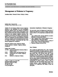
Fig. 1. Protein degradation mediated by ubiquitin–proteasome pathway.
the world. It is interesting to know that up to 30% of the newly synthesized proteins in a cell are broken down via proteasome since they do not pass the cell’s rigorous quality control.
The ubiquitin–proteasome system Degradation of a protein via the ubiquitin–proteasome pathway involves several discrete and successive steps (Fig. 1), mainly directing two events: (a) tagging of the substrate by covalent attachment of multiple ubiquitin molecules; and (b) degradation of the tagged protein by the 26S proteasome complex with release of free and reutilizable ubiquitin. Initially, the ubiquitin-activating enzyme, E1, activates ubiquitin (highly evolutionarily conserved 76-residue polypeptide) in an ATP-requiring reaction (step 1). Several E2 enzymes act as ubiquitin-carrier proteins or ubiquitin-conjugating enzymes (UBCs) and participate in the transferring process (step 2). Activated ubiquitin is transferred to the substrate that is specifically bound to a member of the ubiquitin-protein ligase family, E3 (step 3). The ubiquitin molecule is generally transferred to an NH2 group of an internal lysine residue in the substrate to generate a covelent isopeptide bond. By successively adding activated ubiquitin moieties to internal lysine residues on the previously conjugated ubiquitin molecule, a polyubiquitin chain is synthesized (step 4). This chain is specifically recognized by the downstream 26S proteasome
119 complex (step 5). The 26S proteasome is a large complex (2000 kDa) that exhibits multiple intrinsic protease activities to degrade proteins to small peptides. It is composed of a 20S catalytic core particle and two 19S regulatory particles. Models of proteolysis suggest that polyubiquitin chains of four or more residues on the target protein are recognized by subunits in the 19S; other subunits cleave the polyubiquitin chain for recycling (step 6). Target proteins are subsequently unfolded and guided into the 20S proteolytic core for cleavage to small peptides. In conjunction with E1, E2, and E3, recent studies point to existence of E4 proteins that may catalyse chain formation on particular substrates [3]. The requirement for the addition of multiple ubiquitin molecules may be part of a proof-reading mechanism, as a large number of deubiquitylating enzymes (DUBs) are also found within cells [4]. DUBs remove ubiquitin molecules before a sufficiently large branch structure has been synthesized to activate proteasomal destruction. Alternatively, they also prevent unwanted protein aggregation.
Diversity and specificity of ubiquitins The specificity of protein ubiquitylation resides in the interactions between a specific E2, an E3 and target protein. It appears that there exist more than 40 different Ub conjugating enzymes and 500 different Ub ligases. Even the modest genome of S. cerevisiae encodes thirteen E2-like products, termed Ubc1-13. Ubc4 and Ubc5 are involved in targeting the bulk of short-lived/abnormal/misfolded proteins. Others such as Ubc6 and Ubc7 are involved in degradation of proteins from within the ER [5]. The E3 ubiquitin ligases are very important in the pathway in that they provide selectivity to the ubiquitin process by serving as docking proteins that bring the substrate proteins and the E2 carrier protein with activated ubiquitin together [6]. The E3 ubiquitin ligases have been divided into specific subclasses, including single subunit RING finger proteins, multi-subunit RING finger proteins, HECT domain containing proteins and more recently, U-box containing E3’s [7–9]. The sheer number of these proteins indicates that ubiquitination plays an important role within the vast majority of signaling pathways. In fact, Ub modified proteins are known to have regulatory functions in cell cycle progression, organelle biogenesis, antigen presentation, stress responses, signal transduction, DNA repair, apoptosis, and transcriptional aspects [7, 8]. Much of our knowledge of E3 enzymes has emerged from studies on tumour suppressor protein p53 ubiquitination and degradation. High-risk strains of the human papillomavirus causes uterine cervical carcinoma tumours and degradation of p53 mediated by the HECT domain E3 enzyme E6-AP, was considered as an important mechanism used by the virus to transform cells [10]. The case of Parkinson’s disease (PD) also highlights the complexity of
the involvement of the ubiquitin system in the pathogenesis of neurodegeneration. Mutations in various genes such as α-synuclein, ubiquitin C-terminal hydrolase (UCH-L1, gene encoding the ubiquitin-recycling isoenzyme) and parkin (a RING-finger-containing E3 protein) were appeared to be responsible for the pathogenesis of PD [11]. NF-kB is the beststudied example of how the Ub-proteasome pathway modulates transcription. Latent NF-kB is kept sequestered in the cytoplasm via an interaction with the inhibitory action of IkB proteins [12]. Activation of NFkB is facilitated by IkB kinase, which phosphorylates IkBs. In turn, activation of IkB kinase requires regulation by a RING finger E3, the tumour necrosis factor receptor-associated factor 6 (TRAF6). Phosphorylated IkBs are polyubiquitinated and subsequently degraded by the 26S proteasome involving multi-RING finger E3 complex in association with Ub conjugating enzyme Cdc34 or Ubc5. Degradation of IkB unmasks the nuclear localization signal and the NFkB complex migrates to the nucleus where it regulates the expression of genes harboring NF-kB-response elements. With the many substrates targeted and processes involved, it is not surprising to find that aberrations in the ubiquitin system underlie the pathogenesis of many diseases. The pathological states associated with the ubiquitin system can be classified into two groups: (a) those that result from loss of function, i.e., mutations in a ubiquitin-system or target substrate that result in stabilization of certain proteins; and (b) those that result from gain of function, i.e., abnormal or accelerated degradation of the protein target. Some of the pathologies arising due to aberrations in ubiquitin– proteasome system include malignancies, Liddle’s syndrome, Angelman syndrome, Neurogenerative diseases, cystic fibrosis, immune and inflammatory response and muscle wasting [11].
Insulin signaling molecules misguided to proteasome in Type 2 diabetes From the studies of transgenic animal models and humans, there is evidence that inappropriate levels of signaling molecules strongly affect insulin action. It appears that controlled degradation of specific proteins by the ubiquitin– proteasome system plays an important role in the execution of various biological functions, including insulin signaling. Insulin promotes cellular growth and maintenance by a wide variety of both anabolic and anticatabolic actions, including the inhibition of overall proteolysis [13]. Although insulin administration causes an increase in the synthesis of some specific proteins, the effect of insulin on the level of total cellular protein is attributable almost entirely to decreased protein degradation [14]. In a series of experiments,
120 Bennett et al. [15] have demonstrated that insulin inhibits ATP-and ubiquitin-dependent degradation by the proteasome in vitro and in cultured cells. These findings extend the cellular processes controlled by insulin to include the ubiquitin– proteasome pathway. In contrast, evidence has accumulated over the past few years suggesting that prolonged exposure of cells to insulin promotes insulin receptor substrate-1 (IRS-1) degradation [16, 17]. Zhande et al. [17] have demonstrated that ubiquitin conjugation of IRS-1 is a prerequisite for insulin-induced IRS-1 proteasome degradation and that a structural element in the N-terminal region of IRS-1 specifically target IRS-1 to the ubiquitin–proteasome degradation pathway. It is now well established that IRS proteins are central effectors of the insulin signaling cascade and, on the contrary, is their inhibition or degradation that leads to the inhibition of insulin signaling [18–20]. Proinflammatory cytokines have been shown to promote serine phosphorylation of IRS1 and IRS2 that inhibits coupling to the activated insulin receptor and/or p85 regulatory subunit of phosphatidylinositol-3 kinase [21, 22]. In addition, IRS proteins are decreased in experimental and clinical diabetes. Although several insulinresistance-inducing stimuli, such as hyperinsulinemia, TNFα or other cytokines, glycated molecules and fatty acids have
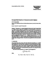
been shown to negatively regulate insulin signaling, the exact molecular mechanisms involved are poorly understood. Recent studies have revealed that IRS proteins are ubiquitinated and subsequently degraded by the 26S proteasome during insulin stimulation or during cellular stress [16, 23]. In an elegant study, White and his colleagues indicated that SOCS-1 and SOCS-3 (a family of proteins called the suppressor of cytokine signaling, SOCS) modulate insulin signaling by targeting IRS-1 & 2, for destruction by the proteasome [18]. Their work revealed two important observations. First, SOCS1 and SOCS3 were shown to have distinct binding sites for IRS1 or IRS2 and for elongin BC ubiquitin ligase. Secondly, SOCS-mediated ubiquitination of IRS1 or IRS2 is a necessary step for inhibition of insulin action in various cell lines and mouse liver. This mechanism is consistent with the function of SOCS1 and SOCS3 as adapter molecules linking tyrosine-phosphorylated proteins to an elongin BC-based E3 ubiquitin-ligase [24]. It appears that the ‘SOCS box’ acts as an adapter to facilitate the ubiquitination of signaling proteins and their subsequent targeting to the proteasome (Fig. 2). The SOCS box contains a elongin C binding motif that binds to elongin C, which in turn associates with a complex consisting of elongin B, a cullin family member, and RING-box protein 1 [25]. This whole protein complex constitutes an E3
Fig. 2. Insulin signaling molecules under proteasomal threat! Left: Normal insulin signaling pathway leading to balanced metabolic and mitogenic actions. Right: SOCS proteins, distal to the IR, mediating ubiquitin–proteasomal degradation of IRS.
121 ubiquitin ligase, which ultimately target proteins for degradation by the proteasome. These studies indicate proteasomal degradation of insulin signaling molecules as one of the molecular mechanisms of insulin resistance. In patients with HCV (Hepatic C Virus) infection, the HCV-associated insulin resistance has been demonstrated as a consequence to proteasomal degradation of IRS1 and IRS2 through ubiquitination involvling upregulation of SOC3 [26]. SOCS5mediated recruitment of ubiquitin complexes and EGFR (epidermal growth factor receptor) has also been recently demonstrated [27]. The existence of several isoforms of SOCS also indicates the possibility of a number of insulin signaling molecules becoming their binding partners for proteasome degradation. Another E3 ubiqutin ligase, (the Casitas b-lineage lymphoma, c-Cbl) has an important role in regulating the degradation of cell surface receptors [28]. It has been shown that c-Cbl-deficient mice have reduced adiposity, higher energy expenditure, and improved peripheral insulin action [29]. Since c-Cbl is an adapter protein with an intrinsic E3 ubiquitin ligase activity and a negative regulator of signaling, its role in insulin-regulated pathways need further investigations. Retinoic acid has been shown to mediate degradation of IRS-1 by the ubiquitin proteasome pathway, via a PKC-dependent mechanism [30]. A recent study also links ubiquitin proteasome pathway with β-cell mass decrease in Type 2 diabetes [31]. In this study, chronic activation of mTOR (mammalian target of rapamycin) by glucose (and/or IGF-1) in β-cells lead to increased Ser/Thr phosphorylation of IRS-2 that targets it for proteasomal degradation, resulting in decreased IRS-2 expression and increased β-cell apoptosis. Direct interaction of the glucose transporters GLUT1 and GLUT4 with members of the ubiquitin family has also been reported to play a role in the control of glucose uptake [32]. In an insulin-infusion study in human skeletal muscle, Rome et al. [33] showed that insulin increased the expression level of 17 mRNAs of the ubiquitin-conjugating enzymes and 11 mRNAs of the proteasome components. Recently, upregulation of ubiquitin specific protease 2 has been demonstrated in streptozotocin-induced diabetic mice [34]. The involvement of the ubiquitin–proteasome system in the regulation of transcription is also well known [35]. Mimnaugh et al. [36] demonstrated insulin-induced increases in the mRNA of USP16, which participates in the deubiquitination of histones H2A and H2B. The ubiquitin–proteasome pathway has also been implicated in the control of the levels of specific transcription factors such as nucelar factor kB1, retinoid X receptor α, peroxisome proliferator-activated receptors α and γ , thyroid hormone receptor, sterol regulatory elementbinding proteins and Forkhead transcription (FOXO1) factors [35, 37–41]. Interestingly, most of these transcription factors have been directly implicated in or related to insulin action [42, 43].
Role in Type 1 diabetes Although Type 1 and Type 2 diabetes are considered to be two distinct diseases in terms of aetiology, genetics and pathogenesis, inflammatory mediators are important in the pathogenesis of both diseases. In particular, beta cell loss in Type 1 and Type 2 diabetes may be a consequence of apoptosis and necrosis induced by inflammatory mediators [44, 45]. SOCS3 has been shown to protect beta cells against IL-1β and IFNγ -mediated toxicity [46]. Quite interestingly in a recent study [47], a SOCS3 mutation in the promoter region appeared as a protective mechanism against the development of diabetes. This study has speculated decreased SOCS3 expression leading to less degradation of IRS1 and IRS2 in insulin-sensitive tissues. Although mutation screening of the human SOCS3 gene identified no variations associated with Type 1 diabetes, SOCS may have indirect influences and cellular consequences. The newly identified SUMO (small ubiquitin-related modifier) genes tag proteins to modulate subcellular localization and/or enhance protein stability and activity and they do so by counteracting ubiquitin and stabilise proteins against degradation by the 26S proteasome [48]. In support of this, a M55V polymorphism in SUMO-4 has been shown differentially activating heat shock transcription factors and conferring susceptibility to Type 1 diabetes [49]. SUMO modifications were also reported to influence NFkB activation and apoptosis in pancreatic β-cells [50, 51]. While ubiquitin-like protein 1 (Ubl1) has been shown to modify IkBα [52], making it resistant to degradation, an increase in Ubl1 seeen in NOD mice [53] could decrease the expression of genes controlled by NFkB, including genes involved in immune and inflammatory responses in Type 1 diabetes.
Endoplasmic reticulum (ER) stress and ubiquitination While ER stress is caused by the accumulation of misfolded proteins in the ER [54], recent observations suggest that chronic ER stress in β-cells plays a role in the pathogenesis of diabetes [55]. ER stress has also been shown to cause β-cell death in the Akita mouse model for diabetes [56, 57]. The Akita mouse is a C57BL/6 mouse that is heterozygous for a mutation in the insulin-2 gene (cysteine 96 to tyrosine), which results in misfolding of the insulin precursor in the ER of β-cells. ER stress elicits stress signaling pathways termed the unfolded protein response (UPR). One component of the UPR, the ER-associated protein degradation (ERAD system) has an important function in the survival of ER stressed cells [58]. It has recently been shown that misfolded insulin produced in Akita mouse is selectively
122 ubiquitinated and degraded by a HRD1-mediated ERAD pathway [59]. Interestingly, HRD1 that is upregulated in the islets of Akita mice encodes an E3 ubiquitin ligase required for the ERAD system [60]. Thus, HRD1 regulation appears to be central to the protection of β-cells from ER stressmediated death. Oxygen-regulated protein 150 (ORP150), a molecular chaperone found in the ER, has also been shown to protect cells from ER stress [61]. ER stress playing a crucial role in the insulin resistance was also recently demonstrated in a study [62] when sense ORP overexpression in the liver of obese diabetic mice significantly improved insulin resistance and markedly ameliorated glucose tolerance. These studies warrant more research related to ER stress and ubiquitination regulatory aspects, which may unravel potential new therapeutic targets for diabetes.
Role in diabetic complications Alterations in protein turnover during diabetes have been previously correlated with increased activity of the ubiquitindependent proteolytic system in skeletal [63, 64] and cardiac [65] muscle. The severity of electroneurographic changes in patients with Type 2 diabetes has been correlated with increased ubiquitin levels [66]. It was hence implied that ubiquitin–proteasome pathway might have a role in the development of diabetic complications. For example, polyubiquitination of IGF-1 receptor and downreguation of heat shock proteins have been shown to be fundamental mechanisms underlying the development of diabetic cardiomyopathy [67]. Although the regulation of GLUT1 levels in retinal endothelial cells subjected to chronic hyperglycemia remains a matter of controversy [68–70], hyperglycemia is reported to impose a significant decrease on the amount of GLUT1 protein without significant changes on the levels of its mRNA. This implies that the decreased levels of GLUT1 may result from an increase on the degradation rate of the protein. Indeed, Fernandes et al. [71] have recently demonstrated the downregulation of retinal GLUT1 in diabetes by a mechanism of ubiquitinylation and hence, ubiquitinylation could be one of the alterations in endothelial cells that transduce pathophysiological changes in diabetic retinopathy. Moreover, many renal abnormalities (loss of muscle mass in chronic renal failure, von Hippel-Lindau disease, Liddle syndrome and ischemic acute renal failure) were reported to be associated with defective protein degradation involving the ubiquitin–proteasome pathway [6].
Future perspectives Ubiquitin-mediated proteolysis controls a variety of cellular processes: regulation of cell cycle and division,
differentiation and development, response to stress and extracellular effectors, morphogenesis of neuronal networks, modulation of cell-surface receptors, ion channels and the secretory pathway, DNA repair, transcriptional regulation, long-term memory and biogenesis of organelles [11]. Recent mining of the human and mouse genomes, use of yeast genetics, and detailed analyses of several biochemical pathways, have resulted in the identification of many new roles for ubiquitin conjugation. Mono- and polyubiquitination have now been shown to be involved in the regulation of a wide variety of important cellular processes [72]. Furthermore, several ubiquitin-like moficications (e.g., Sumoylation, Neddylation, and ISGylation) have been recently identified [73–75]. Because the ubiquitination processes affect the regulation of insulin signaling cascade and insulin action, it appears that there may be several hidden drug targets for diabetes and its complications in the ubiquitin–proteasome system. Considering the large number of transcription factors and Ub ligases present in the cell, it is possible that Ub or Ub-like mediated regulation of transcriptor factor localization will participate in the pathogenesis of diabetes and its complications. As suggested by Ciechanover [11], development of drugs that modulate the activity of the ubiquitin system may be difficult, considering its central role in a broad array of basic cellular processes. However, recent experimental evidence strongly suggests that proteasome inhibitors may indeed be beneficial in certain pathologies, such as in cancer, asthma, brain infarct and autoimmune encephalomyelitis. There may be a future in bioprospecting of natural products. Curcumin, an active component of Indian spice (Turmeric) has been shown to impair ubiquitin–proteasome system [76]. A different approach to drug development can be the development of small molecules that bind and inhibit specific E3s. Alternately, there is much scope in the development of small molecules that are substrate-specific and bind, preferably to specific substrates or to their ancillary proteins, rather than E3 proteins. A new UbE1 inhibitor, himeic acid A was recently isolated from a culture of marine-derived fungus, Aspergillus sp [77]. Smallmolecule drugs that inhibit SOCS action or the components of ubiquitin–proteasome system could also become important for the treatment of diabetes and its associated complications. Finally, much more research is needed to understand the factors controlling the signaling by ubiquitin and ubiquitin-like modifications, as this could lead to better understanding of the clinical implications of defects in this abundant, complex, and highly regulated protein modification machinery in the biological system.
References 1. Franch HA, Sooparb S, Du J, Brown NS: A mechanism regulating proteolysis of specific proteins during renal tubular cell growth. J Biol Chem 276: 19126–19131, 2001
123 2. Price SR, Bailey JL, Wang X, Jurkovitz C, England BK, Ding X, Phillips LS, Mitch WE: Muscle wasting in insulinopenic rats results from activation of the ATP-dependent, ubiquitin–proteasome proteolytic pathway by a mechanism including gene transcription. J Clin Invest 98: 1703– 1708, 1996 3. Grossman SR, Deato ME, Brignone C, Chan HM, Kung AL, Tagami H, Nakatani Y, Livingston DM: Polyubiquitination of p53 by a ubiquitin ligase activity of p300. Science 300: 342–344, 2003 4. Wilkinson KD: Ubiquitination and deubiquitination: targeting of proteins for degradation by the proteasome. Semin Cell Dev Biol 11: 141– 148, 2000 5. Glickman MH, Ciechanover A: The ubiquitin–proteasome proteolytic pathway: destruction for the sake of construction. Physiol Rev 82: 373– 428, 2002 6. Debigare R, Price SR: Proteolysis, the ubiquitin–proteasome system, and renal diseases. Am J Physiol 285: F1–F8, 2003 7. Hershko A, Ciechanover A: The ubiquitin system. Annu Rev Biochem 67: 425–479, 1998 8. Pickart CM: Mechanisms underlying ubiquitination. Annu Rev Biochem 70: 503–533, 2001 9. Hatakeyama S, Nakayama KI: Ubiquitylation as a quality control system for intracellular proteins. J Biochem (Tokyo) 134: 1–8, 2003 10. Scheffner M, Werness BA, Huibregtse JM, Levine AJ, Howley PM: The E6 oncoprotein encoded by human papillomavirus types 16 and 18 promotes the degradation of p53. Cell 63: 1129–1136, 1990 11. Ciechanover A: The ubiquitin proteolytic system and pathogenesis of human diseases: a novel platform for mechanism-based drug targeting. Biochem Soc Trans 31: 474–481, 2003 12. Baldwin AS Jr: The NF-kappa B and I kappa B proteins: new discoveries and insights. Annu Rev Immunol 14: 649–683, 1996 13. Russell-Jones DL, Umpleby M: Protein anabolic action of insulin, growth hormone and insulin-like growth factor I. Eur J Endocrinol 135: 631–642, 1996 14. Fryburg DA, Jahn LA, Hill SA, Oliveras DM, Barrett EJ: Insulin and insulin-like growth factor-I enhance human skeletal muscle protein anabolism during hyperaminoacidemia by different mechanisms. J Clin Invest 96: 1722–1729, 1995 15. Bennett RG, Hamel FG, Duckworth WC: Insulin inhibits the ubiquitindependent degrading activity of the 26S proteasome. Endocrinology 141: 2508–2517, 2000 16. Sun XJ, Goldberg JL, Qiao LY, Mitchell JJ: Insulin-induced insulin receptor substrate-1 degradation is mediated by the proteasome degradation pathway. Diabetes 48: 1359–1364, 1999 17. Zhande R, Mitchell JJ, Wu J, Sun XJ: Molecular mechanism of insulininduced degradation of insulin receptor substrate 1. Mol Cell Biol 22: 1016–1026, 2002 18. Rui L, Yuan M, Frantz D, Shoelson S, White MF: SOCS-1 and SOCS-3 block insulin signaling by ubiquitin-mediated degradation of IRS1 and IRS2. J Biol Chem 277: 42394–42398, 2002 19. Johnston AM, Pirola L, Van Obberghen E: Molecular mechanisms of insulin receptor substrate protein-mediated modulation of insulin signalling. FEBS Lett 546: 32–36, 2003 20. Ueki K, Kondo T, Tseng YH, Kahn CR: Central role of suppressors of cytokine signaling proteins in hepatic steatosis, insulin resistance, and the metabolic syndrome in the mouse. Proc Natl Acad Sci USA 101: 10422–10427, 2004 21. Aguirre V, Werner ED, Giraud J, Lee YH, Shoelson SE, White MF: Phosphorylation of Ser307 in insulin receptor substrate-1 blocks interactions with the insulin receptor and inhibits insulin action. J Biol Chem 277: 1531–1537, 2002 22. Grimble RF: Inflammatory status and insulin resistance. Curr Opin Clin Nutr Metab Care 5: 551–559, 2002
23. Rui L, Fisher TL, Thomas J, White MF: Regulation of insulin/insulinlike growth factor-1 signaling by proteasome-mediated degradation of insulin receptor substrate-2. J Biol Chem 276: 40362–40367, 2001 24. Kamura T, Sato S, Haque D, Liu L, Kaelin WG Jr, Conaway RC, Conaway JW: The Elongin BC complex interacts with the conserved SOCS-box motif present in members of the SOCS, ras, WD-40 repeat, and ankyrin repeat families. Genes Dev 12: 3872–3881, 1998 25. Krebs DL, Hilton DJ: A new role for SOCS in insulin action. Suppressor of cytokine signaling. Sci STKE 11: PE6, 2003 26. Kawaguchi T, Yoshida T, Harada M, Hisamoto T, Nagao Y, Ide T, Taniguchi E, Kumemura H, Hanada S, Maeyama M, Baba S, Koga H, Kumashiro R, Ueno T, Ogata H, Yoshimura A, Sata M: Hepatitis C Virus Down-Regulates Insulin Receptor Substrates 1 and 2 through Up-Regulation of Suppressor of Cytokine Signaling 3. Am J Pathol 165: 1499–1508, 2004 27. Kario E, Marmor MD, Adamsky K, Citri A, Amit I, Amariglio N, Rechavi G, Yarden Y: Suppressors of cytokine signaling 4 and 5 regulate epidermal growth factor receptor signaling. J Biol Chem (in press), 2004 28. Thien CB, Langdon WY: Cbl: many adaptations to regulate protein tyrosine kinases. Nat Rev Mol Cell Biol 2: 294–307, 2001 29. Molero JC, Jensen TE, Withers PC, Couzens M, Herzog H, Thien CB, Langdon WY, Walder K, Murphy MA, Bowtell DD, James DE, Cooney GJ: c-Cbl-deficient mice have reduced adiposity, higher energy expenditure, and improved peripheral insulin action. J Clin Invest 114: 1326– 1333, 2004 30. del Rincon SV, Guo Q, Morelli C, Shiu HY, Surmacz E, Miller WH: Retinoic acid mediates degradation of IRS-1 by the ubiquitin– proteasome pathway, via a PKC-dependant mechanism. Oncogene 23: 9269–9279, 2004 31. Briaud I, Dickson LM, Lingohr MK, McCuaig JF, Lawrence JC, Rhodes CJ: IRS-2 proteasomal degradation mediated by a mTOR-induced negative feedback downregulates PKB-mediated signaling pathway in betacells. J Biol Chem 280: 2282–2293, 2005 32. Giorgino F, de Robertis O, Laviola L, Montrone C, Perrini S, McCowen KC, Smith RJ: The sentrin-conjugating enzyme mUbc9 interacts with GLUT4 and GLUT1 glucose transporters and regulates transporter levels in skeletal muscle cells. Proc Natl Acad Sci USA 97: 1125–1130, 2000 33. Rome S, Clement K, Rabasa-Lhoret R, Loizon E, Poitou C, Barsh GS, Riou JP, Laville M, Vidal H: Microarray profiling of human skeletal muscle reveals that insulin regulates approximately 800 genes during a hyperinsulinemic clamp. J Biol Chem 278: 18063–18068, 2003 34. Yechoor VK, Patti ME, Ueki K, Laustsen PG, Saccone R, Rauniyar R, Kahn CR: Distinct pathways of insulin-regulated versus diabetesregulated gene expression: an in vivo analysis in MIRKO mice. Proc Natl Acad Sci USA 101: 16525–16530, 2004 35. Conaway RC, Brower CS, Conaway JW: Emerging roles of ubiquitin in transcription regulation. Science 296: 1254–1258, 2002 36. Mimnaugh EG, Chen HY, Davie JR, Celis JE, Neckers L: Rapid deubiquitination of nucleosomal histones in human tumor cells caused by proteasome inhibitors and stress response inducers: effects on replication, transcription, translation, and the cellular stress response. Biochemistry 36: 14418–14429, 1997 37. Hauser S, Adelmant G, Sarraf P, Wright HM, Mueller E, Spiegelman BM: Degradation of the peroxisome proliferator-activated receptor gamma is linked to ligand-dependent activation. J Biol Chem 275: 18527–18533, 2000 38. Matsushima-Nishiwaki R, Okuno M, Adachi S, Sano T, Akita K, Moriwaki H, Friedman SL, Kojima S: Phosphorylation of retinoid X receptor alpha at serine 260 impairs its metabolism and function in human hepatocellular carcinoma. Cancer Res 61: 7675–7682, 2001
124 39. Hirano Y, Yoshida M, Shimizu M, Sato R: Direct demonstration of rapid degradation of nuclear sterol regulatory element-binding proteins by the ubiquitin–proteasome pathway. J Biol Chem 276: 36431–36437, 2001 40. Blanquart C, Barbier O, Fruchart JC, Staels B, Glineur C: Peroxisome proliferator-activated receptor alpha (PPARalpha) turnover by the ubiquitin–proteasome system controls the ligand-induced expression level of its target genes. J Biol Chem 277: 37254–37259, 2002 41. Matsuzaki H, Daitoku H, Hatta M, Tanaka K, Fukamizu A: Insulininduced phosphorylation of FKHR (Foxo1) targets to proteasomal degradation. Proc Natl Acad Sci USA 100: 11285–11290, 2003 42. Auwerx J: PPARgamma, the ultimate thrifty gene: Diabetologia 42: 1033–1049, 1999 43. Foretz M, Guichard C, Ferre P, Foufelle F: Sterol regulatory element binding protein-1c is a major mediator of insulin action on the hepatic expression of glucokinase and lipogenesis-related genes. Proc Natl Acad Sci USA 96: 12737–12742, 1999 44. Nerup J, Mandrup-Poulsen T, Helqvist S, Andersen HU, Pociot F, Reimers JI, Cuartero BG, Karlsen AE, Bjerre U, Lorenzen T: On the pathogenesis of IDDM. Diabetologia 37(Suppl 2): S82–S89, 1994 45. Butler AE, Janson J, Soeller WC, Butler PC: Increased beta-cell apoptosis prevents adaptive increase in beta-cell mass in mouse model of type 2 diabetes: evidence for role of islet amyloid formation rather than direct action of amyloid. Diabetes 52: 2304–2314, 2003 46. Karlsen AE, Ronn SG, Lindberg K, Johannesen J, Galsgaard ED, Pociot F, Nielsen JH, Mandrup-Poulsen T, Nerup J, Billestrup N: Suppressor of cytokine signaling 3 (SOCS-3) protects beta-cells against interleukin1beta- and interferon-gamma-mediated toxicity. Proc Natl Acad Sci USA 98: 12191–12196, 2001 47. Gylvin T, Nolsoe R, Hansen T, Nielsen EM, Bergholdt R, Karlsen AE, Billestrup N, Borch-Johnsen K, Pedersen O, Mandrup-Poulsen T, Nerup J, Pociot F: Mutation analysis of suppressor of cytokine signalling 3, a candidate gene in Type 1 diabetes and insulin sensitivity. Diabetologia 47: 1273–1277, 2004 48. Hattori N, Mizuno Y: Pathogenetic mechanisms of parkin in Parkinson’s disease. Lancet 364: 722–724, 2004 49. Bohren KM, Nadkarni V, Song JH, Gabbay KH, Owerbach D: A M55V polymorphism in a novel SUMO gene (SUMO-4) differentially activates heat shock transcription factors and is associated with susceptibility to type I diabetes mellitus. J Biol Chem 279: 27233–27238, 2004 50. Guo D, Li M, Zhang Y, Yang P, Eckenrode S, Hopkins D, Zheng W, Purohit S, Podolsky RH, Muir A, Wang J, Dong Z, Brusko T, Atkinson M, Pozzilli P, Zeidler A, Raffel LJ, Jacob CO, Park Y, Serrano-Rios M, Larrad MT, Zhang Z, Garchon HJ, Bach JF, Rotter JI, She JX, Wang CY: A functional variant of SUMO4, a new I kappa B alpha modifier, is associated with type 1 diabetes. Nat Genet 36: 837–841, 2004 51. Owerbach D, Pina L, Gabbay KH: A 212-kb region on chromosome 6q25 containing the TAB2 gene is associated with susceptibility to type 1 diabetes. Diabetes 53: 1890–1893, 2004 52. Desterro JM, Rodriguez MS, Hay RT: SUMO-1 modification of IkappaBalpha inhibits NF-kappaB activation. Mol Cell 2: 233–239, 1998 53. Eckenrode SE, Ruan Q, Yang P, Zheng W, McIndoe RA, She JX: Gene expression profiles define a key checkpoint for type 1 diabetes in NOD mice. Diabetes 53: 366–375, 2004 54. Harding HP, Calfon M, Urano F, Novoa I, Ron D: Transcriptional and translational control in the Mammalian unfolded protein response. Annu Rev Cell Dev Biol 18: 575–599, 2002 55. Harding HP, Ron D: Endoplasmic reticulum stress and the development of diabetes: A review. Diabetes 51(Suppl 3): S455–S461, 2002
56. Oyadomari S, Araki E, Mori M: Endoplasmic reticulum stressmediated apoptosis in pancreatic beta-cells. Apoptosis 7: 335–345, 2004 57. Wang J, Takeuchi T, Tanaka S, Kubo SK, Kayo T, Lu D, Takata K, Koizumi A, Izumi T: A mutation in the insulin 2 gene induces diabetes with severe pancreatic beta-cell dysfunction in the Mody mouse. J Clin Invest 103: 27–37, 1999 58. Kaneko M, Ishiguro M, Niinuma Y, Uesugi M, Nomura Y: Human HRD1 protects against ER stress-induced apoptosis through ER-associated degradation. FEBS Lett 532: 147–152, 2002 59. Allen JR, Nguyen LX, Sargent KE, Lipson KL, Hackett A, Urano F: High ER stress in beta-cells stimulates intracellular degradation of misfolded insulin. Biochem Biophys Res Commun 324: 166–170, 2004 60. Kikkert M, Doolman R, Dai M, Avner R, Hassink G, van Voorden S, Thanedar S, Roitelman J, Chau V, Wiertz E: Human HRD1 is an E3 ubiquitin ligase involved in degradation of proteins from the endoplasmic reticulum. J Biol Chem 279: 3525–3534, 2004 61. Tamatani M, Matsuyama T, Yamaguchi A, Mitsuda N, Tsukamoto Y, Taniguchi M, Che YH, Ozawa K, Hori O, Nishimura H, Yamashita A, Okabe M, Yanagi H, Stern DM, Ogawa S, Tohyama M: ORP150 protects against hypoxia/ischemia-induced neuronal death. Nat Med 7: 317–323, 2001 62. Nakatani Y, Kaneto H, Kawamori D, Yoshiuchi K, Hatazaki M, Matsuoka TA, Ozawa K, Ogawa S, Hori M, Yamasaki Y, Matsuhisa M: Involvement of endoplasmic reticulum stress in insulin resistance and diabetes. J Biol Chem 280: 847–851, 2005 63. Rodriguez T, Busquets S, Alvarez B, Carb N, Agell N, Lpez-Soriano FJ, Argils JM: Protein turnover in skeletal muscle of the diabetic rat: activation of ubiquitin-dependent proteolysis. Int J Mol Med 1: 971– 977, 1998 64. Galban VD, Evangelista EA, Migliorini RH, do Carmo Kettelhut I: Role of ubiquitin–proteasome-dependent proteolytic process in degradation of muscle protein from diabetic rabbits. Mol Cell Biochem 225: 35–41, 2001 65. Liu Z, Miers WR, Wei L, Barrett EJ: The ubiquitin–proteasome proteolytic pathway in heart vs skeletal muscle:effects of acute diabetes. Biochem Biophys Res Commun 276: 1255–1260, 2000 66. Akarsu E, Pirim I, Capoglu I, Deniz O, Akcay G, Unuvar N: Relationship between electroneurographic changes and serum ubiquitin levels in patients with type 2 diabetes. Diabetes Care 24: 100–103, 2001 67. Shan YX, Yang TL, Mestril R, Wang PH: Hsp10 and Hsp60 suppress ubiquitination of insulin-like growth factor-1 receptor and augment insulin-like growth factor-1 receptor signaling in cardiac muscle: implications on decreased myocardial protection in diabetic cardiomyopathy. J Biol Chem 278: 45492–45498, 2003 68. Badr GA, Tang J, Ismail-Beigi F, Kern TS: Diabetes downregulates GLUT1 expression in the retina and its microvessels but not in the cerebral cortex or its microvessels. Diabetes 49: 1016–1021, 2000 69. Kumagai AK: Glucose transport in brain and retina: implications in the management and complications of diabetes. Diabetes Metab Res Rev 15: 261–273. 1999 70. Tang J, Zhu XW, Lust WD, Kern TS: Retina accumulates more glucose than does the embryologically similar cerebral cortex in diabetic rats. Diabetologia 43: 1417–1423, 2000 71. Fernandes R, Carvalho AL, Kumagai A, Seica R, Hosoya K, Terasaki T, Murta J, Pereira P, Faro C: Downregulation of retinal GLUT1 in diabetes by ubiquitinylation. Mol Vis 10: 618–628, 2004
125 72. Sun L, Chen ZJ: The novel functions of ubiquitination in signaling. Curr Opin Cell Biol 16: 119–126, 2004 73. Hilgarth RS, Murphy LA, Skaggs HS, Wilkerson DC, Xing H, Sarge KD: Regulation and function of SUMO modification. J Biol Chem 279: 53899–53902, 2004 74. Xirodimas DP, Saville MK, Bourdon JC, Hay RT, Lane DP: Mdm2mediated NEDD8 conjugation of p53 inhibits its transcriptional activity. Cell 118: 83–97, 2004, 75. Malakhova OA, Yan M, Malakhov MP, Yuan Y, Ritchie KJ, Kim KI, Peterson LF, Shuai K, Zhang DE: Protein ISGylation
modulates the JAK-STAT signaling pathway. Genes Dev 17: 455–460, 2003 76. Jana NR, Dikshit P, Goswami A, Nukina N: Inhibition of proteasomal function by curcumin induces apoptosis through mitochondrial pathway. J Biol Chem 279: 11680–11685, 2004 77. Tsukamoto S, Hirota H, Imachi M, Fujimuro M, Onuki H, Ohta T, Yokosawa H: Himeic acid A: a new ubiquitinactivating enzyme inhibitor isolated from a marine-derived fungus, Aspergillus sp. Bioorg Med Chem Lett 15: 191–194, 2005