J Appl Physiol 97: 1299 –1308, 2004. First published June 4, 2004; 10.1152/japplphysiol.01350.2003.
Neural coupling between upper and lower limbs during recumbent stepping Helen J. Huang1 and Daniel P. Ferris1,2 Departments of 1Biomedical Engineering and 2Movement Science, University of Michigan, Ann Arbor, Michigan 48109-2214 Submitted 18 December 2003; accepted in final form 28 May 2004
Huang, Helen J., and Daniel P. Ferris. Neural coupling between upper and lower limbs during recumbent stepping. J Appl Physiol 97: 1299 –1308, 2004. First published June 4, 2004; 10.1152/japplphysiol. 01350.2003.—During gait rehabilitation, therapists or robotic devices often supply physical assistance to a patient’s lower limbs to aid stepping. The expensive equipment and intensive manual labor required for these therapies limit their availability to patients. One alternative solution is to design devices where patients could use their upper limbs to provide physical assistance to their lower limbs (i.e., self-assistance). To explore potential neural effects of coupling upper and lower limbs, we investigated neuromuscular recruitment during self-driven and externally driven lower limb motion. Healthy subjects exercised on a recumbent stepper using different combinations of upper and lower limb exertions. The recumbent stepper mechanically coupled the upper and lower limbs, allowing users to drive the stepping motion with upper and/or lower limbs. We instructed subjects to step with 1) active upper and lower limbs at an easy resistance level (active arms and legs); 2) active upper limbs and relaxed lower limbs at easy, medium, and hard resistance levels (self-driven); and 3) relaxed upper and lower limbs while another person drove the stepping motion (externally driven). We recorded surface electromyography (EMG) from six lower limb muscles. Self-driven EMG amplitudes were always higher than externally driven EMG amplitudes (P ⬍ 0.05). As resistance and upper limb exertion increased, selfdriven EMG amplitudes also increased. EMG bursts during selfdriven and active arms and legs stepping occurred at similar times. These results indicate that active upper limb movement increases neuromuscular activation of the lower limbs during cyclic stepping motions. Neurologically impaired humans that actively engage their upper limbs during gait rehabilitation may increase neuromuscular activation and enhance activity-dependent plasticity.
currently available for individuals that have experienced stroke or spinal cord injury is manually assisted locomotor training (4, 24, 51). This therapy uses a harness to help support the body weight of patients and manual assistance to help patients practice stepping on a treadmill. Multiple therapists supply manual assistance to the patients’ lower limbs, promoting normal stepping kinematics. The task-specific motor practice improves gait control and functional ability. More recently, a number of research groups have developed and begun testing robotic devices for providing physical assistance during locomotor training (11, 25, 38). A drawback to both manually assisted and robotic locomotor training is that patients may allow the external assistance to do most of the mechanical work and become less active during training. For motor learning and recovery to occur, the patient
must provide sufficient effort to promote activity-dependent plasticity (34, 53). It is obvious that to maximize activitydependent plasticity the patient must be as active as possible during his/her therapy. Encouraging patients to be active during therapy increases neuromuscular recruitment and should therefore accelerate motor recovery. Ideally, the amount and timing of physical assistance should complement the subject’s exertion by assisting the motion but not dominating it. Physical therapy methods that rely on external assistance may cause patients to fight the assistance or become less active. Both situations are undesirable. One way to minimize inappropriately timed assistance and/or increased patient activity is to put the patient in control of the physical assistance. This would encourage patients to be more active and allow them to practice stepping without the need for constant therapist intervention. During gait rehabilitation, patients could use their upper limbs to assist their lower limbs. Self-assistance (i.e., using upper limbs to assist lower limbs) could potentially be beneficial during gait rehabilitation due to inherent interlimb neural coupling (14, 56). There are neural connections in the spinal cords of cats that coordinate muscle activation between the forelimbs and hindlimbs (30, 31). Although similar functional connections in humans have not been irrefutably proven, humans do adopt phase-locked coordination patterns between their upper and lower limbs during many locomotor tasks such as walking, creeping, and swimming (48). Humans also demonstrate neural coupling in reflex responses between upper and lower limbs during walking (15, 20, 23, 55). Last, anecdotal evidence suggests that reciprocal arm swing can help to initiate lower limb muscle activation during stepping in subjects with incomplete spinal cord injury (5). These observations support the premise that upper limb neuromuscular activation may affect lower limb neuromuscular activation during cyclic stepping movements. The purpose of this study was to determine whether evidence of neural coupling between the upper limbs and lower limbs exists in healthy subjects during a simplified stepping task. To constrain movement kinematics, we studied subjects as they exercised on a recumbent stepper that mechanically coupled upper and lower limb motion. We compared muscle activation during self-driven and externally driven stepping motions. Self-driven refers to an individual using only his/her upper limbs to drive his/her relaxed lower limbs through the stepping motion. Externally driven refers to a separate individual driving the subject’s relaxed upper and lower limbs through the stepping motion. We hypothesized that self-driven stepping motion will result in greater lower limb muscle activity compared with exter-
Address for reprint requests and other correspondence: H. J. Huang, Human Neuromechanics Laboratory, 1206A CCRB, 401 Washtenaw Ave., Univ. of Michigan, Ann Arbor, MI 48109-2214 (E-mail:
[email protected]).
The costs of publication of this article were defrayed in part by the payment of page charges. The article must therefore be hereby marked “advertisement” in accordance with 18 U.S.C. Section 1734 solely to indicate this fact.
gait; locomotion; rehabilitation; interlimb coordination; electromyography
THE MOST PROMISING GAIT REHABILITATION THERAPY
http://www. jap.org
8750-7587/04 $5.00 Copyright © 2004 the American Physiological Society
1299
1300
NEURAL COUPLING BETWEEN UPPER AND LOWER LIMBS
pression load cells (LCWD-1000, Omegadyne, Sunbury, OH). We also measured joint angles using electrogoniometers (Biometrics, Ladysmith, VA). All subjects were given time to practice stepping using each combination of upper and lower limb exertions. For each trial, we collected data for 10 s after subjects verbally confirmed that they were comfortable with the test condition. We recorded two trials for each test condition and randomized trial order for each subject to minimize potential learning effects. Subjects also rested for ⬃20 s between trials and had the option to rest longer. Experimental Design
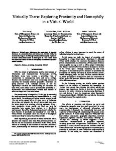
Fig. 1. NuStep TRS 4000 recumbent stepper (NuStep, Ann Arbor, MI).
nally driven stepping motion. We based this hypothesis on evidence that the upper and lower limbs of humans appear to be neurally coupled during locomotor movements (5, 14, 15, 20, 23, 48, 55, 56). METHODS
Subjects Twenty-four healthy subjects (14 men and 10 women; age range 19 –29 yr) participated in this study. The University of Michigan Medical School Institutional Review Board granted approval for this project, and all subjects gave informed, written consent. Recumbent Stepper The NuStep recumbent stepper (TRS 4000, NuStep, Ann Arbor, MI) is a commercially available exercise machine that is a hybrid between a recumbent bicycle and a stair stepper. It has two handles and two pedals that are all coupled to one another, allowing users to exercise with upper and/or lower limbs (Fig. 1). The right handle and left foot pedal move together, as does the left handle and right foot pedal. The contralateral handle-pedal pairs are 180° out of phase with each other so that when the right upper limb and left lower limb are fully extended, the left upper limb and right lower limb are fully flexed. This mechanical coupling results in a simplified one degree of freedom stepping motion that preserves normal walking interlimb phase relationships. The recumbent stepper also has 10 unitless levels of resistance, created via an eddy-current disk brake. A mechanical lever allows the user to move an array of magnets closer and farther from the spinning conductive disk, thus increasing or decreasing, respectively, the resistive forces.
We conducted two separate experiments. In the first experiment, the upper and lower limb motions were mechanically coupled as dictated by the stepper’s design. In the second experiment, we used two recumbent steppers to test a similar protocol but with the upper and lower limb kinetics decoupled. In this second experiment, we placed one recumbent stepper behind another and modified the front stepper with long handle extensions (Fig. 2). This allowed subjects to drive the front stepper with their upper limbs while sitting in the back stepper. Decoupling the upper and lower limb kinetics in this manner prevented subjects from pushing with their lower limbs to lessen the work performed by the upper limbs. Experiment 1. Subjects (n ⫽ 20) stepped using four different combinations of upper and lower limb exertions. We classified these exertions as “active” and “passive” in reference to the subject’s conscious intent. There was one condition in which subjects stepped using both their upper and lower limbs (active arms and legs). This condition provided us with information about the normal timing of lower limb muscle activation during active recumbent stepping. There were three conditions in which subjects attempted to totally relax their lower limbs (i.e., passive lower limbs). For the simplest passive condition, subjects rested their hands on their laps as another person drove their lower limbs through the stepping motion (externally driven). For a second passive condition, subjects’ hands and feet were fixed to the handles and pedals using Velcro mitts and straps, respectively, as another person drove their relaxed upper and lower limbs through the stepping motion (externally driven arms and legs). We tested this condition to determine whether passive movement of the upper limbs made a difference in the results. For the last passive condition, subjects actively used their arms to drive their relaxed lower limbs through the stepping motion (self-driven).
General Procedure Subjects adjusted the stepper’s handle and seat positions so that their knees did not reach full knee extension. Velcro straps secured the subject’s feet to the stepper’s pedals and the subject’s torso to the stepper’s seat. A digital metronome set to a frequency of 1.25 Hz (75 beats/min) helped subjects maintain a constant stepping frequency. We recorded electromyography (EMG) from six muscles on each lower limb (Konigsberg Instruments, Pasadena, CA). We measured the pedal force for each foot during stepping using single-axis comJ Appl Physiol • VOL
Fig. 2. Schematic of the kinetically decoupled setup used in experiment 2. Subjects used the handle extensions to drive the front NuStep. Because subjects were instructed to relax their lower limbs, another person had to drive the subject’s lower limbs through the stepping motion. In this setup, subjects could not use their lower limbs to ease the upper limb task (i.e., cheat). Modified from Ref. 26.
97 • OCTOBER 2004 •
www.jap.org
NEURAL COUPLING BETWEEN UPPER AND LOWER LIMBS
1301
We examined three levels of resistance for the self-driven passive condition: easy, medium, and hard. These resistance levels were determined before testing for each subject. Each subject’s hard resistance level corresponded to the maximum resistance that he/she could maintain using just his/her arms moving at the designated frequency for at least 20 s. The medium resistance level corresponded to ⬃60% of the subject’s hard resistance level. The easy resistance level was the lowest resistance setting on the recumbent stepper. Because the level of resistance was not important for the externally driven conditions, the resistance was set at easy for all externally driven trials. Experiment 2. The main difference between this experiment and the first experiment was that a second recumbent stepper was used to decouple the kinetics of the pedals and handles (Fig. 2). We tested four healthy male subjects under three conditions: 1) externally driven, 2) upper limb exertion (easy), and 3) upper limb exertion (hard). In the externally driven condition, subjects were instructed to attempt to totally relax their upper and lower limbs, as other people drove the subject’s upper and lower limbs through the range of motion at the designated frequency. In the upper limb exertion conditions, subjects were instructed to actively use their upper limbs to drive the front stepper at the designated frequency while relaxing their lower limbs as another person drove their legs through the motion. We instructed the person driving the subject’s lower limbs to match the subject’s upper limb movement. For the upper limb exertion conditions, we examined two resistance levels: easy and hard. These resistance levels were determined before testing for each subject. Each subject’s hard resistance level corresponded to the maximum resistance that he/she could step at using just his/her arms while maintaining the designated frequency for at least 20 s.
zeroed as subjects stood in a neutral position. From these data, we identified the beginning and end of step cycles. We also used goniometer data to define flexion and extension phases. Pedal forces. For experiment 1, we measured the pedal force for each foot during stepping using a set of three compression load cells (LCWD1000, Omegadyne). These load cells were coplanar, positioned in a tripod formation between two custom-shaped aluminum plates for each foot. The summation of the three load cells for each foot provided a measure of the force between the foot and pedal. Before each data collection, we calibrated each load cell within the aluminum plates with known weights. We summed the three load cell measurements for each foot plate and then filtered the data with a fourth-order low-pass Butterworth filter with zero phase shift (cutoff frequency of 6 Hz). We calculated the mean forces during lower limb flexion and extension for six step cycles per condition. We then normalized the forces to the maximum mean force across conditions for each subject. Last, we averaged the forces across all subjects for the flexion and extension phases. We were only able to collect pedal forces on 10 subjects.
Data Acquisition and Analysis
Experiment 1: One NuStep with Upper and Lower Limb Kinetics Coupled
In the first experiment, we collected EMG, joint angle (hip, knee, and ankle), and pedal force data during each trial. In the second experiment, we collected EMG and joint angle (knee and elbow) data during each trial. For both experiments, a personal computer sampled each data channel at 1,000 Hz via an analog-to-digital board. EMG. For experiments 1 and 2, we measured muscle activity from six muscles on each lower limb [vastus lateralis (VL), vastus medialis (VM), medial hamstrings (MH), medial gastrocnemius (MG), tibialis anterior (TA), and soleus (SO) muscles] using surface electrodes (Konigsberg Instruments). For each electrode site, we shaved and cleaned the area with alcohol. We then placed EMG surface electrodes (diameter, 1.1 cm; interelectrode distance, 3.5 cm) in the middle of the muscle belly along its long axis. For the SO, however, we placed the electrodes over the lateral distal third of the muscle belly rather than the center to avoid potential cross talk from the gastrocnemius. We then visually examined each EMG signal for noise and cross talk (52). We placed tape over all electrodes and covered the subject’s lower limbs with elastic stockings to minimize mechanical artifacts. To compare muscle activation across conditions, we calculated the averaged normalized root mean square (RMS) EMG amplitude for each condition and muscle for flexion and extension phases. We processed EMG with a fourth-order high-pass Butterworth filter with zero phase shift (cutoff frequency of 20 Hz) and then full-wave rectified the signals. For each phase, condition, muscle, and subject, we averaged RMS amplitudes for six step cycles. We then normalized the averaged RMS amplitudes to the maximum RMS amplitude calculated for each muscle across conditions for each subject to reduce intersubject variability (54). To compare EMG timing across conditions, we used cross-correlation analysis to calculate R values and lag times. Joint angles. For experiment 1, we measured joint angles of the hip, knee, and ankle on both lower limbs using twin-axis electrogoniometers (Biometrics), placed along the sagittal plane. For experiment 2, we measured knee joint angles on both lower limbs and elbow joint angles on both upper limbs using twin-axis electrogoniometers (Biometrics) placed along the sagittal plane. Electrogoniometers were J Appl Physiol • VOL
Statistical Analyses For each experiment, we used a repeated-measures ANOVA to determine whether there were significant differences across conditions. For the ANOVA analyses of EMG in the six muscles, we set P ⬍ 0.0083 for the significance level (Bonferroni correction). When the ANOVA indicated a significant difference, we used Tukey’s honestly significant difference (THSD) post hoc test to determine which conditions were significantly different (P ⬍ 0.05). RESULTS
Self-driven stepping motion resulted in greater lower limb muscle activation compared with externally driven stepping motion. Externally driven EMG raw data showed little EMG activity for all six muscles, whereas distinctive rhythmic EMG bursts were visible in self-driven (hard) EMG raw data (Fig. 3). Group-averaged EMG profiles indicated that lower limb muscle activation for the self-driven (hard) condition had similar patterns compared with the active arms and legs condition (Fig. 4). Onset of the EMG bursts during self-driven (hard) and active arms and legs conditions occurred at nearly the same time in the step cycle. Lag times at maximum correlation between self-driven (hard) and active arms and legs were close to zero (Table 1). This confirmed that self-driven (hard) and active arms and legs EMG bursts had similar timing. Crosscorrelation results comparing medium and easy self-driven conditions to active arms and legs also showed similar timing in muscle activation (not shown). For all six muscles during both flexion and extension, the normalized EMG amplitudes for the three self-driven conditions were greater than the amplitudes for the externally driven and externally driven arms and legs conditions (Fig. 5). Across all six muscles, self-driven EMG amplitudes for the flexion phase were ⬃35, 42, and 55% for easy, medium, and hard resistances, respectively. In contrast, externally driven and externally driven arms and legs conditions had EMG amplitudes of 30% across all six muscles. Similarly, self-driven EMG amplitudes averaged for all six muscles for the extension phase were 38, 46, and 55% for easy, medium, and hard resistances, respectively. Externally driven and externally driven arms and legs conditions had EMG amplitudes of 32 and 31%, respectively, averaged for all six muscles. Statistical
97 • OCTOBER 2004 •
www.jap.org
1302
NEURAL COUPLING BETWEEN UPPER AND LOWER LIMBS
Fig. 3. Single subject raw electromyography (EMG), joint angle, and pedal force data for externally driven (left) and self-driven (hard) (right) conditions. Self-driven (hard) EMG has distinct bursts and greater muscle activity compared with externally driven EMG, which has little activity. Joint profiles for the hip, knee, and ankle show that all 3 joints have a triangular pattern and reach their range of motion end points simultaneously. VL, vastus lateralis; VM, vastus medialis; MH, medial hamstrings; MG, medial gastrocnemius; SO, soleus; TA, tibialis anterior.
analyses indicated that, for both flexion and extension phases, self-driven (hard) and active arms and legs were significantly different (THSD, P ⬍ 0.05) from externally driven and externally driven arms and legs conditions for a majority of the muscles (Fig. 5).
During self-driven stepping, greater upper limb exertion led to greater lower limb muscle activation. Self-driven (easy), self-driven (medium), and self-driven (hard) EMG amplitudes increased in a stepwise manner (Fig. 5). Self-driven (medium) was not significantly different from self-driven (easy) for any of the muscles (THSD, P ⬎ 0.05). Self-driven (hard) was significantly different from self-driven (easy) for the MH, SO, and TA muscles during flexion and for the VL, VM, MH, SO, and TA during extension (THSD, P ⬍ 0.05). Self-driven (hard) was significantly different from self-driven (medium) for the TA and SO muscles during flexion (THSD, P ⬍ 0.05). Pedal force profiles were similar for self-driven and active arms and legs stepping (Fig. 6A). Both conditions had higher peak forces during the extension phase than during the flexion phase, and the two conditions demonstrated similar timing of Table 1. Self-driven (hard) vs. active arms and legs EMG cross-correlation analysis
Fig. 4. Group-averaged EMG bursts for active arms and legs, self-driven (hard), and externally driven in experiment 1. The timing of the self-driven (hard) passive leg EMG patterns is similar to the timing of the active leg EMG patterns in the active arms and legs condition (see Table 1 for more analysis). J Appl Physiol • VOL
Muscle
R Value
Lag Time, % cycle
VL VM MH MG SO TA
0.84⫾0.10 0.84⫾0.09 0.89⫾0.06 0.90⫾0.06 0.90⫾0.06 0.88⫾0.10
⫺1⫾11 ⫺3⫾14 4⫾12 1⫾5 ⫺2⫾13 2⫾14
Values are means ⫾ SE. A positive lag time indicates that self-driven (hard) follows active arms and legs. A negative lag time means that self-driven (hard) begins before active arms and legs. Mean lag times for all muscles are ⬍4% of a step cycle from zero, indicating that self-driven (hard) and active arms and legs have similar timing. EMG, electromyography; VL, vastus lateralis; VM, vastus medialis; MH, medial hamstrings; MG, medial gastrocnemius; SO, soleus; TA, tibialis anterior.
97 • OCTOBER 2004 •
www.jap.org
NEURAL COUPLING BETWEEN UPPER AND LOWER LIMBS
1303
Fig. 5. Averaged normalized root mean square (RMS) EMG with standard error bars for all subjects for flexion and extension phases in experiment 1. White columns represent externally driven conditions, whereas gray columns indicate self-driven conditions. Black columns correspond to the active condition. *Significantly different from the externally driven condition on the far left [Tukey’s honestly significant difference (THSD), P ⬍ 0.05]. For both flexion and extension, active arms and legs and selfdriven (hard) RMS EMG amplitudes were significantly different from both externally driven conditions for a majority of the muscles (THSD, P ⬍ 0.05). Externally driven and externally driven arms and legs RMS EMG amplitudes were not significantly different for any of the 6 muscles in either phase (THSD, P ⬎ 0.05).
force increase during the start of the extension phase. In contrast, the externally driven pedal force profile had a different pattern (Fig. 6A). Overall, the externally driven condition had lower peak forces during the extension phase than during the flexion phase. There was a slight rise in pedal force at the end of extension and the beginning of flexion to decelerate the lower limb. Self-driven mean flexion pedal forces were 45, 42, and 40% for easy, medium, and hard resistances, respectively (Fig. 6B). Externally driven mean flexion pedal forces were 50% for both conditions (externally driven, externally driven arms and legs). For the extension phase, self-driven mean pedal forces were 39, 44, and 53% for easy, medium, and hard conditions, respectively (Fig. 6C). Both externally driven and externally driven arms and legs mean extension pedal forces were 41%. Self-driven and externally driven mean pedal forces J Appl Physiol • VOL
were not significantly different from each other for either phase (THSD, P ⬎ 0.05). Active arms and legs mean extension force (93%) was significantly different from externally driven mean extension force (THSD, P ⬍ 0.05). Joint angle data indicated that ranges of motion for the hip, knee, and ankle were similar for each subject across all conditions. Hip, knee, and ankle profiles during recumbent stepping were regular and synchronized, having a triangular pattern (Fig. 3). The average hip angle excursion for all subjects was 73– 43° of hip flexion (for all 3 joints, 0° was standing posture). The average knee angle excursion was 75–23° of knee flexion. The average ankle angle excursion was 19° of dorsiflexion to ⫺3° of plantarflexion. There was no significant difference in range of motion between conditions (ANOVA, P ⬎ 0.05).
97 • OCTOBER 2004 •
www.jap.org
1304
NEURAL COUPLING BETWEEN UPPER AND LOWER LIMBS
condition (Figs. 7 and 8). Group-averaged EMG showed that the coupled and decoupled protocols resulted in similar EMG patterns (Fig. 7). Externally driven EMG profiles were fairly constant with no distinct bursts in any of the muscles. Upper limb exertion (hard) EMG profiles had distinct EMG bursts for the VL, VM, and TA muscles. Both upper limb exertion conditions had greater normalized EMG RMS amplitudes compared with the externally driven condition for both flexion and extension phases (Fig. 8). Statistical analyses indicated that upper limb exertion (hard) was significantly different (THSD, P ⬍ 0.05) from externally driven for a majority of the muscles during both flexion and extension phases. DISCUSSION
Fig. 6. A: mean low-pass filtered (cutoff frequency ⫽ 6 Hz) pedal forces for active arms and legs, self-driven (hard), and externally driven conditions. Timing was similar for self-driven (hard) and active arms and legs filtered force profiles. The externally driven filtered force profile, however, has a different pattern. B: averaged normalized mean pedal force for flexion phase. White columns represent externally driven conditions, whereas gray columns indicate self-driven conditions. Black columns correspond to the active condition. There was no significant difference (THSD, P ⬎ 0.05) between externally driven and any self-driven condition or the active arms and legs condition. However, self-driven (medium) and self-driven (hard) were both significantly different from active arms and legs. Mean flexion force for active arms and legs stepping was 53 N. C: averaged normalized mean pedal force for extension phase. The active arms and legs extension force (marked with an asterisk) was the only condition significantly different from externally driven extension force (THSD, P ⬍ 0.05). There were no significant differences between the 3 self-driven and 2 externally driven conditions. Mean extension force for active arms and legs stepping was 75 N.
Experiment 2: Two NuSteps with Upper and Lower Limb Kinetics Decoupled Upper limb exertion conditions resulted in greater lower limb muscle activation compared with the externally driven J Appl Physiol • VOL
Our main finding is that rhythmic upper limb activity increases lower limb neuromuscular recruitment when healthy subjects attempt to relax their lower limbs. In experiment 1, self-driven conditions had greater EMG amplitudes compared with externally driven and externally driven arms and legs conditions. In experiment 2, upper limb exertion conditions had greater EMG amplitudes compared with the externally driven condition. Thus upper limb activity resulted in greater lower limb muscle activity regardless of whether upper limb and lower limb kinetics were coupled or decoupled. An important aspect of this finding was that lower limb muscle activation during the self-driven conditions was dependent on upper limb effort. As resistance level increased for the upper limbs, EMG amplitudes for the lower limb muscles also increased. At all resistance levels, the timing of the self-driven EMG bursts in the lower limbs was similar to the timing of active stepping EMG bursts. Muscles were primarily active during the concentric phase of the stepping motion. For the VL, VM, and SO, this was the extension phase. For the TA, this was the flexion phase. The biarticular MH and MG showed some activation during both flexion and extension phases. The concentric timing of the uniarticular muscles indicates that the increased muscle activation during self-driven stepping was probably not a result of stretch reflexes. If the stretch reflexes had caused the muscle activation, the EMG bursts would have occurred during and/or slightly after the muscles were stretched in their eccentric phase. One potential neural mechanism responsible for lower limb muscle activation during self-driven stepping is spinal connections in locomotor neural networks. Humans have spinal connections coordinating left and right lower limbs during stepping (19) in a manner similar to cats (18). Cats also have spinal connections between cervical and lumbar locomotor networks coordinating forelimb and hindlimb muscle activation patterns (27, 31, 41). Indirect evidence indicates that humans also coordinate upper limb and lower limb movements during locomotion through spinal connections, reviewed in Refs. 14 and 56. Humans demonstrate natural frequency locking between upper limb movement and lower limb movement during walking, crawling, and swimming (48). Mechanical and electrical perturbations to the lower limbs result in short-latency reflex responses in the upper limbs during walking but not during standing or sitting (15, 23). Furthermore, near-infrared spectroscopic topography reveals that there is little brain activation in presumed arm areas of primary sensorimotor cortex during human walking (32). In contrast, active arm swinging
97 • OCTOBER 2004 •
www.jap.org
NEURAL COUPLING BETWEEN UPPER AND LOWER LIMBS
1305
Fig. 7. Group-averaged EMG bursts for self-driven (hard) and externally driven in the kinetically coupled setup (left) and upper limb exertion (hard) and externally driven in the kinetically decoupled setup (right). Both self-driven (hard) and upper limb exertion (hard) conditions had greater EMG activity than the externally driven conditions.
during standing activated cortical areas not activated during gait (lateral and rostral parts of primary sensorimotor cortex) (32). These findings point to humans having similar spinal interlimb connections as cats and other quadruped vertebrates. A second potential neural mechanism that could explain our findings is neural cross talk above the spinal cord level. When humans perform muscle contractions greater than a certain force threshold, unintended muscle activity frequently occurs in other contralateral and ipsilateral muscles (1, 2, 10, 17, 21, 57). When the unintended muscle activity occurs in contralateral homologous muscles, it has traditionally been referred to as mirror movements or contralateral irradiation. Several observations suggest a cortical origin of the unintended activation, potentially related to a decrease in inhibition along the corpus callosum (29, 42). Other studies have found evidence for a different type of neural cross talk that occurs when humans perform rhythmic upper limb movements combined with rhythmic ipsilateral lower limb movements (3, 9, 28, 44), reviewed in Ref. 43. These studies indicate that the nervous system has a natural tendency to prefer in-phase movements of ipsilateral limbs rather than out-of-phase movements. These observations of supraspinal neural cross talk do not appear to be relevant to our findings, however. The lower limb muscle activation during self-driven stepping had a timing pattern similar to active stepping (ipsilaterally out of phase). Thus the unintended lower limb muscle activity did not occur at the same time as ipsilateral upper limb muscle activity. Both of the neural cross-talk forms identified above would be associated with concurrent activation of upper and lower limb muscles. On the basis of the timing of the lower limb muscle activity, it seems likely that the neural signal responsible for activating lower limb motoneurons was filtered through neural networks responsible for active stepping. The TA demonstrated the greatest EMG amplitudes of all the muscles during self-driven conditions. The mean TA EMG J Appl Physiol • VOL
during self-driven (hard) was even higher than during the active-stepping condition. It is possible that the subjects’ strong intent to inhibit lower limb muscle activation may have decreased inhibition onto TA motoneurons via reciprocal inhibition from the plantarflexors (12, 13, 35, 36). This is only speculation, however, and future electrophysiological studies should yield more insight. A limitation of this study was the necessity to rely on subjects’ intent to not use their lower limbs during the selfdriven conditions. Subjects did not have prior knowledge of the study purpose or hypothesis. We instructed all subjects to completely relax their lower limbs before each trial collection. Subjects verbally confirmed that they understood the instructions and did not intentionally use their lower limbs to push the pedals. They may have been tempted to cheat if fatigued, but our protocol limited that possibility because of the short datacollection periods and frequent rest breaks. On average, there was also ⬃20 s of rest between each 15-s trial. All trials were randomized, reducing the probability that subjects would have to work actively for extended periods of time. Furthermore, we analyzed step cycles from the middle of the data-collection period, when subjects were most likely not fatigued. Most importantly, the results from experiment 2 indicate that increased lower limb recruitment still occurs with upper limb exertion even when pedals and handles have been kinetically decoupled. Cheating in experiment 2 could not decrease the work performed by the upper limbs. We conclude that any muscle activity during self-driven conditions was indeed unintentional. Regardless of mechanism, the apparent neural coupling between upper limbs and lower limbs during cyclic stepping movements could be useful during rehabilitation. We only tested subjects using one exercise device (i.e., a recumbent stepper), but it seems feasible that the neural coupling effect would also occur during other types of rhythmic upper and
97 • OCTOBER 2004 •
www.jap.org
1306
NEURAL COUPLING BETWEEN UPPER AND LOWER LIMBS
Fig. 8. Averaged normalized RMS EMG with standard error bars for all subjects in the kinetically decoupled setup with 2 NuSteps for flexion and extension phases. *Significantly different from the externally driven condition on the far left (THSD, P ⬍ 0.05). Upper limb exertion (hard) RMS EMG amplitudes were significantly different from externally driven condition for a majority of the muscles in either phase (THSD, P ⬍ 0.05).
lower limb movements (e.g., upper and lower limb cycling). If neurologically impaired subjects demonstrate similar neural coupling, repetitive upper and lower limb motor tasks may help improve muscle coordination compared with lower limb motor tasks alone. The specificity of learning hypothesis (40) implies that the upper and lower limb motor task should be as close to possible to normal walking to provide the greatest functional benefit for overground locomotion. However, locomotor neural networks appear to be active during other cyclic lower limb motor tasks such as cycling (8, 37, 45– 47). A recent study on spinally transected rats (6) compared the effects of manually assisted treadmill stepping and mechanically assisted cycling. J Appl Physiol • VOL
The study found that cycling produced similar or better recovery of locomotor function than treadmill stepping (6). In addition, Werner et al. (49) demonstrated that upright use of an elliptical-type trainer produces improvements in overground walking ability comparable to improvement with manually assisted treadmill stepping. These findings indicate that rhythmic stepping tasks are likely to contribute to gait rehabilitation even if they do not exactly follow the same movement dynamics as walking. When recumbent stepping is compared with walking, there are some obvious differences between the dynamics of the two motor tasks. First, the hip joint does not hyperextend at the end of the extension phase during recumbent stepping as it does during walking. Second, the bottom of the foot is continually in contact with the pedal during lower limb flexion while recumbent stepping, unlike lower limb flexion while walking. Third, the ankle and knee joints only flex and extend once during a complete cycle in recumbent stepping but flex and extend twice during each cycle in walking. Fourth, recumbent stepping uses a reclined posture instead of the vertical posture characteristic to walking. These four differences between recumbent stepping and walking clearly produce discrepancies in sensory feedback. In particular, the lack of hip flexor muscle stretch at the end of lower limb extension and the presence of cutaneous pressure during lower limb flexion are potentially critical to gait rehabilitation (7, 16, 19, 22, 33, 34, 39, 41, 50). Future research is needed to determine how critical these discrepancies would be on neurorehabilitation. There are some benefits to recumbent stepping that partially offset limitations in task specificity and make it appealing as an alternative and/or supplemental gait rehabilitation therapy. Self-assisted recumbent stepping offers a way to give patients control over the amount and timing of physical assistance as they practice stepping. Currently, locomotor training for gait rehabilitation relies on external assistance from therapists or robots to induce stepping in neurologically impaired patients (4, 11, 24, 25, 38, 51). Self-assisted gait rehabilitation devices eliminate these expensive manual labor costs. Additionally, self-assisted devices may provide more appropriately timed and scaled physical assistance for stepping. Another advantage of recumbent steppers or other future self-assisted gait rehabilitation devices is that they offer neurologically impaired individuals a means to practice stepping at home. Because individuals could practice whenever they choose, they could complete more training sessions compared with therapies that are only available at clinics. Performing a stepping task that is only 50% similar to walking daily may be more beneficial to gait rehabilitation than performing a stepping task that is 95% similar to walking once a week. More generally, it appears that self-assisted stepping devices of some design may be able to capitalize on neural coupling between the upper and lower limbs during rhythmic locomotor-like tasks. ACKNOWLEDGMENTS The authors thank NuStep for loaning the recumbent stepper for the research and members of the University of Michigan Human Neuromechanics Laboratory for helpful comments on a draft of the manuscript. The authors also thank Pei-Chun Kao for assistance during data collections. GRANTS This research was supported in part by a grant from the Christopher Reeve Paralysis Foundation (to D. P. Ferris).
97 • OCTOBER 2004 •
www.jap.org
NEURAL COUPLING BETWEEN UPPER AND LOWER LIMBS REFERENCES 1. Aranyi Z and Rosler KM. Effort-induced mirror movements—a study of transcallosal inhibition in humans. Exp Brain Res 145: 76 – 82, 2002. 2. Armatas CA, Summers JJ, and Bradshaw JL. Mirror movements in normal adult subjects. J Clin Exp Neuropsychol 16: 405– 413, 1994. 3. Baldissera F, Cavallari P, and Civaschi P. Preferential coupling between voluntary movements of ipsilateral limbs. Neurosci Lett 34: 95–100, 1982. 4. Barbeau H, Norman K, Fung J, Visintin M, and Ladouceur M. Does neurorehabilitation play a role in the recovery of walking in neurological populations? Ann NY Acad Sci 860: 377–392, 1998. 5. Behrman AL and Harkema SJ. Locomotor training after human spinal cord injury: a series of case studies. Phys Ther 80: 688 –700, 2000. 6. Bose P, Li XY, Parmer R, Sen S, O’Steen W, Anderson DK, Reier PJ, and Thompson FJ. Treadmill vs. cycle locomotor training for SCIspasticity and locomotor recovery. In: Christopher Reeve Paralysis Foundation Spinal Cord Symposium. Oak Brook Hills, IL: Christopher Reeve Paralysis Foundation, 2004, p. 60. 7. Bouyer LJ and Rossignol S. Contribution of cutaneous inputs from the hindpaw to the control of locomotion. II. Spinal cats. J Neurophysiol 90: 3640 –3653, 2003. 8. Brooke JD, Cheng J, Collins DF, McIlroy WE, Misiaszek JE, and Staines WR. Sensori-sensory afferent conditioning with leg movement: gain control in spinal reflex and ascending paths. Prog Neurobiol 51: 393– 421, 1997. 9. Carson RG, Goodman D, Kelso JAS, and Elliott D. Phase-transitions and critical fluctuations in rhythmic coordination of ipsilateral hand and foot. J Mot Behav 27: 211–224, 1995. 10. Cernacek J. Contralateral motor irradiation— cerebral dominance. Arch Neurol 4: 165–172, 1961. 11. Colombo G, Wirz M, and Dietz V. Driven gait orthosis for improvement of locomotor training in paraplegic patients. Spinal Cord 39: 252–255, 2001. 12. Crone C, Hultborn H, Jespersen B, and Nielsen J. Reciprocal Ia inhibition between ankle flexors and extensors in man. J Physiol 389: 163–185, 1987. 13. Crone C and Nielsen J. Central control of disynaptic reciprocal inhibition in humans. Acta Physiol Scand 152: 351–363, 1994. 14. Dietz V. Do human bipeds use quadrupedal coordination? Trends Neurosci 25: 462– 467, 2002. 15. Dietz V, Fouad K, and Bastiaanse CM. Neuronal coordination of arm and leg movements during human locomotion. Eur J Neurosci 14: 1906 – 1914, 2001. 16. Dietz V and Harkema SJ. Locomotor activity in spinal cord-injured persons. J Appl Physiol 96: 1954 –1960, 2004. 17. Dimitrijevic MR, McKay WB, Sarjanovic I, Sherwood AM, Svirtlih L, and Vrbova G. Co-activation of ipsi- and contralateral muscle groups during contraction of ankle dorsiflexors. J Neurol Sci 109: 49 –55, 1992. 18. Duysens J and Pearson KG. Inhibition of flexor burst generation by loading ankle extensor muscles in walking cats. Brain Res 187: 321–332, 1980. 19. Ferris DP, Gordon KE, Beres-Jones JA, and Harkema SJ. Muscle activation during unilateral stepping occurs in the nonstepping limb of humans with clinically complete spinal cord injury. Spinal Cord 42: 14 –23, 2004. 20. Frigon A, Collins DF, and Zehr EP. Effect of rhythmic arm movement on reflexes in the legs: modulation of soleus H-reflexes and somatosensory conditioning. J Neurophysiol 91: 1516 –1523, 2004. 21. Gandevia SC, Macefield VG, Bigland-Ritchie B, Gorman RB, and Burke D. Motoneuronal output and gradation of effort in attempts to contract acutely paralysed leg muscles in man. J Physiol 471: 411– 427, 1993. 22. Grillner S and Rossignol S. On the initiation of the swing phase of locomotion in chronic spinal cats. Brain Res 146: 269 –277, 1978. 23. Haridas C and Zehr EP. Coordinated interlimb compensatory responses to electrical stimulation of cutaneous nerves in the hand and foot during walking. J Neurophysiol 90: 2850 –2861, 2003. 24. Harkema SJ. Neural plasticity after human spinal cord injury: application of locomotor training to the rehabilitation of walking. Neuroscientist 7: 455– 468, 2001. 25. Hesse S and Uhlenbrock D. A mechanized gait trainer for restoration of gait. J Rehabil Res Dev 37: 701–708, 2000. J Appl Physiol • VOL
1307
26. Hildebrandt MD, Sutton TA, and Sarns SW. US Patent 6,666,799. Recumbent Total Body Exerciser. Washington, DC: NuStep, 2003. 27. Kato M, Murakami S, Yasuda K, and Hirayama H. Disruption of foreand hindlimb coordination during overground locomotion in cats with bilateral serial hemisection of the spinal cord. Neurosci Res 2: 27– 47, 1984. 28. Kelso JAS and Jeka JJ. Symmetry-breaking dynamics of human multilimb coordination. J Exp Psychol Hum Percept Perform 18: 645– 668, 1992. 29. Mayston MJ, Harrison LM, and Stephens JA. A neurophysiological study of mirror movements in adults and children. Ann Neurol 45: 583–594, 1999. 30. Miller S, Reitsma DJ, and van der Meche FG. Functional organization of long ascending propriospinal pathways linking lumbo-sacral and cervical segments in the cat. Brain Res 62: 169 –188, 1973. 31. Miller S, Van Der Burg J, and Van Der Meche F. Coordination of movements of the hindlimbs and forelimbs in different forms of locomotion in normal and decerebrate cats. Brain Res 91: 217–237, 1975. 32. Miyai I, Tanabe HC, Sase I, Eda H, Oda I, Konishi I, Tsunazawa Y, Suzuki T, Yanagida T, and Kubota K. Cortical mapping of gait in humans: a near-infrared spectroscopic topography study. Neuroimage 14: 1186 –1192, 2001. 33. Muir GD and Steeves JD. Phasic cutaneous input facilitates locomotor recovery after incomplete spinal injury in the chick. J Neurophysiol 74: 358 –368, 1995. 34. Muir GD and Steeves JD. Sensorimotor stimulation to improve locomotor recovery after spinal cord injury. Trends Neurosci 20: 72–77, 1997. 35. Petersen N, Christensen LO, Morita H, Sinkjaer T, and Nielsen J. Evidence that a transcortical pathway contributes to stretch reflexes in the tibialis anterior muscle in man. J Physiol 512: 267–276, 1998. 36. Petersen N, Morita H, and Nielsen J. Modulation of reciprocal inhibition between ankle extensors and flexors during walking in man. J Physiol 520: 605– 619, 1999. 37. Raasch CC and Zajac FE. Locomotor strategy for pedaling: muscle groups and biomechanical functions. J Neurophysiol 82: 515–525, 1999. 38. Reinkensmeyer DJ, Lum PS, and Winters J. Emerging technologies for improving access to movement therapy following neurologic injury. In: Emerging and Accessible Telecommunications, Information and Healthcare Technologies: Engineering Challenges in Enabling Universal Access, edited by Winters J, Robinson C, Simpson R, and Vanderheiden G. Arlington, VA: RESNA, 2002, p. 136 –150. 39. Rossignol S, Bouyer L, Langlet C, Barthelemy D, Chau C, Giroux N, Brustein E, Marcoux J, Leblond H, and Reader TA. Determinants of locomotor recovery after spinal injury in the cat. Prog Brain Res 143: 163–172, 2004. 40. Schmidt RA and Lee TD. Motor Control and Learning: A Behavioral Emphasis. Champaign, IL: Human Kinetics, 1999. 41. Sherrington CS. Flexion reflex of the limb, crossed extension-reflex, and reflex stepping and standing. J Physiol 40: 28 –121, 1910. 42. Shinohara M, Keenan KG, and Enoka RM. Contralateral activity in a homologous hand muscle during voluntary contractions is greater in old adults. J Appl Physiol 94: 966 –974, 2003. 43. Swinnen SP. Intermanual coordination: from behavioural principles to neural-network interactions. Nat Rev Neurosci 3: 348 –359, 2002. 44. Swinnen SP, Dounskaia N, Verschueren S, Serrien DJ, and Daelman A. Relative phase destabilization during interlimb coordination: the disruptive role of kinesthetic afferences induced by passive movement. Exp Brain Res 105: 439 – 454, 1995. 45. Ting LH, Kautz SA, Brown DA, and Zajac FE. Contralateral movement and extensor force generation alter flexion phase muscle coordination in pedaling. J Neurophysiol 83: 3351–3365, 2000. 46. Ting LH, Kautz SA, Brown DA, and Zajac FE. Phase reversal of biomechanical functions and muscle activity in backward pedaling. J Neurophysiol 81: 544 –551, 1999. 47. Ting LH, Raasch CC, Brown DA, Kautz SA, and Zajac FE. Sensorimotor state of the contralateral leg affects ipsilateral muscle coordination of pedaling. J Neurophysiol 80: 1341–1351, 1998. 48. Wannier T, Bastiaanse C, Colombo G, and Dietz V. Arm to leg coordination in humans during walking, creeping and swimming activities. Exp Brain Res 141: 375–379, 2001. 49. Werner C, Von Frankenberg S, Treig T, Konrad M, and Hesse S. Treadmill training with partial body weight support and an electromechanical gait trainer for restoration of gait in subacute stroke patients: a randomized crossover study. Stroke 33: 2895–2901, 2002.
97 • OCTOBER 2004 •
www.jap.org
1308
NEURAL COUPLING BETWEEN UPPER AND LOWER LIMBS
50. Wernig A, Muller S, Nanassy A, and Cagol E. Laufband therapy based on ‘rules of spinal locomotion’ is effective in spinal cord injured persons. Eur J Neurosci 7: 823– 829, 1995. 51. Wernig A, Nanassy A, and Muller S. Laufband (treadmill) therapy in incomplete paraplegia and tetraplegia. J Neurotrauma 16: 719 –726, 1999. 52. Winter DA, Fuglevand AJ, and Archer SE. Crosstalk in surface electromyography: theoretical and practical estimates. J Electromyogr Kinesiol 4: 15–26, 1994. 53. Wolpaw JR and Tennissen AM. Activity-dependent spinal cord plasticity in health and disease. Annu Rev Neurosci 24: 807– 843, 2001.
J Appl Physiol • VOL
54. Yang JF and Winter DA. Electromyographic amplitude normalization methods: improving their sensitivity as diagnostic tools in gait analysis. Arch Phys Med Rehabil 65: 517–521, 1984. 55. Zehr EP, Collins DF, Frigon A, and Hoogenboom N. Neural control of rhythmic human arm movement: phase dependence and task modulation of Hoffmann reflexes in forearm muscles. J Neurophysiol 89: 12–21, 2003. 56. Zehr EP and Duysens J. Regulation of arm and leg movement during human locomotion. Neuroscientist 10: 347–361, 2004. 57. Zijdewind I and Kernell D. Bilateral interactions during contractions of intrinsic hand muscles. J Neurophysiol 85: 1907–1913, 2001.
97 • OCTOBER 2004 •
www.jap.org