FURTHER RESEARCHES ON PARASITIC PROTOZOA FOUND IN CANCEROUS TUMOURS. By M. ARMAND RUFFERand H. G. PLIMMER.
(PLATES I. TO IV.) From the Conjoint Laboratories of the Royal Colleges of Physicians (Lond.) and Surgeons (Eng.).
SECTION 11.
WE have seen in the preceding section that the protozoa infecting the cancerous cells of the breast1 occur in the nucleus, but are to be found mainly in the protoplasm; hence the necessity for cutting a large number of sections in order to find the parasite inside the nucleus. The comparative rarity of the protozoon in this situation may account for the fact that Sawtschenko, Fo&, and others, have not observed it in the nucleus, though it is probable that some of the bodies found by Thoma, and especially by Steinhaus, in the nucleus of the cancer cell were really parasites. The structure of the parasite, as it is observed in the infected epithelial cell of breast cancers, must now be discussed a t length. The central, more darkly-staining part of the parasite we have shortly called the nucleus, because, when seen in hardened specimens, it resembles in structure the nuclei of other protozoa; but it is probable that this represents but a small part of the nucleus. It differs, however, from the nuclei of the epithelial cells in its micro-chemical reactions, as also in its structure. I n the first place, the nucleus of the parasite-for brevity’s sake. we shall refer to it in future as “the nucleus”4oes not stain easily with the ordinary nuclear dyes. This aversion to nuclear dyes is found in other protozoa also, after these have been submitted to the action of hardening reagents, and some light is thrown on this question by the Since the first section of the paper was written, we have completed the examination of fifty cases of cancer of the breast, in all of which we have found the above-described protozoa. We do not include in these fifty eases several metastases of breast cancers in the glands and internal organs, nor a case of cancer of the male breast, all of which ’yere similarly affected. JL. OF PATH.-VOL.
IS.
1
AL ARMAND RUFF3R AND N; G. PLIMMzR.
4
reaction observed in the coccidia infesting the biliary ducts of the rabbit, the nuclei of these parasites also having no affinity for nuclear dyes. R. Pfeifferl has made a similar observation, for, in his paper on the coccidia of the rabbit, he states : I‘ Der Kern ist schwer zu sehen, und erscheint, wenn er erkennbar ist, wie eine hellere rundliche Vacuole ohne jede Struktur. Bei FLrbungen mit den gewohnlichen Kernfiirbungsmitteln farbt sich das Protoplasma dieser Korperchen schwach. Der W U C ~ C U ~ bleibt ganx ungefarbt wnd stellt sich dahcr als yelntiv grossc, ricnde Liicke dar, i n deren inncren ein stark gefafbter sehr gyosscr, viilliy runder Nucleolus hewortritt.” Similarly, it is extremely difficult to stain the nucleus of the cancer parasite with h e m a t o x y h , and Burchardt,3 working under Recklinghausen, is of opinion that the parasites described by him in one case of colloid cancer are “absolut refraktar” towards hematoxylin. Personally we would not go quite so far. It is quite true that hamatoxylin stains such parasites only with great difficulty, nevertheless it is possible to do so in specimens fixcd in osmic acid solution, or in Flemming’s fixing fluid, especially if Delafield‘s staining solution be employed. But even in successful cases the nucleus as a rule takes the same colour as the protoplasm of the cell, and is always sharply differentiated from the nucleus of the epithelial cell. I n other cases it shows the peculiar phenomena of metachromatosis, described by Soudakewitch and by Ruffer and Walker, the nucleus of the epithelial cell remaining blue and the nucleus of the parasite violet-coloured. Ruffer arid Walker 4 had already drawn attention to the fact that the protoplasmic substance of the large parasites in a case of cancer of the stomach often stained Cambridge-blue with the Ehrlich-Bioncli mixture, whilst the nucleus stained red, the nucleolus of the epithelial cell also assuming the same colour. I n the breast such marked metachromatism of the protoplasm of the parasite is but rarely observed. The nucleus, however, nearly always stains more or less brilliantly red, as it takes up the acid fuchsine contained in the solution. A similar reaction may sometimes be obtained with Loffler’s blue in osmic acid preparations. The iiucleus of the epithelial cell is of a faint, dirty-green colour, the nucleus of the’parasite dark blue. If, on the other hand, sections be made from a cancer hardened in chromic acid and spirit, and stained for twelvehours in eosin and then in aniline blue for a few minutes, washed in water, dehydrated in alcohol, and mounted in balsani, senmdum ar.tem, the parasite remains ,intensely blue, even when all the aniline blue has been washed out, both from R. Pfeiffer, “ Beitrige ziir Protozoen-ForscliiuIg, Hcft 1. Die Coccidien Kranklieit der Kaninchen,” 1892, p. 5 . The italics are ours. Riirchardt, see preceding section, p, 125. M. Armand Ruffer and J.’ Herbert Walker, Jourical of W t h o l o g y and B m - i ~ l o g ~ , vol. i. p. 205.
‘
PARASITIC‘ PBOTOZOA IN CANCER0 US TUMO URS.
5
the protoplasm and from the nucleus of the cancer cell (see Plate II., Fig. 35). It is possible to obtain a similar differentiation in sections fixed in chromicised spirit, by first staining by Gram’s method. After the usual iodine solution has been allowed to act, the section is stained for a sufficient length of time in a 1 per cent. solution of acid fuchsine. It is then passed through alcohol, aniline-xylol, xylol, and mounted in Canada balsam. In sections carefully prepared in this manner, the nuclei of the epithelial cells are purple, the protoplasm rosy-red, the parasite and the fibrous tissue red (see Plate I., Fig. 2). Another method which we have used successfully in order to obtain a differentiation is as follows :-Fix in chromicised spirit, or Flemming’s solution, and harden in alcohol of gradually increasing strength. Soak the sections in a saturated solution of iron-alum for twelve hours, a t a temperature of 38” C., wash well in water, stain in 1 per cent. aqueous solution of hEmatoxylin until the section turns black, place in hydrochloric acid solution, 1 to 500, until the section turns yellow, and then in saturated solution of carbonate of lithium until it becomes blue. Now stain with a saturated solution of cochineal in water, wash out with alcohol, clear in clove oil or xylol, and mount in xylol-balsam. I n sections so treated, the parasite and the fibrous tissue stain a brownred with the cochineal, whilst the nucleus of the epithelial cell and the nucleolus stain blue with the htematoxylin. Should cochineal not be used, and the section be deeply stained in aqueous haematoxylin alone, the nucleus of the parasite retains the hEmatoxylin and shows well-marked metachroniatosis. It seizes upon the cochineal at once if this be allowed to act afterwards, and the colour of the parasite will depend on the concentration of the cochineal stain. As one of us1 has already pointed out, the nucleus of the parasite retains the acid fuchsine in specimens fixed in Flemming’s fluid and stained with methyl green and acid fuchsine. The nucleus of the parasite is therefore in many respects different in its micro-chemical reactions from the nucleus of the cancer cell ; a distinction not without importance, as we shall perceive presently. To some extent it reacts to aniline dyes, like the nucleolus of the epithelial cell, as Ruffer and Walker have already pointed out, but the reactions, neyertheless, differ in several particulars. Thus, with haematoxylin and cochineal, the nucleolus of the epithelial cell stains blue, whilst the parasitic nucleus takes up the cochineal. A more striking example ol this is seen in specimens fixed in picric acid, or chromicised spirit, and stained with eosin, and afterwards with aniline blue. If the section be now washed in water, and the process be watched under the microscope, the first structure which, as the blue dissolves out, appears through the confused mass of staining matter, and which is stained intensely with eosin, is the nucleolus of the epithelial cell ; whereas the nucleus of the Rnffeer and Wal,ker, Zoc. cit.
6
M ARMAND RUFFER AND H G. PLIMMER.
parasite retains the blue, when this has been dissolved out from every other structure except the fibrous tissue. The nucleus is, in hardened specimens, perfectly homogeneous as a rule, but with very high powers a lighter spot (vacuole?) can most frequently be observed in it, especially when hamatoxylin and cochineal are used as staining reagents. We have never been able to detect anything like a membrane or any karyokinetic figures when fission occurs in the parasite. We would here draw attention to the fact that these parasites can be plainly made out in cover-glass preparations, fixed with the vapour of hydrocyanic acid or sublimate solution, and stained with EhrlichBiondi’s reagent. In such preparations the nuclei stain red. The nucleus in the living protozoon is larger than in the same organisms subjected to the action of fixing fluids. I n fresh scrapings, examined in a 7 5 per cent. salt solution, the parasite can be made out surrounded by its capsule and lying in the interior of the cell ; the nucleus appears as a round clear vacuole, undergoes spontaneous movements, and changes its shape during the course of observation. Unfortunately, our work with fresh specimens has for various reasons been delayed, and is not advanced enough for publication. There is but little to add concerning the protoplasm of the parasite. The radiated appearance, previously described, can be almost always observed as soon as the parasite attains a certain size. When the parasite, for any reason or other, has shrunk within its capsule, these rays start froin the periphery of the protoplasm and not froin the capsule itself. The capsule of the parasite requires a few more words of comment, if it were only to set at rest a question left unanswered by Ruffer and Walker in their first paper on the subject. When discussing the mode of formation of the capsule, these observers wrote as follows:1“ W e are inclined to believe that this capsule is secreted by the invaded cell, and not by the enclosed parasites, as it is continuous with the protoplasm of the cell, and is often quite distinct from the parasite, which sometimes, as we have seen, is perfectly free only in the interior of the cyst.” They felt inclined to regard the capsule a s a kind of protecting membrane, thrown out by the cell to defend itself against the invading protozoon. The question can only be settled by examining the protozoon freed from any surrounding cellular structures, so it is at present still iinpossible to say whether this opinion be correct or not, but, when formed, the capsule is certainly part and parcel of the protozoon. This is easily demonstrated when, for some reason or other, the capsule shrinks in the interior of the epithelial cell. (See Plate 11.)Fig. 19 a, Fig. 22 a ; Plate I., Fig. 7 a, etc.) I n Plate II., Fig. 19 a, for instance, the parasite, capsule and all, lies in a clear space in the epithelial cell, whereas the pseudo-capsule which surRuffer and Walker, Zoc. cit. p. 206.
P A R A S I T I C PROTOZOA IN C A N C E R O U S TUMOURS.
7
rounds the pseudo-parasite of Wickham, is always closely applied t o the surrounding cell. (Plate III., Fig. 50 a, Fig. 46 a, Fig. 52 a, Fig. 59 a, etc.) In other cases the capsule may be wrinkled and folded over, thus clearly showing its independence. Lastly, and this may be mentioned here, although the observation was made on an abdominal cancer, we have observed the parasite lying free in a cancer-alveolus, still surrounded by its capsule, but without any trace of a cell around it. The protoplasm of the parasite is either quite homogeneous, or contains a few granules and the rays above mentioned. Sometimes, however, these granules arrange themselves in a beautifully regular manner, close t o the capsule of the cell (see Plate I.,Fig. 17 ; Plate II., Figs. 23, 24), or at any rate at the periphery of the protoplasm of the parasite (Fig. 23). On first examination one may feel inclined t o assume that the granules so formed are stages of reproduction of the parasite, so beautifully symmetrical is at first their arrangement, and SO equal their size. Such a conclusion is not warranted, for in a further stage the nucleus of the parasite undergoes a similar kind of breaking up (Plate I., Fig. 9 ; Plate II., Fig. 23), whilst the rays above mentioned often become much plainer (Plate I., Fig. l?),and other granules appear scattered in an extremely regular fashion throughout the protoplasm of the parasite (Plate II., Fig. 24, and Plate I., Fig. 11). Later on, the whole protoplasm becomes converted into a mass of granules (Plate I., Fig. 9). On using a very high power, however (Zeiss Apochr. 1.5 mm. comp. Oc. IS), these granules are found to be by no means always regular in shape, or in arrangement (Plate III., Fig. 36 a). Moreover, they never seem to develop into anything more, never burst the capsule, never surround theniselves with protoplasm or set themselves free, so that although unable to give an altogether satisfactory explanation of the appearances, we cannot allow at present that they have anything t o do with the reproduction of the parasite ; especially as the possibility of their being artificial products, due t o the action of the coagulating reagent, cannot be excluded. We are unable also to account satisfactorily for appearances such as are shown in Plate I., Fig. 17, and Plate II., Fig. 33. These granules arc much coarser, much more irregular, and more deeply stained than the nuclei formed during the stages of reproduction of the protozoon. The parasites vary greatly in size, and somp, attain very large dimensions, but their size varies in one section from 0-004mm. to 0.04 mm., or even larger. When they attain this latter magnitude, their capsule becomes niuch thicker, wrinkled, or variously distorted, and they are rarely perfectly spherical on section, but rather oval or even slightly irregular in shape (Plate II., Fig. 34, and Plate I., Fig. lo), reseiiibling then t o a remarkable extent the coccidia found in the rabbit’s liver arid sheep’s intestine,l The resemblance becomes even more 1 Ed, Nocard, “ Coccidial Tumonrs from the Small Intestine of the Sheep,” Jozrr~zal of PatAology a?zd Bacteriology, vol. I. p. 404.
8
M: ARMAND RUFTER AND H G. PLIMMER.
striking when the protozoon, having attained its iizaximum size, no longer fills the capsule, but lies in the middle, or on one side, floating probably in some albuminous fluid, and staining slightly with the ordinary protoplasmic dyes (see Plate I., Figs. 7, 10 ; H a t e II., Fig. 34; Ylate 111.)Fig. 36, etc.). I n such cases it not unfreqnently breaks up into large round homogeneous clumps of varying size (see Plate I., Fig. 10 a),the physiological meaning of which is still not cletw.
SECTION 111. We now pass to what is perhaps one of the most important parts of our subject, namely, the phenomena of division of these parasites, as they have been observed by us in cancer of the breast. At the discussion which took place before the Pathological Society in London, we briefly indicated the mode of reproduction of the parasites of cancer, and a very short abstract of our researches appeared in the Contpt. rend. de I’Acad. des Sciences for April 1893; and in the Conipt. r e d . de In SOC.de Biologic for the same month. We may now give our observations in detail, prefacing our remarks by saying that although all the illustrations and descriptions are derived from carcinomata of the mamma, yet similar stages have been observed by us in cancer of the intestinal tract, as well as in epithelioinata of the skin. I n the first place, let us see what has been written on this question by observers who have been working a t the same subject :-Wickham i! has described in some of these cysts which he regards as parasitic, a number of sniall bodies, flattened against each other. We shall return to this description in the last part of our paper, and simply say here that Borrel3 has already, in our opinion, shown the fallacy of Wickham’s opinion. Nils Sjtibring’ states that in the parasites which he has described, segmentation takes place into a great many spores, whilst, at the same time, a capsule is developed around the parasites, SO that a sporocyst is formed. The spores are a t first of the shape of small curved rods with swollen ends, and possess a membrane ; later on, they leave this membrane, and become converted into small round bodies, which then invade another cell. We cannot say that our observations bear out Sjobring’s description, although possibly the first stage described in his paper may agree with some of the appearances observed by us. hf. Aimand Ruffer c t H. G. Plinimer, “Sur le Mode dc reproduction des parasites du cancer,” Con~pt.rend. de la SOC.de Biologie, 15 Avril 1893, and Coin@. rend. de I’Aead. des Sciemes, 17 Avril 1893. Wickham, “ Anatomie e t Nature de la Slaladie de Paget du Mamelon,” Arch. de 1)~6d. cxp, et d’mtat. path. 1890, vol. ii. p. 49. A. Uorrel, “ Sur la signification dcs figures d6crites coinme coccidies dans les epithbliomes,” ibid. 1890, 1’. 786. Nils Sjobring, “ Ehl parasitirer Protozoon-artiger organistnus in carcinomen,” li’ortschr. d. Medicin, 1890, No. 16.
PARASITIC PROTOZOA IN CANCEROUS TUMO URS.
9
Soudakewitch,l in his second paper, speaks as follows of falciform bodies observed by him :--“ Some of these inclusions presented a mass, for the most part spherical, of colourless protoplasm, on the surface of which two, three, or four intensely staining falciform bodies were arranged. As a rule, the falciform bodies were two in number, and their concave borders were turned against each other a t an acute angle, or more frequently they were arranged parallel to each other. The space between the corpuscles contained a structureless and colourless snbstance. These inclusions were far smaller than a blood corpuscle. The smallest inclusions of this category had the appearance of small protoplasmic masses with a chromatophile granule. (‘These forms of inclusions just described were nearly always contained in cells, but in the same tumours I found other forms which, although not contained in cells, had some characteristics in common with the inclusions described. These forms were considerably larger, and their colourless protoplasm was somewhat granular. Chromatophile granules of various forms and shapes were disseminated sometimes on its surface and sometimes through the interior. Some of them showed the falciform appearance already described. The study of the various forms of the inclusions of this kind (faleiform bodies) enabled us to establish a certain relationship. These inclusions had but few features in common with the degenerative products of cell protoplasm or cell nuclei. The existence of a well-marked capsule around some of these enclosed bodies seems to be an argument in favour of their parasitic nature.” Let us see what other observers have to say on some of these (‘falciform bodies,” and other like structures occurring in cancer. Podwyssoszki and Sawtschenko have described the protozoa filled with ‘‘ characteristic falciform embryos.” FoAs states that in the course of the development of the parasite, the central corpuscle constantly increases and becomes a large lobulated mass, from which a number of homogeneous, highly refractive, smaller corpuscles, which he believes to be spores, become detached. Fogs paper is but a preliminary note, so that we may be allowed to postpone criticism until his full paper is published. We may, however, remark that our observations apparently agree in many particulars, but differ in others. Sawtschenko has described in his second paper clustcrs of sniall parasites, which arise Parasitisme intra-cellulaire des n6oplasies cancbreuses,” Annales dc See also Strehe’s “ Referat ” in Centralbl. .f. Path. Anat. ‘16. ally. Path. 1891, Bd. ii. p. 460. * Pod wyssoszki and Sawtschcnko, “ Parasitisnius bei Carcinomen nebst Beschreibung einiger schmarotzenden Sporozoen,” Centralbl. f. Bakteriologic u. Parasitenhkunde, 1892, 13d. xi. p. 560. Pi0 Foh, Sol paressiti del cancro,” Estratto della Gazcttn ilferlicn di T o r i ~ o1893, , Anno xliv. No. 3, and Brit. Med. Jozmz. “Epitome of Current Literature,” 26th Feb. 1893, p. 32. Sawtsckonko, (‘Weitere Untcrsuchungen ueber schmarotzenden Sporozoen in den Iirebsgescliwulsten,” Cestralbl. f.Bakferiologic21. Parnsitewln~nde,1892, Bd. xii. p 17. 1
Soudakewitch,
“
rrllstittLt Pastcur, 1892, tome vi. p. 553.
10
M. ARMAND RUFFER AND H. G. PLIMMER.
through a process of segmentation, starting from the periphery of a larger parasite which then loses its capsule. I n other cells,-the so-called physaliphora,-Sawtschenko states that he has fouiicl falciform bodies representing, as he thinks, the falciform spores of sporozoa. Metchnikoff,l on the other hand, writes as follows concerning these falciforni bodies :-(‘ MM. Strmbe, Fodwyssoszki, and Sawtschenko, and quite recently M. Soudakewitch, have discovered falciform bodies in several cases of cancer. From all that I have been able to observe up to the present, as well as from all the statements of the authors whom I have just quoted, the formations taken by them for falciform bodies, or (what is the same thing) for stages of the crescent, can in no way be compared with the corresponding productions of coccidia or of sporozoaria in general. . . . I look upon the latter (falciform bodies, etc.) as chromatic degenerations of the nucleus of cancer cells. They may be designated as pseudo-crescents, just as in cancers (especially in epitheliomata) it is necessary to distinguish pseudo-cocci&a, so often confounded with formations redly analogous to sporozoaria.” L. Pfeiffer,‘ describes the zoospores of cancer as follows :-“ This form of zoospore is present in two sizes; the larger is more polymorphous, the snialler is rounded or angular. Both forms have the characteristics of epithelial cells of the type of gland cells, and have a granular protoplasm ; their nucleus is large, coarsely granulated with distinct nucleolus, often with large vacuoles. I n the larger cells karyokinetic figures occur in variable numbers ; these karyokinetic figures are found sometimes in the centre of young cancerous nests, and sometimes a t the periphery. Single direct division of the large cells is the rule. The horny masses in the alveoli and the older parts of the tumour which are already arranged in layers around a centre, have no mitoses. ‘(The smaller form is found especially in the neighbourhood of large cancer alveoli. The infiltration of small cells described up to the present in the neighbourhood of such cancerous alveoli rests, in our opinion, wholly, or to il large extent, at any rate, on the migration of such young zoospores; the smaller form grows into the larger, which is able to divide. . . .” And further on he states, “ A distinction between the parasite cell and the epithelial cell is not possible even a t the present time.” Alexis Korotneff? under the name of Bhoj7alocephaltls carcirtoMetclinikoff, “Remarks on Carcinomata and Coccidia,” Brit. Hd.Jozdmt. 10th Dec. 1892, p. 1273. L. Pfeiffer, “ Untersuchungen iibcr den &ells, Die Zrll-Erlrra~lkl~l~gen und die Geschwulstbilrl~uigendnruh Sporoxcen,” Jena, 1893, p. 98. Alexis IZorotneff, “ Rhopalocephalus carcinomatosas n. g. und sp. Kor. (lirebsparasite),” Cegltralbl. f.Bcckteriologie u. ParasiteiLkunde, March 1893, Bd. xiii. p. 373. Korotnef in his paper makes the following remark :-”Oft sind besondere Bildungen zwischen den Carciiionuellen zu finden, die cine Agglomeration von Alveoleli mit stark lichtbrechenden Kontnren (Winden) vorstellen ; das inncrc dcr Alvcolen ist schleilnig und
PARASITIC PROTOZOA IN CANCEROUS TUMOURS.
11
i m t o s u s , represents structures which he regards as parasitic, and which, according to him, develop from an amceba. The description given by this author applies, as far as we can gather, to carcinoma of the lip only, and is, in many respects, so remarkable that we must defer its discussion to a later paper, which will treat more particularly of these cancers. We would state at once that, up to the present, we have not been able to confirm many of KorotnefT‘s statements, which appear to us to be for the most part erroneous. Lastly, Jackson Clarke has described in several communications before the Pathological Society of Londoii various appearances illustrating, in his opinion, the formation of spores in the parasites of cancer. We have had occasion to express our opinion on this point before this learned Society, and we can only add that our criticisms on the observer’s work have been practically endorsed by the Morbid Growths Committee of the Society. We must state again, however, that neither Dr. Sims Woodhead (in whose laboratory our researches have been carried out, and who has seen a large number of our preparations) nor ourselves have seen in Mr. Clarke’s preparatioiis more than a single structure resembling even remotely those described by Walker and ourselves. I n a further communication, Mr. Clarke showed some structures1 which he had found in a tumour of the eat’s lip, and which he designated as psorospermial growths. Professor Boyce and one of us (R.) contended a t the meeting that the bodies shown had nothing in common with psorosperms, and Professor Boyce stated that they were probably eggs of nematodes, resembling those found in flukes.
The parasite of cancer, according to our observations, either divides into two, or into multiples of two, the simple division into two parts being the more frequent form of multiplication. I n such cases the nucleus of the parasite first elongates a little so as to become somewhat oval in shape. The nucleus then divides into two absolutely equal parts, a fissure making its appearance exactly in the centre, and gradually deepening (Plate II., Fig. 27, and following). The two nuclei thus formed then gradually separate, though they remain connected for a long time by very fine, delicate, and somewhat granular threads. The capsule of the parasite shows no changes a t first, and the time faibt sich ganz schwach (fig. 15), es sind leere Cysten von Sporozooiden, die von den1 Plasma inhalte verlassen sind nnd gewohnlich von lymphatischen Zellen eingenommen werden ; ich finde daher die Meinung, die iieulich in der Litteratnr ausgesprochen ist (Ruffer and Walker) dass es gestorbene Parasiten sind, nnhaltbar.” I must reinark that this criticism is evidently a misunderstanding on Professor Korotneff’s part, as the dead parasites we describcd vere not present between t h e “ carcinomzellen ” but were contaiued in the cells themselves. In the first plate of our paper, Metchnikoff has depicted such a parasite in the interior of a cancer-cell (% A. R.). I. 1 Pathological Society, reported in LaiLcet and Brit. Meed. Joitrn. Satnrday, 6thMay 1893.
12
M. ARMAND RUIFER AND H; G. PLIMMEK.
at which it undergoes division appears to vary somewhat even in the same cancer. As a rule, however, it begins to divide when the two nuclei are separated from each other, but are still connected by the threads above mentioned (Plate 11.)Fig. 28). A septum is gradually formed by a prolongation thrown out from either side of the capsule until the two prolongations meet, but even a t that time, when the septum is formed, the threads connecting the two nuclei are still plainly seen. It may interest those engaged in microphotographic researches to know that this last stage was first revealed to us by a photograph. In a later stage the threads disappear; the two parasites lie with their inner parts flattened against one another, then gradually become rounded off until, finally, they separate as two young parasites. The stage in development at which this division by fission takes place varies greatly, it may be in very small as well as in extremely large parasites. In the large majority of cases, however, it occurs in those of medium size, as illustrated in Plate 11.)Fig. 27, and the following sketches. I n other cancers, more especially abdoniina1 carcinomata, we have observed divisions in some very large parasites. It is not rare to see in some of the cells several parasites undergoing division a t one and the same time, and in this way the cell may include a large number of small parasites. This form of division of the parasite is the most common, and may be observed in every cancer of the breast; but sometimes, instead of dividing into two, it subdivides into four, six, eight, sixteen, or as many as thirty-two young parasites. In this form parts of the nucleus become fragmented off, and arrange themselves a t the periphery of the parasite, whilst a t the same time, or shortly afterwards, a process of segmentation takes place in the capsule. The fragments of the nucleus thus separated again subdivide into several parts, the division of the capsule generally following suit. I n this way a body is produced, resembling, to a great extent, the form en Tosacc of the parasite of malaria, as described by Laveran and others. Our friend, Professor Metchnikoff, who examined our preparations, had the kindness to paint Plate 111.)Fig. 39, for us, which partly illustrates our statement. The cell which he has depicted here contained one more parasite in the act of dividing, which, however, was obscured by the nucleus lying above it, a d which, for this reason, was not included in the painting. W e beg to thank him for giving us his assistance as one of the most competent of zoologists, and for allowing us to make use of this painting. Not unfrequently a small part of the nucleus lying in the centre remains behind, and seems to take no part in the division. I t resembles, and is possibly identical with, the corps de w&ipwat described in sporozoa. It is often difficult to make out whether the fragmentation of the nucleus precedes or follows the segmentation of the capsule, but, on the whole, it is probable that the former process is the rule. This does
P A R A S I T I C PROTOZOA IN C A N C E R O U S T U M O URS.
13
not take place a t one stroke, but often in several stages, according to the size of the parasite. After the periphery has already divided, the centre again subdivides, until the whole of the parasite (except perhaps the c o q x de relipmt) has been used up for the forniation of the new parasites. One may often see this appearance in the clusters of young parasites which are formed in this way (Plate III., Figs. 38, 39, 40, 41, 42, 43, 45,47, and 49). This mode of division does not result in a number of parasites all contained in the same capsule, i.e. a, sporocyst, but each young parasite is surrounded by its own independent capsule, and from henceforth leads its own independent life. Each one increases in size and separates from the others, or it may leave the cell to infect another one. We have diligently sought in each one of the fifty cancers of the breast examined by us for anything resembling crescentic spores, such as are characteristic of sporozoa. We can only say that, although we have observed structures resembling those described by Strebe, Soudakewitch,’ Podwyssoszki, and Sawtschenko, we have never been able to trace their formation from the structures we describe as parasites. We therefore fully endorse Metchnikoff’s opinion, previously quoted. On two occasions, however, we have seen, in preparations fixed in Flemming’s solution and stained with methyl green and acid fuchsine, crescentic bodies, such as Soudakewitch has depicted in his second paper. So like were these figures that, mutatis mutandis, Soudakewitch’s2 drawings might have been taken from m e of our preparations. But although .these bodies were for the most part crescentic in shape, containing a darker nuclear (?)centre, surrounded by a protoplasm, yet their irregular size and arrangement, and the want of definiteness in the contour of their protoplasm, led us to believe that we were most likely dealing with a product of degeneration, and not, as we had hoped, with ti mode of reproduction of the parasite of cancer. SECTION 1v.3
It is our intention in this section to discuss briefly various structures See Soudakewitch, Zoc. cit. plate xix. fig. 5. See Soudakewitch s second paper, plate iii. fig. 5 . In “ A Preliminary Note on some Parasitic Protozoa fonnd in Cancerous Tumonrs.” puhlished in the Brit. Med. Joum. July 16, by Dr. Ruffer and myself, three of the plates illustrating it (figs. 4, 6, and 7 ) were wrongly described as containing parmites instead of pseudo-parasites. The error is wholly mine. Dr. Ruffer being abroad did not see the proofs, and therefore my hurried and somewhat careless description of these figures was allowed to go to press uncorrected. My whole time latterly having been given to entirely difIerent work, I have been unable to follow closely the discusions on the subject of protozoa in cancers. I find that it is rapidly becoming one of the “burning quevtions of the day,” and, in consequence, wish to clear away any misconstructions, which might easily arise from the fact that we described the same figures as one thing in our preliminary notes, and as another thing in our more detailed paper. My correction coming so long after the error, I wish to make it the more complete. I must repeat, therefore, that Dr. Ruffer was
14
M. ARMAND RUPFER AND H G. PLIMMER.
present in carcinomata of the breast, which have been described as parasites-erroneously in our opinion-by various observers, or which we think might be mistaken for parasites. The difficulties of such criticisni are obvious, as, not having seen the prepartitions of other observers, we must trust ourselves oiily to descriptions and illustrations, although, on the other hand, some of the papers already enumerated by us have been beautifully illustrated. At the same time, we shall seize the opportunity of answering the criticisms which have been launched against Ituffer and Walker by various observers. It has been known for some time, especially since the researches of Arnold, that the nuclei in various normal and pathological structures of men and animals undergo a process which Arnold has described under the name of segmentation and fragmentation.l Indeed, he states that when this fragmentation of the nucleus takes place, small cells may arise through endogenous formation, around the part of the nucleus which is fragmented off from the rest of the nucleus. Vitalis Miiller? a pupil of Arnold, has tried to prove that the bodies described by RuEer and Walker were nothing more than fornis of endogenous cells derived in that way. W e will now discuss Miiller’s contention in detail, as far, at any rate, as it applies to cancer of the breast. In the first place, Muller assullies as proved the formation of endogenous cells in cancer described by Arnold. H e might have added, however, that this formation of endogenous cells is accepted by very few competent microscopists, and that Arnold‘s observations have been contradicted by inore than one eoiiipetent observer. Deny: for instance, who has repeated Rriiold’s work, denies that this. process occurs, a t any rate, as far as the bone-niarrow is concerned. Cornil? working independently, is intensely sceptical. Demarquaix 5 is of opinion that the appearances described by Arnold are simply post%vhollyuncoiiscious of the mistake until it appeared in print ; bat as our full paper was t o appear so slior tly in the JozmLal of Pathology and Baelwiology, ivc unfortunately omitted to point out the error, At the time of the publication of this preliiiiinsry note the original draiiings and paintings of these same figuies with their tine descriptions were in the hands of Dr. Sims Woodliead [That is the case-ED.], the Editor of the Journal of patholog?j and Bacteriology. He had frequently seen all our preparations, and had discussed with us the scopc of o w p p e r , and t h e question of what illnstrdtions should accompany it. J. HERUEXT WALKER,&LA. (Oxon.) Strebe Kerntheilung nnd Riese~r~elleiiliildung in Geuchwiilsten und in Knochenmark,” Eeitragc cur allgenwi~w?~ P ~ ~ t l ~ UIUI o l opatliologische~c ~~ A m t o m i c , 1890, vol. vii. I). S43j has given an excellent account of the state of this question. Yiic7~ozo’sArchiw, 1892, Vitalis &fuller, “ Ueber crllnlare Vorgixiige in Gesch~~~ulsteii,” Bd. iv. p. 512. Denys, “ L a cyitodi6rbse des celliiles gPantcs et des petites celloles incolor6es de la moclle des os,” La Cellzilc, tome ii. Cornil, “ Sur la mnltiplication des cellules de la nioelle des GS par division indirecte dans l’inflammatiou,” Arch. dc pkysiol. norm. el pffith. 1887. Demarquaix, “ Qnelqnes rcmarques propos du dernicr travail $Arnold sur la fra,mentation indirecte,” LffiCellirle, 1889, tome v. ( ‘ I
P A R A S I T I C PROTOZOA IN CANCBROUS TUMO URS.
15
mortem changes, whilst Kolliker 1 is not prepared to accept Arnold‘s explanations. Lukjanow,2 discussing the question in his work on the pathology of the cell, asks himself the question: “Kann man in pathologischen Fiillen irgend welche Daten 3 zu Gunsten der Hypothese von der endogenen Vermehrung finden ? ”-surely a sceptical frame of mind. Strebe? working under Nauwerk‘s direction, has repeated Arnold’s work on the bone-marrow, and has come to the conclusion that the figures described by Arnold under the name of indirect fragmentation of the nucleus are perfectly accurate. With regard to the division of the protoplasm in the giant cells of bone-marrow, however, he is far more cautious, saying : “ Anch Anzeichen von Yrotoplasmatheilung ltabe ich an den Eiesenzellen x u sehen geglnubt, besonders Einfurschungen vom Rande her.” With regard to cancer cells (carcinoma and sarcoma) he has found figures belonging to the scheme of direct and indirect fragmentation of the nucleus as described by Arnold. But with regard to the formation of endogenous cells in other cancer cells, Strebe is again extremely cautious. He says : Wie Arnold, konnte ich in einer allerdings beschrankten Anzahl von Fallen E l d e r sehen, welche darauf hinxndezcten schienen, dass derartige isolirte, wandstandig liegende Kerne sich mitsamnit dem anliegeliden Protoplasma vom alten Zell-leib abfurchten und lostrennten. Eanc Bildung von Tochtemellen im Innem dey iV1utterzelle habe ich nicht geselun.” Such being the case,it was only to be expected that before attacking other observers’ work, Miiller would first place Arnold’s contentions on a more satisfactory basis, but we have in vain looked in his paper for a single fact complementing Arnold‘s observations. Unable to describe any new facts, Muller tries to bolster up Arnold‘s contentions by the work of other observers without waiting . t o consult their latest publication. Since the beginning of our researches, we have directed our special attention to this question, and we have seen most of the appearances of the nuclei described by Arnold in his numerous papers. For this purpose we have examined bone-marrow and tumours fixed whilst still alive in corrosive sublimate, osmic acid, Fogs solution, Flemming’s solution, and absolute alcohol, and stained with the most varied nuclear and protoplasmic colouring reagents-hematoxylin, carnine, saffranin, nuclear black, methyl blue, methyl green, rose bengale, acid fuchsine, eosin, cochineal, etc. Neither in the bone-marrow nor in the malignant tumours did we find any appearances which gave any support to the theory of the endogenous formation of cells, nor indeed to the idea. of the direct division of cells. Moreover, Arnold in his descriptions has not excluded the possibility of these so-called endogenous forms being invnginated cells or leucocytes which have been absorbed by the giant cells Liikjanoa, ‘‘ Die Pathologie der Zelle.” 1 Kolliker, ‘‘ Handbuch,” quoted by Strcebe. I‘
The italics in this and other quotations are ours.
4
LOC. C i t .
16
i l l AXMAND RUFFEX AND H G. PLIMMER.
of bone-iiiarrow or have penetrated into cancer cells, although their presence in both kinds of structures has been recognised by many observers, as well as by ourselves. However, all the appearances described by Arnold can be accounted for in a different and far more probable manner. Xheridan Delepine has fallen into the same mistake as Arnold, and has described, as a product of endogenous formation, cells which are clearly invaginated, and which resemble, as he rightly observes, the bodies described by Darier, Albarran, Wickhaal, Hutchinson, and Howlby, none of which, by the way, do we consider bear any relation to the parasites described by us. Any doubt as to Professor DelQpine’s inistake in this matter may be dispelled by reference to p. 681, where he repeats and emphasises his statement. We must discuss here the appearances presented by iiivaginated cells, especially as they have by some observers becii mistaken for parasites, whilst others, DelEpine for instance, have described them as endogenously formed cells. The fact that certain cells appear to be contained in others was first discovered in 1853 by Virchow, who included in the same category a great many heterogeneous structures ; but as early as 1868 an assistant of Volknian, Steuclener, showed that this appearance was caused by part of some cellular elements being forced into others, so as to be partly surrounded by the latter. As Steudener has proved, they are not really contained in other cells, for by teasing or by exaniiiiing fresh in salt solution or by other means, the two are easily separated, and the supposed daughter cell is seen to lie in part only in the mother cell. We have, in Plates 111. and IV.,Figs. 44,4G, 45,50,51,52,53, and 59, represented some invaginated cells, after fixing them by various methods and staining with different staining reagents. But practically they all exhibit the Sitme character. They consist of a dark nucleus, stained with the greatest ease with all nuclear dyes, surrouiidecl by avarying quantity of protoplasm. Even when the enclosed cell degerierttes (Plate 111.)Figs. 48 and SO), the remains of chromatin and the coarse protoplasm are sufficiently characteristic to enable us to niake the diagiiosis. A difficulty arises in the fact that the enclosing cell, through pressure or otherwise, fornis a dark border simulating a capsule around the criclosed cell, so that the whole niay resemble a parasite lying inside the cyst (Plate IV., Fig. 52; Plate 111.)Fig. 46, etc.). Moreover, it not unfreqnently happens that one of the invaginated cells is pressed into a cell, the nucleus of which is undergoing, or has undergone, the so-called direct fragiiientntion of Arnold. What evidence have we that cells, such as are figured in Plate IV., Figs. 59 n and 60 a, are not due to simple invagination, and not t o a hypothetical endogenous formation, for which there is not the shadow of a proof ? That such cells have been mistaken for parasites is undoubtedly Sheridan DelBpine, “Protozoa and Carcinoma” (fig. I ) , Brig. Ned. Joum 1892, vol. ii. 11. 6 f 4 .
PARASITIC PROTOZOA IN CANCEROUS TUMOUKS.
17
true, and we cannot do better than analyse in this respect the paper by Wickham, which has appeared in the A?&. de m&d. exp. d‘annt. et path. 1890. I n doing so, our criticisms apply not only to Wickham’s work, but also to that of Darier, in whose laboratory Wickham worked, and whose paper he endorsed, and to that of Malassez, who shortly afterwards claimed for himself the priority of the supposed discovery of the supposed parasite of Molluscuat contagiosunz, and explained how he, Malassez, first denionstrated the so-called coccidia of Molluscum colztagiosum t o Darier.l If we insist on this fact, it is because in this paper Malassez, speaking of Darier’s work on Paget’s disease, says: “J’ai bien examine ses prdparations, mais je n’ai eu qu’a confirmer ce qu’il y avait vu;)’ and so apparently endorses Darier’s views; whereas three years later, in a criticism of our work: the same observer expresses himself as follows: “ I 1 est vrai de dire que je suis toujours rest4 sur une grande reserve touchant la nature de ces formes cellulaires, ne m’avangant rkellement qu’au sujet de celles qni ressemblaient le plus & quelqu’une des formes parasitaires bien eonnues et dont, par suite, la nature dtait plus Bvidente et plus certaine ; telles sont celles que j’ai trouvdes dam I’epithAliome du maxdlaire de M. Albarran et dans la psorospermose folliculaire de M. Darier, formes que ces distinguks observateurs ont parfaiteinent d6crites depuis,” and does not mention Darier’s work on the breast. We are all the more unable to understand Malassez’s position in this matter as, in the sentence previous to the oiie first quoted, he attributes to Darier the discovery of the supposed coccidia of Paget’s disease. We would notice in passing that up to the present moment we have been unable to satisfy ourselves as to the presence of parasites in Molluscum contagiosum. Now,with regard to the body depicted by Wickham as a typical coccidium (see Wickham, Plate 11.)Fig. 4), it might have been copied from Plate III.,Fig. 51 of our paper. It contains a hard, darkly staining nucleus, with a certain amount of protoplasm around. The capsule corresponds exactly to the pseudo-capsule we have described around these pseudoparasitic cells. Similarly, Plate III., Fig. 13 D.B., might have been parasitic, copied in Fig. 62 of our Plate. In Plate II., Fig. 5, Wickham has drawn a parasite (1) in which the protoplasm incompletely retracted, is still adherent to the cyst wall by nieans of filaments.” One might feel inclined to think that possibly these elements might correspond to the radiations described by us, were they not almost identical with the figtires described by Steinhaus and others-and in our opinion correctly -as ‘*Carcinomzellen invaginationen.” That the bodies described by us can be sharply distinguished from the 1 hfalassee, Snr les nouvelles psorospermoses chez l’homme” ( Note &ctificnti~), drch. de mid. exp. et d’mat. path. 1890, tome ii. p. 301. 2 JIalassez, L‘Surles parasites du cancer,” Compt. rend. de la SOC. de Biologic, 1893, p. 443. 3 Steinhaw, I ‘ Weitere Beobachtungen iiber Carcinomeinschliisse,” Yirchow’s Archiu, Ed. cxxvii. p. 173.
18
M ARMAND RUFIER AND H. G. PLIMMER.
pseudo-parasite of Wickham is shown in Plate IT., Fig. 53, in which one of Wickham’s pseudo-parasites ( a ) contains one of the bodies which we regard as parasitic (6). W e would also remark that Borrel,’ who long ago demonstrated the numerous fallacies contained in Wickham’s paper, has found in epithelioniata, bodies closely resembling some of those described by us, of which he speaks as follows I1 y a la des formations spbciales qu’il est impossible de rattacher 2~ 1’6volution cellulaire. 11 ne peut &,re question ici de formations cellulaires endoghes, de d6gdnerations de leucocytes introduits dans la cellule,” etc. As we are of opinion that the typical coccidium described by Wicldiani is nothing but a n invaginated epithelium cell, we need not discuss further the other appearances described by Wickhani as evidence of multiplication, etc. We may add that such bodies may not uiifrequently be found in the perfectly healthy skin of man and the ox, and that this appearance can be artificially produced by cutting oblique sections of the epithelium covering the healthy cornea. Although compelled to express our disagreement with Wickham’s explanation of the appearances found in his preparations, y e gladly bear testimony to the impulse given by Malassez to the study uf parasites in tnmours. Indeed, it appears certain that Malassez saw a great c ’ a l more than did either Darier or Wickham, as Barrel? who exami,ed his preparations and who overthrew Wickham’s work, expresses nimself as follows :-“ M. Malassez me fit l’honneur de me montrer ses preparations et les figures qui lui avaient suggBr6 sa remarquable hypoth&se. J e fus vivement frappe des figures qu’il me nioiitra ; ce n’etaient pas du tout celles que j’avais crii devoir critiquer.” For the same and other reasons we are quite unable to agree with the views expressed by I,. Pfeiffer, concerning the appearances which he regards as young parasitcs. We may, after this digression, return to Muller’s objections, and point out that this author nowhere gives us any inkling as to how we are to distinguish these invaginated cells from the cells which he supposes to have formed endogenously; and we would remark, by the way, that the figures with which Mdlier illustrates his paper hardly support his own contentions, and that, quite apart from the parasitic theory, a totally different explanation may be given of all his figures. His technique, like that of Professor Boyce, who trusted to alcohol hardening and hmiatoxylin stainiiig, is extremely deficient. His preparations are overstained, and nowhere have we seen any iiuclei of such funereal blackness as are represented in his drawings. They are indeed clad in (‘the trappings and the suits of woe.” Now, although we, like most other observers, have not been able t o satisfy ourselves as to the formation of endogenous cells, we would remark that, even allowing that this process occasionally takes place, the :-I‘
Borrel,
Evolution Cellulaire et parasitisme daiis I’kpithelioma,” p. 24. Bfontpellicr,
1892.
Borrel, loc. cit. p. 12.
PARASITIC PROTOZOA IN CANCER0 US TUMO URS.
19
parasites described by us are totally different from the endogenous cells described by Arnold, or, in fact, from any tissue-cells either formed endogenously or invaginated. The nuclei of the so-called endogenous cells have all the reactions of the nuclei of ordinary cells. They stain darkly with any nuclear dye, after having been hardened in almost anything (Plate IV., Figs. 55, 56, 57, 59, and 60). On the other hand, the nucleus of the parasite is almost “absolut refraktar ” to nuclear dyes and even to hematoxylin. True, it may take up hzmatoxylin when quite young, as Ruffer and FOB have shown, but even then it frequently shows the phenomenon of metachromatism ; and when the section is stained with saffranin and hematoxylin, after fixation in Herrmann’s fluid, the nucleus of the cell takes up saffranin, the parasite retaining the hzmatoxylin. As soon as the parasite increases in size, it prefers protoplasmic dyes, even when it divides and subdivides. True, by over-staining, etc., it is possible to stain even the parasite with hEmatoxylin, but such over-coloration would spoil all possible differentiation, as may be well illustrated by examinhg t?e pictures accompanying Miiller’s paper. Moreover, in the large majority of cancers of the breast, the parasite is a t no time in the nucleus, or even connected with the nucleus, the two being independent from first to last. It would also be interesting to know how Arnold and his pupils would explain the forms of division which we have illustrated in Plate II., Figs. 26, 27, 28, 29, and Plate III., Fig. 39, etc., or such forms as seen in Plate III., Figs. 40, 41, 42, 45, 47,49, or even such groups as in Plate I., Figs. 5 and 6. It is quite true, we admit, that one may find in cancer nuclei resembling those described by Arnold, but nowhere have we seen any figures illustrating the supposed formation of daughter cells by endogenous formation. A fact which in our opinion militates very strongly against Arnold‘s theory is that, in many cases, the protoplasm of the cells containing such nuclei shows marked degenerative changes instead of changes of a progressive nature, though, on the other hand, we admit that they are often present near actively growing points. As a matter of fact, therefore, Arnold‘s theory is not proven, and even if proven, the arguments which his pupil Miiller has brought forward in no way invalidate the observations of Ruffer and Walker, simply because the bodies described by Miiller are not identical with those depicted by the latter observers. We now wish to draw attention to certain structures often met with in cancer, and also, we believe, in normal tissues, which are of particular interest because they have, in our opinion, been mistaken for stages in the life history of cancer protozoa. We believe that they resemble greatly some bodies found in physiological tissues, such as those described by Nicolas,2 who considers them Bnrchardt, Zoc. eit. We were unfortunately unable to obtain Nicolas’ original paper. JL. OF PATH.-VOL.
11.
2
20
M ARMAND RUFFER AND H. G. PLIMMER.
as a special secretion product of the cells, and by Bizozzero,l who regards them as stages of degeneration of leucocytes. Similar and probably identical bodies have been described by Heidenhain in the columnar epithelial lining of the intestinal tract of various animals. Heidenhain also suggests that their origin may be in degenerated leucocytes. The bodies referred to are small round structures lying in the interior of the protoplasm, and consisting of a chromatic part, which stains well with the ordinary nuclear dyes, surrounded by a layer of protoplasm which takes up the counter-stain (Plate IV., Figs. 64,66,67, 68 a). Not unfrequently several small chromatin bodies are scattered through t h h body, whilst a t other times the chromatin has a falciform appearance. They resemble in an extraordinary degree, and are, iii our opinion, identical with, some of the bodies described as falciforni spores by Soudakewitch and Sawt~chenko.~They also appear to us to resemble some of the structures described by Podwyssoszki and Sawtschenko, but as these observers adopted staining methods very different from our own, we throw out this hypothesis in a tentative fashion only. They are often met with near degenerated parts of the tumour, but also not unfrequently in cells undergoing karyokinetic division, SO that possibly they may be found to be aberrant fragments of chromatin. They also resemble some of the bodies depicted in so beautiful a manner by Steinhaus 4 in his first paper, and some described by Klebs as degenerated white corpuscles? Their signification is to us somewhat obscure. I n spite of many endeavours, we have not been able t o trace them t o leucocytes, and we cannot therefore accept the theory that they are degenerated leucocytes, though we have little doubt that they have been more than once so mistaken. The small size of their nuclei, the little chromatin globules scattered through them, and other characteristics, differentiate them in many respects from leucocytes undergoing cellular digestion. We have had occasion t o compare them with a whole series of preparations from various healthy and pathological tissues (bone-marrow, granulation tissue, intestinal tract, etc.) in which this intracellular digestion of leucocytes is apparent, but at no stage did we see anything absolutely identical with the chromatin bodies just described. Are they, then, some product of the degeneration of epithelial Bizozzero, “ Ueber die schlauchformigen Driisen des Magendarm Kanals und die Beziehungen ihres Epithels zii dem Oberfiachenepithel der Schleimhaut,” Archivf. mikr. Annt. 1892, Bd. Ix. Heft 3, pp. 360, 361. a Heidenhain, ‘‘Beitrage zur Histologio und Physiologie der Driisenschleiml~aut,” -4rchivf.d. ges. Physiol. 1888, Bd. xliv. Supplementheft, p. 22. See Soudakewitch, Zoe. cit. plate xii. fig. 18. Sawtschenko, Zoe. cit. Taf. i. fig. 19. Steinhalls, “ Ueber Carcinom-Einschliisse,” Vi~chow’sArchiw, Bd. cxxvi. p. 533. Klebs, “ Allgemeine Pathologie,” Bd. ii. p. 525.
PARASITIC PROTOZOA IN CANCEROUS TUMOURS. 21
cells ? The fact that they are not unfrequently found near degenerated parts is in favour of this contention; on the other hand, we must remember that they are often seen in perfectly healthy cells, and even in dividing cells. They may possibly be small chromatin bodies thrown off during karyokinesis, which afterwards lead a separate existence. But whatever be their true signification, one thing appears to us certain, namely, that they have nothing to do with the parasites which we have described. They may be found in large numbers in cancers which contain few parasites and, conversely, the cancer examined by us which showed most parasites did not contain a single one of them. When both parasites and these bodies were present in the same cancer they were hardly ever present together in the same cell, and we never saw one of these bodies within the cyst wall surrounding the parasite. Moreover, the presence of a corpuscle staining so easily with any nuclear dye, is quite enough to differentiate them sharply from the parasites described by us. We must now discuss certain other appearances which may be found in cancer, and which resemble to some extent the parasites which form the primary subject of this paper. We refer more particularly to the appearances described as hyperchromatosis of the nuclei, and to degenerative changes in the nuclei and in the cell-protoplasm. Under the term of hyperchromatosis, Klebs has described an appearance met with in cancer and sarcoma cells in which the nucleus contains a large number of chromatic granules of different sizes. He is of opinion that this hyperchromatosis is due t o the breaking up of leucocytes and the absorption of the nuclear substance of the latter into the chromatin of the cancer cells. We need not here repeat the objections which have been brought against this mode of interpretation of these phenomena ;1 it should be observed, however, that hyperchromatic nuclei are by no means always present in carcinoma, and although they may be demonstrated in tissues hardened in Flemming’s solution, they are perhaps best seen in fresh cover-glass preparations appropriately stained, But the hyperchromatosis is far from being a change of a progressive character, and in this we agree with Strcebe, who thinks that it is rather a sign of degeneration. Nevertheless, the contents of these nuclei, consisting of bodies staining intensely with aniline dyes, might be mistaken for parasites, if their intense coloration and their very irregular size and arrangement were not sufficient to distinguish them from an intranuclear stage of a parasite. We would in this respect draw special attention to the observations of Steinhaus, though we are inclined to believe that some of the bodies described by him were of parasitic origin. It sometimes happens that a body forms in the lumen of an alveolus surrounded by cancer cells, and this body may become surrounded by a pseudo-membrane. One can easily see that this body is merely a sort 1
See Strcebe, Zoc. cit. p. 25 ; also Ruffer and Walker, Zoc.
Cit.
a2
M ARMAND RUFJER AND 27. G. PLIMMER.
of cast containing the excretions of the cancer cells. It contains homogeneous granules of varying size, which have been formed in the cells and are thrown off into the centre space of the alveolus (Plate IV., Figs. 54 and 61 u). Should the central cells of the alveolus also degenerate, a granular mass is formed which may be perfectly round, very sharply separated from the healthy tissue around, and which will stain very deeply with saffranin for instance. W e have little doubt that some of the bodies described as parasites by certain authors have been formed in this way. We have purposely abstained from entering into the question of the parasites as etiological faCtor8, reserving this part of our subject until we have finished the examination of the cancer material a t our disposal, from both man and animals. DESCRIPTION OF PLATES.
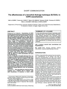
PLATEI. in Fogs solution. Cancer cell, overstained with hematoxylin, containing 3 cancer parasites. VBrick Oc. 1, Obj. Oil Imm. 7%.
FIG. 1.-Fixed
FIG. 2.-Fixed
in chromicised spirit, and stained with gentian violet and acid fuchsine. Cancer cell containing 2 parasites. VBriclc Oc. 1, Obj. Oil Imm. &.
FIG. 3.-Hematoxylin Imm.
and cochineal staining.
a. parasite.
Vhrick Oc. 1, Obj. Oil
A.
FIG. 4.-Methyl green and Biondi staining. Cancer cell containing a parasite a. VBrick Oc. 1, Obj. Oil Imm. A.
FIG. 5.-Several parasites contained in one cell. Obj. Oil Imni. &.
Cochineal staining.
YBrick Oc. 1,
FIG. &-Fixed in Foh's solution, and acid fuchsinc staining. A group of parasites in a single canccr cell. The nucleus of the cell could be seen bclow but was not VBrick OC. 1, Obj. Oil Inim. ;$. painted in.
FIG. 7.-Fixed
in chromicised spirit, and stained with hzmatoxylin and cochineal. a. Two parasites. The nucleus of the ccll is not shown in the figure. YBrick Oc. 1, Obj. Oil Imm. &.
FIG. 8.-Four
parasites from an unstained preparation fixcd in chromicised spirit. Verick Oc. 1, Obj. Oil Imm. &.
FIG. 9.-Fixed
in chromicised spirit ; gentian violet and acid fuchsine staining. At a, a parasite breaking into a mass of round granules, without showing any other signs of division. Vdrick Oc. 1, Obj. Oil Imm. A.
FIG.10.-Fixed
in chromicised spirit ; hzniatoxyliu and cochineal staining. a. Parasite which has broken up into a number of coarse homogeneolls clumps. YMck Oc. 1, Obj. Oil Imm. &,
FIG.11.-Fixed
in chromicised spirit, stained with hsmatoxylin and cochineal. a. Large parasite undergoing fragmentation without any sign of division. b. Small parasite. It. Nucleus of cancer cell. VCick Oc. 1, Ohj. Oil Imm. &.
FIG.l2.-Stained with hiematoxylin and cochineal. a. Small parasite. Obj. Oil Imm. &.
FIG.13.-Hiematoxylin
and cochineal staining. Yerick Oc. 1, Obj. Oil Imni. A.
VBrick Oc. 1,
Cancer cell containing-a.
Parasite.
JOURNAL OF PATROLOGY. -VoL. 2
PLATE I
PLATE I1
PARASITIC PROTOZOA IN CANCER0 US TUMO URS.
23
FIG. 14.-Hardened
in chromicised spirit, and stained with hsmatoxylin and cochineal. a. Large parasite. Vkrick Oc. 1, Obj. Oil Imm. A.
FIG.15.-Fixed
in FoYs solution, stained with gentian violet and acid fuchsine. parasites, stained with fuchsine. Vkrick Oc. 1, Obj. Oil Imm. &-.
Two
FIG. 16.-Cancer cell containing a parasite.
a. The nucleus of the parasite has broken up into several fragments, but there are no indications of a division. VCick OC. 1, Obj. Oil Imm. h.
FIG.17.-Fixing in chromicised spirit. HEematoxylinand cochineal staining. a. Parasite. VBrick Oc. 1, Obj. Oil Imm.
A.
PLATE11.
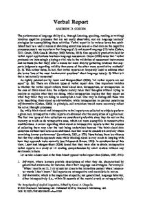
FIG.18.-Fixed in chromicised spirit. HEematoxylin and cochineal staining. The parasite shows the peripheral arrangement of protoplasmic granules. Obj. Oil Imm. 6. FIG.
FIGS.
19.-Fixation, staining and magniecation as before. has shrunk in the interior of the cell.
Vkrick OC. 1,
The capsule of the parasite (a)
20, %-Two parasites from an unstained preparation, fixed in chromicised spirit. VBrick Oc. 1, Obj. Oil Imm. A.
FIG.22.-Fixed
in chromicised spirit, stained with hamatoxylin and cochineal. a. Parasite with wrinkled capsule. VBrick Oc. 1, Obj. Oil Imm. &.
FIG.23.-Hardened in chromicised spirit, stained with gentian violet and acid fuchsine. Peripheral arrangement of granules. No sign of division. The nucleus of the cell has not been painted in. VQick Oc. 1, Obj. Oil Imm. &. FIG.24.-Fixed in Fogs solution, stained with hsmatoxylin and cochineal. a. Parasite with symmetrical arrangement of granules a t the periphery. No sign of division. VIrick Oc. 1, Obj. Oil Imm. 2;.
FIG.25.-A parasite from an unstained preparation fixed in chromicised spirit. FIG. 26.-Cancer parasite undergoing division. The two parts of the nucleus are still joined by threads. V6rick Oc. 1, Obj. Oil Imni. &. with harnatoxylin and cochineal. a. Parasite undergoing simple division. The nucleus is fully divided, the capsule only just beginning to dividc. Zeiss 00.comp. 4, Obj. Oil Imm. &.
FIG.27.-Stained
FIG,28.-Hsematoxylin
and cochineal staining. Cancer cell containing-a. going division. VBrick Oc. 1, Obj. Oil Irnm. &.
Parasite under-
FIG.29.--Cancer cell containing<. Parasite undergoing division. VBrick Oc. 1, Obj. Oil Imm. &. FIG. 30.-Stained with hamatoxylin and cochineal. a. Parasite which has just undergone division. VBrick Oc. 1, Obj. Oil Imm. &. FIG.31.-Last stage of division. Two parasites still partly joined together. Eosin and aniline blue staining. VBrick Oc. 1, Obj. Oil Imm. &. FIG. 32.-Fixed in osmic acid, and stained with hamatoxylin. Multiple division and subdivision of parasite. At a tho subdivision is not complete. VBrick Oc. 1, Obj. Oil Imm. A. FIG.33.-Fixed in BOB'Ssolution, gentian violet and acid fuchsine staining. a. Parasite, probably undergoing degeneration. Vkrick Oc. 1, Obj. Oil Imm. A. FIG.34.-Fixed in chromicised spirit. Hsmatoxylin and cochineal staining. Cancer cell containing parasite. VBrick Oc. 1, Obj. Oil Imm. &.
24
M. ARMAND RUFFER AND N: G. PLZMMER.
FIU. 35.-Fixed in chromicised spirit, eosin and auiline blue staining. ?a. Nucleus of cancer cell. Zeiss, comp. Oc. 4, Obj. Oil Inim. T$.
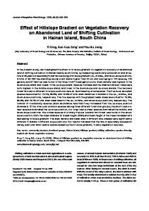
PLATE111. FIG. 36.-Compare No. 9. Very high magnification. Zeiss Oc. 12, Obj. Oil Imm. 23. FIG. 37.-Hsmatoxylin and cochineal staining. Cancer cell containing-a. Parasite in first stage of division. Vhick Oc. 1, Obj. Oil Imm. =$. FIG.38.-Fixing in Foa’s solution, gentian violet and acid fuchsine staining. Segmentation of parasite. The nucleus of tho cell is not shown in the figure. VMck Oc. 1, Obj. Oil Imm. h. FIQ. 39.-From a painting by Dr. Metchnikoff. Cancer cell containing 8 parasites, 5 of which are undergoing division and segmentation. Zeiss comp. Oc. 8, Obj. Oil Imni. &. FIG. 40.-Cochineal and hieinatoxylin staining. The nucleus of the cell is not shown in the figure. a. Pragmentatioii of the nucleus of the parasite. At the periphery the capsule is seen to divide also. Verick Oc. 1, Obj. Oil Imm. &. FIU. 41.-Fixing in chromicisedspirit ; gentian violet and acid fuchsine staining. Cell containing a parasite undergoing fragmentation. The capsule has not yet begun to divide. Vkrick Oc. 1, Obj. Oil Imm. &. FIU. 42.-Fixed in chromicised spirit, and stained with hsematoxylin and cochineal. Cancer cell containing a segmenting parasite. Zeiss Oc. 1, Obj. Oil Imm. &.
FIG.43.-Fixed in chromicised spirit ;cochineal staining. A cancer cell containing-a. Small parasite ; b. a large parasitc undergoing division and segmentation ; c. a small intranuclear parasite. Vkrick Oc. 1, Obj. Oil Imni. =$. FIG. 44.-Fixed in Fleniming’s solution ; methyline green and Biondi staining. a. Invaginated cell, or pseudo-parasite. V6rick Oc. 1, Obj. Oil Imm. &. FIU. 45.-Fixed in chromicised spirit, and stainad with hsmatoxylin and cochineal. a. Division and segmentation of parasite. VBrick Oc. 1, Obj. Oil Imm. 75. FIG. 46.-Fixing in chroniicised spirit ; hiematoxylin and cochineal staining. Pseudo-parasite identical with parasite of Wickham. a. Points to the pseudo-capsule. Y6rick Oc. 1, Obj. Oil Imm. A. FIU. 47.-Parasite undergoing division and segmentation of capsule. VBrick 00. 1, Obj. Oil Imm. =%.
FIG. 48.-Fixing
in chromicised spirit ; hsmatoxylin and cochineal staining. a. Degenerating invaginated cell or parasite of Wickham. VBrick Oc. 1, Obj. Oil Imm.
FIG. 49.-One
parasite dividing and subdividing into a number of younger parasites. At a the division is not yet complete. Hsmatorylin and rochineal staining. Vkrick Oc. 1, Obj. Oil Iinm. ri.
FIU.50.-Fixed in chroniicised spirit, and stained with hzmatoxylin and cochineal. a. An invrrgiiiated and degenerating cancer cell or parasite of Wickham. V6rick Oc. 1, Obj. Oil Imm. &. FIG.51. -Fixing in chroniicised spirit ; hamatoxylin and cochineal staining. a. Invaginated cell, identical with parasite of Wickham. VBrick Oc. 1, Obj. Oil Imm. &.
FIG. 52.-Fixcd
in osmic acid and stained with hrematoxylin. A compound invagjnation of cells. a. Innermost iiivaginated cell, simulating a parasite. VBrick Oc. 1, Obj. Oil Inim. A,
.rOURNe4L OF PATHOLOGY -VOI.
-?
PLATE111
JOURNAL OF PATHOLOCY.--VOL 2 .
PLATEIX
PARASITIC PROTOZOA I N CANCER0 US TUMO URS. 25 PLATEIV.
FIG. 53.-Methyl green and Biondi staining. Cancer cell a, containing an invaginated cell or pseudo-parasite b, which contains real parasite c. VBrick Oc. 1, Obj. Oil h i m . A. FIG. 54.-Fixed in chromicised spirit, and darkly stained with hsematoxylin and cochineal. Part of cancer alveolus, containing a sharply defined degenerated area a. VBrick 00. 1, Obj. Oil Inim. A. FIG.55.-Fixing in Flemming’s solution ; methyl green and Biondi staining. Beginning of direct fragmentation of nucleus. VBrick Oc. 1, Obj. Oil Imm. A.
FIG.56.-Hardening in Flemming’s solution ; methyl green and Biondi staining. Nucleus undergoing direct fragmentation. VBrick Oc. 1, Obj. Oil Imm.
A.
FIG.57.-Hardening in Flemming’s solution ; methyl green and Biondi staining. Nucleus undergoing direct fermentation. Virick Oc. 1, Obj. Oil Imm. A. FIG.58.-From a preparation by Dr. H. Snow. A hypertrophic, hyperchromatic, and fragmenting nucleus. Ykrick Oc. 1, Obj. Oil Imm. A. FIG.59.-Fixed in osmic acid, andstained with haematoxylin. A cancer cell. the nucleus of which has undergone extreme direct fragmentation. a. An endogenous (1) cell. VBrick Oc. 1, Obj. Oil Imm. A. FIG.60.-From a preparation by Dr. H. Snow. A large hypertrophied cell containing a hypertrophic nucleus n, and one or more invaginated cells a. Ydrick Oc. 1, Obj. Oil Imm. &.
FIQ.61.-Fixed in chromicised spirit, and stained with hcematoxylin and cochineal. Part of cancer alveolus : n. represents a cast formed by the degeneration of cancer cells ; b. a granule on the point of being discharged. Vkrick Oc. 1, Obj. Oil Imm. A. FIQ. 62.-Stained with methyl green and rose bengale. a. An invaginated cancer cell simulating a parasite. VBrick Oc. 1, Obj. Oil Imm. &. FIQ.63.-Fixed in osmic acid, and stained with haematoxylin. Marked hypertrophy and degeneration and hydrops of the cell. Direct fragmentation of the nucleus a t a. Vkrick Oc. 1, Obj. Oil Imm. ri. FIG. 64.-Stained with methyl green and fuchsine. a. Pseudo-parasite. n. Nucleus of cell. Y6rick Oc. 1, Obj. Oil Imm. A. FIG. 65.-Fixing in chromicised spirit ; eosin and aniline blue staining. a. Hyperchromatic nucleus lying in an hypertrophied cell. Compare with b, normal cancer cells. Vdrick 00.1, Obj. Oil Imm. A. PIG. 66.-From 06.
a cancer hardened in alcohol, containing a t a, chromatine bodies. 1, Obj. Oil Imm. A.
Vdrick
FIG. 67.-Fixed in Flemming’s solution, and stained with methyl green and Biondi‘s reagent. Beginning fragmentation of the nucleus. YBrick Oc. 1, Obj. Oil Imm. A.
FIG.6S.-Fixed
in Flemming’s solution and stained with methyl green and Biondi‘s reagent.
A t a are two chromatine granulcs, mistaken by some authors for parasites. Vkrick 00. 1, Obj. Oil Inini. A.