BR A I N R ES E A RC H 1 0 8 5 ( 2 00 6 ) 1 9 5 –1 98
a v a i l a b l e a t w w w. s c i e n c e d i r e c t . c o m
w w w. e l s e v i e r. c o m / l o c a t e / b r a i n r e s
Short Communication
Excitotoxic hippocampal neuron loss following sustained electrical stimulation of the perforant pathway in the mouse Friederike Kienzler a , Peter Jedlicka a , Mario Vuksic a,b , Thomas Deller a,1 , Stephan W. Schwarzacher a, ⁎,1 a
Institute of Clinical Neuroanatomy, Johann Wolfgang Goethe-Universität, D-60590 Frankfurt am Main, Germany Croatian Institute for Brain Research, School of Medicine, University of Zagreb, 10000 Zagreb, Croatia
b
A R T I C LE I N FO
AB S T R A C T
Article history:
Prolonged electrical stimulation of the perforant pathway in the rat evokes epileptiform
Accepted 13 February 2006
discharges in dentate granule cells and irreversibly damages hilar neurons. In this in vivo
Available online 3 April 2006
study, we demonstrate that similar perforant path stimulation in C57Bl/6 mice causes the same pattern of hippocampal neuron loss. Therefore, this mouse model of seizure-induced
Keywords:
hippocampal injury can be used for a wide variety of studies in genetically altered
Epilepsy
conditions not available in rats.
Seizure
© 2006 Elsevier B.V. All rights reserved.
Epileptic brain damage Dentate gyrus Hilus
Temporal lobe epilepsy (TLE) with hippocampal sclerosis is a common form of medically refractory human epilepsy (Falconer, 1974; Engel, 2001). Prolonged electrical stimulation of the main afferent pathway to the hippocampus in rats produces the pattern of selective neuron loss that characterizes human hippocampal pathology in TLE (Sloviter, 1983, 1987, 1991a; Sloviter and Damiano, 1981; Sloviter et al., 2003). In this study, we demonstrate that the features of this experimental model of seizure-induced brain damage can be adapted to the mouse, which will make it possible to use transgenic mouse resources to study a wide variety of mechanisms underlying excitotoxicity, selective vulnerability, and epileptogenesis. Experiments were performed in accordance with German laws governing the use of laboratory animals. Care was taken to follow the experimental protocol established in the rat (Sloviter, 1983; Sloviter, 1987). Adult C57Bl/6 mice (49–87 days
old, 20–30 g; Charles River, Sulzfeld, Germany) were anesthetized with urethane (1.9 g/kg body weight, 125 mg/ml saline, s.c.; supplemental doses of 5–10 mg every 8–12 h as needed). Subcutaneous administration of urethane maintained surgical anesthesia for more than 24 h, followed by full recovery. Animals were positioned in a stereotaxic apparatus. The position of electrodes was chosen using a mouse brain atlas (Franklin and Paxinos, 1997) and on the basis of previous studies of perforant path stimulation in the mouse in vivo (Namgung et al., 1995). For local anesthesia, mepivacain hydrochloride (1%, Astra, Wedel, Germany) was injected s.c. in the area surrounding the incision prior to surgery. Holes were drilled in the skull and a bipolar stimulation electrode (NE-200, 0.5 mm tip separation; Rhodes Medical, Summerland, CA, USA) was positioned in the angular bundle of the perforant pathway (2.1 mm lateral and 3.8 mm posterior to the bregma, 1.8 mm from the brain surface). NaCl-filled glass recording
⁎ Corresponding author. Fax: +49 69 6301 6425. E-mail address:
[email protected] (S.W. Schwarzacher). 1 Contributed equally to this work. 0006-8993/$ – see front matter © 2006 Elsevier B.V. All rights reserved. doi:10.1016/j.brainres.2006.02.055
196
BR A I N R ES E A RC H 1 0 8 5 ( 2 00 6 ) 1 9 5 –19 8
electrodes were placed bilaterally in the granule cell layers of the dentate gyrus (0.5–0.8 mm lateral and 1.5 mm posterior to the bregma, 1.9–2.1 mm from the brain surface). Supramaximal biphasic current pulses (15–20 V, 0.1-ms duration) were generated by a Grass S88 stimulator with a Grass stimulus isolation unit (Quincy, Massachusetts, USA). Potentials were amplified by a Grass preamplifier and displayed on a Mac Lab computer system (Wiss Tech, Spechbach, Germany). In sham-operated animals, electrodes were positioned with low-voltage pulses and no stimulation was performed. Four days post-stimulation, animals were anesthetized (Isoflurane, Abbott, Wiesbaden, Germany) and transcardially perfused with 4% paraformaldehyde. The brains were removed and kept in the same fixative for 4–12 h followed by phosphate buffer. Coronal vibratome sections (50 μm) were stored in cryoprotection solution at −20 °C. Sections were immunocytochemically stained for the neuronal marker neuron-specific nuclear protein (NeuN, mouse monoclonal antibody, diluted 1:1000, Chemicon, standard ABC-protocol with diaminobenzidin as chromogen) and Nissl staining (cresyl violet). Unless stated otherwise, chemicals were obtained from Sigma (Deisenhofen, Germany). In order to estimate the extent of neuronal loss following stimulation, the number of hilar NeuN-immunoreactive somata was counted with a camera lucida system on 5–8 sections per animal (4 days after 24 h stimulation, n = 3; sham operation, n = 3; control, n = 3). Statistical analysis was performed using Student's paired t test (significance: P < 0.01).
Unilateral stimulation of the perforant path evoked characteristic potentials in the granule cell layer (Andersen et al., 1966) bilaterally (Fig. 1A). The onset of contralateral evoked potentials (4.65 ± 0.20 ms) and population spikes (6.82 ± 0.77 ms) was delayed in comparison to ipsilateral potentials (1.98 ± 0.57 ms) and spikes (4.90 ± 0.64 ms). In addition, evoked potentials and population spike amplitudes were consistently smaller on the contralateral side. The delayed onset of contralateral potentials can be attributed to a longer distance to the stimulation electrode and possibly reflects a direct contralateral excitation of granule cells, although tracing studies revealed only a very limited projection from the perforant path to the contralateral dentate gyrus in C57Bl/6 mice (van Groen et al., 2003). Another explanation could be that contralateral granule cell potentials were evoked by commissural fibers between the right and the left entorhinal cortex, which have been demonstrated previously in rats (Köhler, 1986, 1988; Deller et al., 1996). Paired-pulse stimulation at 2 Hz, with an interpulse interval of 40 ms, decreased the amplitude of the second of two afferent stimuli (Fig. 1A), as shown previously in the rat (Sloviter, 1991b) and mouse (Jones et al., 2001). Intermittent 20Hz stimulation (continuous 2 Hz paired-pulse stimulation and 10 s 20 Hz trains once per minute) evoked bilateral increase of population spikes (Figs. 1B, C) followed by epileptiform discharges (Fig. 1D; ipsilateral, n = 20 of 20 cases; contralateral, n = 3 of 5 cases). Following 20-Hz train stimulation, pairedpulse inhibition changed to paired-pulse facilitation (Fig. 1E),
Fig. 1 – Unilateral stimulation of the perforant path induces bilateral epileptiform discharges in dentate granule cells. (A) 2 Hz paired-pulse stimulation (interpulse interval 40 ms) inhibits the amplitude of the second of two responses (arrows). Note the delayed onset of contralateral granule cell responses in comparison to the ipsilateral recordings. (B,C) 10 s trains of 20 Hz are superimposed on the 2 Hz paired-pulse protocol. At the start of a 20 Hz train the population spikes are small (B). Towards the end of a 20 Hz train, each stimulus evokes a large amplitude population spike (C). (D) Immediately after a 10 s-20 Hz stimulus train, epileptiform activity is observed bilaterally. (E) 20 s after a 20 Hz train, 2 Hz paired-pulses evoke facilitation of the second of two population spikes (arrows).
197
BR A I N R ES E A RC H 1 0 8 5 ( 2 00 6 ) 1 9 5 –1 98
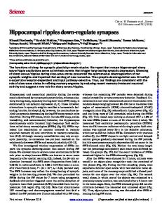
as an apparent consequence of decreased paired-pulse inhibition (Sloviter, 1983). Mice were tested for 4 days following 24 h of intermittent 20-Hz stimulation. During this time period, we did not register indications for the development of spontaneous seizures. Four days after perforant pathway stimulation, loss of NeuNimmunoreactive neuronal somata was apparent in the hilus of the ipsilateral dentate gyrus, whereas the contralateral side was less or minimally affected (Fig. 2). Nissl staining of adjacent sections revealed a loss of large neuronal somata and an apparent increase in the number of small cell bodies with the morphological characteristics of microglia and astrocytes (data not shown). The number of NeuN-immunoreactive somata in the ipsilateral hilus was reduced by 87% (12.6 ± 5.88% in comparison to control and sham-operated animals (Table 1). The results of this study demonstrate that the main physiological, pathophysiological, and pathological features of prolonged perforant pathway stimulation in the rat (Sloviter, 1983, 1987, 1991b) can be reproduced in normal mice using the same in vivo stimulation methods. The ability to produce a highly selective and reproducible pattern of seizure-induced hippocampal damage in mice without evoking behavioral seizures or producing widespread brain damage demonstrates the feasibility of adapting this experimental model for future studies in genetically modified mouse strains. The use of a standardized method for producing limited hippocampal injury may be useful for studies addres-
Table 1 – Loss of hilar neurons after 24-h intermittent perforant path stimulation Cells/10 000 μm2 Control Sham ipsi Sham contra Stim. ipsi Stim. contra
11.50 11.08 11.22 1.41 10.01
± ± ± ± ±
0.10 2.15 1.62 0.66 ⁎ 2.95
Percent of control 100 99.36 109.35 12.60 89.83
± ± ± ± ±
16.68 19.25 14,53 5.88 ⁎ 26.49
NeuN-immunoreactive somata were counted in coronal sections of the dentate gyrus. 4 days after 24 h perforant path stimulation (stim.; n = 3), ipsilateral (ipsi) hilar neurons were significantly reduced, whereas the contralateral (contra) hilus was not impaired compared to control (n = 3) or sham-operated (n = 3) animals. ⁎ Significantly different from control, P < 0.01 (Student's paired t test).
sing the vulnerability of hippocampal neurons in different mouse strains (Schauwecker, 2003) and the roles of cell loss, synaptic reorganization, and granule cell dispersion (Bouilleret et al., 2000) in the process of epileptogenesis.
Acknowledgments The authors thank Anke Biczysko for the excellent technical assistance. This work was supported by the Deutsche
Fig. 2 – Following 24 h of 20 Hz perforant path stimulation, a significant reduction of neurons occurs in the ipsilateral hilus (H) of the dentate gyrus (DG). (A,B) In 24 h sham-operated animals neuronal cell somata appear to be unaffected in NeuN-stained sections. (C,D) In contrast, four days after 24 h intermittent (trains of 20 Hz, 10 s/min) stimulation, a significant reduction of neurons in the ipsilateral hilus of the dentate gyrus can be detected in neuron-specific nuclear protein (NeuN)- immunoreactive sections (D), whereas the contralateral side does not show a significant reduction of hilar neurons (C). CA3, hippocampal subfield CA3; GCL, granule cell layer; ML, molecular layer. Scale bars: 100 μm.
198
BR A I N R ES E A RC H 1 0 8 5 ( 2 00 6 ) 1 9 5 –19 8
Forschungsgemeinschaft (Graduiertenkolleg Neuronale Plastizität to PJ and MV; DE 551/8-1 to TD).
REFERENCES
Andersen, P., Holmqvist, B., Voorhoeve, P.E., 1966. Entorhinal activation of dentate granule cells. Acta Physiol. Scand. 66, 448–460. Bouilleret, V., Loup, F., Kiener, T., Marescaux, C., Fritschy, J.M., 2000. Early loss of interneurons and delayed subunit-specific changes in GABA(A)-receptor expression in a mouse model of mesial temporal lobe epilepsy. Hippocampus 10, 305–324. Deller, T., Adelmann, G., Nitsch, R., Frotscher, M., 1996. The alvear pathway of the rat hippocampus. Cell Tissue Res. 286, 293–303. Engel, J., 2001. Mesial temporal lobe epilepsy: what have we learned? Neuroscientist 7, 340–352. Falconer, M.A., 1974. Mesial temporal (Ammons Horn) sclerosis as a common cause of epilepsy—Etiology, treatment, and prevention. Lancet 2, 767–770. Franklin, K.B.J., Paxinos, G., 1997. The Mouse Brain in Stereotaxic Coordinates. Academic Press, San Diego. Jones, M.W., Errington, M.L., French, P.J., Fine, A., Bliss, T.V., Garel, S., Charnay, P., Bozon, B., Laroche, S., Davis, S., 2001. A requirement for the immediate early gene Zif268 in the expression of late LTP and long-term memories. Nat. Neurosci. 4, 289–296. Köhler, C., 1986. Intrinsic connections of the retrohippocampal region in the rat brain: II. The medial entorhinal area. J. Comp. Neurol. 246, 149–169. Köhler, C., 1988. Intrinsic connections of the retrohippocampal region in the rat brain: III. The lateral entorhinal area. J. Comp. Neurol. 271, 208–228.
Namgung, U., Valcourt, E., Routtenberg, A., 1995. Long-term potentiation in vivo in the intact mouse hippocampus. Brain Res. 689, 85–92. Schauwecker, P.E., 2003. Genetic basis of kainate-induced excitotoxicity in mice: phenotypic modulation of seizure-induced cell death. Epilepsy Res. 55, 201–210. Sloviter, R.S., 1983. “Epileptic” brain damage in rats induced by sustained electrical stimulation of the perforant path: I. acute electrophysiological and light microscopic studies. Brain Res. Bull. 10, 675–697. Sloviter, R.S., 1987. Decreased hippocampal inhibition and a selective loss of interneurons in experimental epilepsy. Science 235, 73–76. Sloviter, R.S., 1991a. Feedforward and feedback inhibition of hippocampal principal cell activity evoked by perforant path stimulation: GABA-mediated mechanisms that regulate excitability in vivo. Hippocampus 1, 31–40. Sloviter, R.S., 1991b. Permanently altered hippocampal structure, excitability, and inhibition after experimental status epilepticus in the rat: the “dormant basket cell” hypothesis and its possible relevance to temporal lobe epilepsy. Hippocampus 1, 41–66. Sloviter, R.S., Damiano, B.P., 1981. Sustained electrical stimulation of the perforant path duplicates kainate-induced electrophysiological effects and hippocampal damage in rats. Neurosci. Lett. 24, 279–284. Sloviter, R.S., Zappone, C.A., Harvey, B.D., Bumanglag, A.V., Bender, R.A., Frotscher, M., 2003. “Dormant basket cell” hypothesis revisited: relative vulnerabilities of dentate gyrus mossy cells and inhibitory interneurons after hippocampal status epilepticus in the rat. J. Comp. Neurol. 459, 44–76. van Groen, T., Miettinen, P., Kadish, I., 2003. The entorhinal cortex of the mouse: organization of the projection to the hippocampal formation. Hippocampus 13, 133–149.