Cognitive Brain Research 24 (2005) 190 – 198 www.elsevier.com/locate/cogbrainres
Research report
EEG evidence for mirror neuron dysfunction in autism spectrum disorders Lindsay M. Obermana,b,T, Edward M. Hubbarda,b, Joseph P. McCleeryb, Eric L. Altschulera,b,c, Vilayanur S. Ramachandrana,b,d, Jaime A. Pinedad,e a Center for Brain and Cognition, UC San Diego, La Jolla, CA 92093-0109, USA Department of Psychology, UC San Diego, 9500 Gilman Drive, La Jolla, CA 92093-0109, USA c Department of Rehabilitation Medicine, Mt. Sinai School of Medicine, New York, New York 10029, USA d Department of Neurosciences, UC San Diego, La Jolla, CA 92093-0662, USA e Department of Cognitive Science, UC San Diego, La Jolla, CA 92093-0515, USA b
Accepted 13 January 2005 Available online 9 March 2005
Abstract Autism spectrum disorders (ASD) are largely characterized by deficits in imitation, pragmatic language, theory of mind, and empathy. Previous research has suggested that a dysfunctional mirror neuron system may explain the pathology observed in ASD. Because EEG oscillations in the mu frequency (8–13 Hz) over sensorimotor cortex are thought to reflect mirror neuron activity, one method for testing the integrity of this system is to measure mu responsiveness to actual and observed movement. It has been established that mu power is reduced (mu suppression) in typically developing individuals both when they perform actions and when they observe others performing actions, reflecting an observation/execution system which may play a critical role in the ability to understand and imitate others’ behaviors. This study investigated whether individuals with ASD show a dysfunction in this system, given their behavioral impairments in understanding and responding appropriately to others’ behaviors. Mu wave suppression was measured in ten high-functioning individuals with ASD and ten age- and gender-matched control subjects while watching videos of (1) a moving hand, (2) a bouncing ball, and (3) visual noise, or (4) moving their own hand. Control subjects showed significant mu suppression to both self and observed hand movement. The ASD group showed significant mu suppression to self-performed hand movements but not to observed hand movements. These results support the hypothesis of a dysfunctional mirror neuron system in high-functioning individuals with ASD. D 2005 Elsevier B.V. All rights reserved. Theme: Disorders of the nervous system Topic: Developmental disorders Keywords: Mirror neurons; Autism spectrum disorders; EEG; Mu rhythm
1. Introduction Disorders on the autism spectrum are characterized by deficits in social and communicative skills, such as imitation, pragmatic language, theory of mind, and empathy [7,19,20]. Elucidating the underlying neural bases of these deficits has been a challenge because the behavioral T Corresponding author. 9500 Gilman Drive, La Jolla, CA 92093-0109, USA. Fax: +1 858 534 7190. E-mail address:
[email protected] (L.M. Oberman). 0926-6410/$ - see front matter D 2005 Elsevier B.V. All rights reserved. doi:10.1016/j.cogbrainres.2005.01.014
manifestations of this disorder vary both in severity (lowand high-functioning) as well as in expression (autistic disorder, Asperger’s disorder, pervasive developmental disorder—not otherwise specified). The recent discovery of bmirror neuronsQ in macaque monkeys by Rizzolatti and colleagues [16], however, may provide a basis for explaining some of the behavioral deficits seen in individuals with autism spectrum disorders (ASD). Mirror neurons are primarily thought to be involved in perception and comprehension of motor actions [46], but they may also play a critical role in higher order cognitive processes such
L.M. Oberman et al. / Cognitive Brain Research 24 (2005) 190–198
as imitation [43,46], theory of mind [23], language [43,44], and empathy [12], all of which are known to be impaired in individuals with autism spectrum disorders [6,8,20,30,47]. Single unit studies indicate that mirror neurons in area F5 of the macaque premotor cortex, which are indistinguishable from neighboring neurons in terms of their motor properties, also discharge in response to observed actions [16] (for a review, see Ref. [46]). That is, when a monkey observes another individual performing an action that is part of its own motor repertoire, mirror neurons fire in the premotor cortex, revealing a type of observation/ execution matching system. These single unit physiology studies also showed that mirror neuron activity was selective for goal-directed actions [24]. This observation/ execution matching system may allow the monkey to perform both an on-line automatic execution of the action or an off-line internal simulation of the observed action. It has been speculated that this simulation may play a critical role in one’s ability to understand other individuals’ movements, an ability that is especially critical for social interaction [23]. Although individual neurons cannot be directly studied in the same way in humans, the existence of an analogous system in the homologous brain region (Broca’s area, Brodmann’s area 44) has been supported by indirect population-level measures such as transcortical magnetic stimulation (TMS) [17], positron emission tomography (PET) [39], and functional magnetic resonance imaging (fMRI) [29]. Fadiga and colleagues [17] found that motor evoked potentials in response to TMS over motor cortex were enhanced when the subject observed another individual performing an action relative to when the subject detected the dimming of a light. They concluded that this enhancement was a result of activity of mirror neurons in the prefrontal cortex influencing the motor response. Parsons et al. [39] used PET to map the brain areas that were active during observation of biological movements. Frontal, parietal, and cerebellar regions, including the inferior premotor cortex (Brodmann’s area 44), were found to be active during actual movement, imagined movement, and observed movement. Iacoboni and colleagues [29], using fMRI, found increased blood flow in Brodmann’s area 44 during both observed and performed actions. Several recent studies have uncovered activations with similar properties in the parietal cortex [10,39], as well as the superior temporal sulcus [11,40]. These results suggest that the frontal mirror neuron system may be one part of a broader action observation/execution network [18,38]. Previous studies in our laboratory [1] and other laboratories [35,36] have investigated the mirror neuron system in humans through analysis of electroencephalography (EEG) mu frequency band oscillations. At rest, sensorimotor neurons spontaneously fire in synchrony [25], leading to large amplitude EEG oscillations in the 8– 13 Hz (mu) frequency band. When subjects perform an action, these neurons fire asynchronously, thereby decreas-
191
ing the power of the mu-band EEG oscillations [41,48]. Because the motor properties of the mirror neurons are indistinguishable from those of neighboring premotor, motor, or sensorimotor neurons, mu wave suppression during self-performed actions is likely to be a result of activation of several neuronal systems in the area of the premotor and sensorimotor cortices. During observed hand actions, however, the mirror neuron system is the only network that has been identified to be active in this area of cortex. This suggests that mu wave suppression to observed actions could conceivably be used as a selective measure of mirror neuron system functioning. Various properties of the mu rhythm directly link it to frontal mirror neuron activity. First, mu power recorded from electrodes on scalp locations C3, Cz, and C4 is reduced in normal adults by self-initiated movement, imagined movement, and observed movement [5,14,26,42]. Second, similar to previous fMRI studies of the mirror neuron system [10], more recent studies have found that the mu rhythm is also modulated by object-directed actions [36]. Taken together, these findings suggest that monitoring levels of mu suppression might provide an inexpensive, non-invasive method to study mirror neuron functioning [2,36]. Since the mu rhythm is generated by activity in sensorimotor areas [25] and mirror neurons are located in the premotor areas [16], it has been hypothesized that the mu rhythm may specifically index downstream modulation of primary sensorimotor areas by mirror neurons [36]. In addition to its involvement in motor observation and production, the human mirror neuron system has been implicated in a variety of higher-level cognitive processes that are known to be impacted in ASD, including imitation, language, theory of mind, and empathy (for a review, see Ref. [54]). Specifically, it has been suggested that mirror neurons have the capacity to associate the visual representation of an observed action with the motor representation of that action which, in humans and some higher order primates, could lead to imitative behaviors [46]. While imitation is common when humans and other primates interact, multiple experimental studies have found that individuals with autism show significant deficits in imitation [47,52]. Once another individual’s actions are represented and understood in terms of one’s own actions, it is possible to predict the mental state of the observed individual, leading to theory of mind abilities [23]. Furthermore, it has been proposed that theory of mind is the core deficit in autism, which leads to the inability to understand others’ thoughts and behaviors [7,8,20]. Similarly to theory of mind, empathy requires the ability to understand another individual’s internal mental state. Studies suggest that individuals with autism fail to respond to or often even attend to another person in distress, reflecting an impairment in spontaneous empathetic behavior [6]. Facial expressions, similar to other actions, also may activate the mirror neuron system. Therefore, empathy may critically depend on one’s ability
192
L.M. Oberman et al. / Cognitive Brain Research 24 (2005) 190–198
to understand the observed facial expression in terms of one’s own motor representations. Lastly, the DSM-IV-R [3] names language impairment as one of the main diagnostic criteria for autistic disorder, and experimental studies find that individuals with autism show significant impairments on a battery of standardized language tests [30]. Correspondingly, it has been theorized that the observation/execution system that mirror neurons provide is an ideal candidate for the evolution of language from an earlier gestural communication system [43,44]. It is also important to note that the human homologue to area F5 is Broca’s area, which is largely thought of as a language production area. Because of the correspondence between the behavioral deficits seen in ASD and the theorized functions of the mirror neuron system, many have suggested that individuals with ASD may have mirror neuron system impairments [2,22,37,43,54]. Williams and colleagues [54] were the first to propose a detailed model outlining this relationship. They suggested that a dysfunctional development of the mirror neuron system, possibly as a result of a combination of genetic and environmental factors, could lead to impaired self-other representations and imitation. This, in turn, could lead to impaired social and communication abilities such as theory of mind, joint attention, empathy, and language, which are the defining features of autism. The goal of the current study was to test whether individuals with ASD would show dysfunctional mirror neuron activity as reflected by mu suppression. Mu suppression was measured in a sample of high-functioning individuals with ASD and age- and gender-matched typically developing controls. Subjects performed four tasks: (1) moving their own hand, (2) watching a video of a moving hand, (3) watching a video of two bouncing balls (non-biological motion), and (4) watching visual white noise (baseline). Based on previous results, we hypothesized that control subjects would show mu suppression in the observed hand movement condition, whereas the ASD subjects would show a lack of suppression during this condition, indicating an impairment in mirror neuron functioning. However, as there is no reason to believe that other motor systems in the area of sensorimotor cortex are impaired in ASD, oscillations in the mu frequency band should be suppressed in both typically developing and ASD subjects in the self-movement condition. Furthermore, because mirror neuron activity seems to be selective to biological motion [45], we predicted no mu suppression in either group to watching bouncing balls.
2. Materials and methods 2.1. Subjects Our original sample consisted of 11 individuals with ASD and 13 age- and gender-matched control subjects. All
subjects in the study were male. The ASD group was composed of ten individuals diagnosed with autism and one individual diagnosed with Asperger’s syndrome. One subject with autism and two control subjects were excluded prior to analysis due to excessive movement artifacts that resulted in an inability to obtain sufficient EEG data. One additional control subject was excluded prior to analysis due to a technical malfunction in the EEG system. Therefore, our final sample consisted of 10 individuals with ASD and 10 age- and gender-matched controls. Subjects ranged in age from 6–47 years (ASD: M = 16.6, SD = 13.0; Control: M = 16.5, SD = 13.6; t(18) = 0.017, P N 0.98). One individual was left handed in the ASD group, while in the control group 3 individuals were left-handed. ASD subjects were recruited through the Cure Autism Now Foundation, the San Diego Regional Center for the Developmentally Disabled, and the Autism Research Institute. Control subjects were recruited through the UCSD Center for Human Development subject pool and the local community. Individuals were included in the ASD group if they were diagnosed with either autism or Asperger’s syndrome by a clinical psychologist. Subjects met DSMIV criteria for a diagnosis of Autistic disorder or Asperger’s disorder [3]. In addition, subjects in the ASD group exhibited the following diagnostic behaviors at the time of testing, including, but not limited to, awkward use of pragmatics, intonation, and pitch in communication, lack of initiation of social interactions, and obsessive preoccupation with the order and specific details of the study. All subjects were considered high-functioning, defined as having age appropriate verbal comprehension and production abilities and an IQ greater than 80 as assessed by either school assessments or psychometric evaluations from a clinician. Subjects without age appropriate verbal comprehension and production abilities were excluded from the study. Subjects were given age-appropriate consent/assents (for subjects under the age of 18). In addition, in order to ensure that subjects understood the procedure and the tasks involved, a picture board was created and the study was fully explained, in age-appropriate language, prior to the subjects’ participation. This project was reviewed and approved by the UCSD Human Research Protections Program. 2.2. Procedure EEG data were collected during four conditions: (1) Moving own hand: Subjects opened and closed their right hand with the fingers and thumb held straight, opening and closing from the palm of the hand at a rate of approximately 1 Hz. Subjects watched their hand at a comfortable viewing distance, the hand held at eye level. (2) Watching a video of a moving hand: Subjects viewed a black and white video of an experimenter opening and closing the right hand in the same manner as subjects moved their own hand. Videos were presented at a viewing distance of 96 cm, and the hand subtended 58 of visual angle when open and 28 when closed.
L.M. Oberman et al. / Cognitive Brain Research 24 (2005) 190–198
The hand was medium gray (8.6 cd/m2) on a black background (3.5 cd/m2). (3) Watching a video of two bouncing balls: two light gray balls (32.9 cd/m2) on a black background (1.0 cd/m2) moved vertically towards each other touched in the middle of the screen then moved apart to their initial starting position. This motion was visually equivalent to the trajectory taken by the tips of the fingers and thumb in the hand video. The ball stimulus subtended 28 of visual angle when touching in the middle of the screen and 58 at its maximal point of separation. (4) Watching visual white noise: full-screen television static (mean luminance 3.7 cd/m2) was presented as a baseline condition. All videos were 80 s in length and both the ball and hand videos moved at a rate of 1 Hz. All conditions were presented twice in order to obtain enough clean EEG data for analyses and the order of the conditions was counterbalanced across subjects, with the constraint that the self-movement condition always followed the watch condition so that the subjects had a model on which to base their movement. To ensure that subjects attended to the video stimuli during the watching hand movement and bouncing balls conditions, they were asked to engage in a continuous performance task. Between four and six times during the 80-s video, the stimuli stopped moving for one cycle (a period of 1 s). Subjects were asked to count the number of times stimuli stopped moving and report the number of stops to the experimenter at the end of the block. 2.3. EEG data acquisition and analysis Disk electrodes were applied to the face above and below the eye and behind each ear (mastoids). The mastoids were used as reference electrodes. Data were collected from 13 electrodes embedded in a cap, at the following scalp positions: F3, Fz, F4, C3, Cz, C4, P3, Pz, P4, T5, T6, O1, and O2, using the international 10–20 method of electrode placement. Following placement of the cap, electrolytic gel was applied at each electrode site and the skin surface was lightly abraded to reduce the impedance of the electrodeskin contact. The impedances on all electrodes were measured and confirmed to be less than 10 KV both before and after testing. Once the electrodes were in place, subjects were seated inside an acoustically and electromagnetically shielded testing chamber. EEG was recorded and analyzed using a Neuroscan Synamps system (bandpass 0.1–30 Hz). Data were collected for approximately 160 s per condition at a sampling rate of 500 Hz. EEG oscillations in the 8–13 Hz frequency recorded over occipital cortex are influenced by states of expectancy and awareness [31]. Since the mu frequency band overlaps with the posterior alpha band and the generator for posterior alpha is stronger than that for mu, it is possible that recordings from C3, Cz, and C4 might be affected by this posterior activity. Therefore, the first and last 10 s of each block of data were removed from all subjects to eliminate the possibility of attentional transients
193
due to initiation and termination of the stimulus. A 1-min segment of data following the initial 10 s was obtained and combined with the other trial of the same condition, resulting in one 2-min segment of data per condition. Eye blink and eye and head movements were manually identified in the EOG recording and EEG artifacts during these intervals were removed prior to analysis. Data were coded in such a way that the analysis was blind to the subjects’ diagnosis. Data were only analyzed if there was sufficient bcleanQ data with no movement or eye blink artifacts. For each cleaned segment, the integrated power in the 8–13 Hz range was computed using a Fast Fourier Transform. Data were segmented into epochs of 2 s beginning at the start of the segment. Fast Fourier Transforms were performed on the epoched data (1024 points). A cosine window was used to control for artifacts resulting from data splicing. Two measures of mu suppression were calculated. First, we calculated the ratio of the power during the observed hand movement and self hand movement conditions relative to the power during the baseline condition. Second, we calculated the ratio of the power during the observed and self hand movement conditions relative to the power in the ball condition. A ratio was used to control for variability in absolute mu power as a result of individual differences such as scalp thickness and electrode impedance, as opposed to mirror neuron activity. The ratio to the ball condition was computed in order to control for the attention to counting or any effects due to stimulus stopping during the continuous performance task and processing of directional motion. Since ratio data are inherently non-normal as a result of lower bounding, a log transform was used for analysis. A log ratio of less than zero indicates suppression whereas a value of zero indicates no suppression and values greater than zero indicate enhancement.
3. Results 3.1. Behavioral performance To ensure that the subjects were attending to the stimuli, during the hand and ball conditions, they were asked to count the number of times the stimuli stopped moving. Since all subjects performed with 100% accuracy on this continuous performance task, we infer that any differences found in mu suppression are not due to differences in attending to the stimuli. 3.2. Mu suppression Power in the mu frequency at scalp locations corresponding to sensorimotor cortex (C3, Cz, and C4) during the selfinitiated action and watching action conditions was compared to power during the baseline (visual white noise) condition by forming the log ratio of the power in these
194
L.M. Oberman et al. / Cognitive Brain Research 24 (2005) 190–198
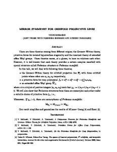
Fig. 1. Mu suppression in control and ASD participants. Bars represent the mean log ratio of power in the mu frequency (8–13 Hz) during the watching balls condition (light gray), watching hand movement condition (medium gray), and moving own hand condition (dark gray) over the power in the baseline condition for scalp locations C3, CZ, and C4 for typically developing individuals (A) and individuals with ASD (B). Error bars represent the standard error of the mean. For all values, a mean log ratio greater than zero indicates mu enhancement; a mean log ratio less than zero indicates mu suppression. Significant suppression is indicated by asterisks, *P b 0.05, **P b 0.01, ***P b 0.005.
conditions for both groups (Figs. 1A, B). Although data were obtained from electrodes across the scalp, mu rhythm is defined as oscillations measured over sensorimotor cortex, thus only data from C3, Cz, and C4 are presented. The control group (Fig. 1A) showed significant suppression from baseline in mu oscillations at each electrode during both the self-initiated hand movement condition (C3 t(9) = 3.97, P b 0.002; Cz t(9) = 2.85, P b 0.01; C4 t(9) = 4.00, P b 0.002) and observed hand movement condition (C3 t(9) = 3.99, P b 0.002; Cz t(9) = 3.21, P b 0.005; C4 t(9) = 2.78, P b 0.01). The ASD group (Fig. 1B) also showed significant mu suppression during the self-initiated hand movement condition (C3 t(9) = 2.27, P b 0.03; Cz t(9) = 1.91, P b 0.05; C4 t(9) = 2.50, P b 0.02). Unlike controls, the ASD group did not show significant suppression during the observed hand movement condition (C3 t(9) = 0.64, P N 0.25; Cz t(9) = 0.98, P N 0.15; C4 t(9) = 0.74, P N 0.20). The failure to find suppression in the ASD group was not due to differences in baseline mu power (C3 t(9) = 0.99, P N 0.30; Cz t(9) = 0.69, P N 0.50; C4 t(9) = 0.47, P N 0.50). Lastly, neither group showed significant suppression from baseline during the non-biological motion (bouncing balls) condition (ASD: C3 t(9) = 0.73, P N 0.20; Cz t(9) = 0.49, P N 0.65; C4 t(9) = .25, P N 0.40; Control: C3 t(9) = 1.45, P N 0.08; Cz t(9) = 0.54, P N 0.30; C4 t(9) = 0.00, P N 0.50).1 1 The control group had a significantly greater amount of clean data; hence, the analysis was reconducted using equal length segments. Segments from the control group were recut through random removal of small epochs of the EEG resulting in a total amount of clean data that was equal to that of the ASD group. In these analyses, the same main findings held: selfinitiated hand movement condition (C3 t(9) = 3.51, P b 0.004; Cz t(9) = 2.77, P b 0.02; C4 t(9) = 3.88, P b 0.002) and observed hand movement condition (C3 t(9) = 4.03, P b 0.002; Cz t(9) = 3.19, P b 0.006; C4 t(9) = 2.97, P b 0.008).
Additional ratios were calculated comparing the power during the observed hand movement and self hand movement conditions to that of the ball condition for both groups. Results were consistent with the baseline ratios. The control group still showed significant suppression in the self hand movement condition (C3 t(9) = 2.84, P b 0.01; Cz t(9) = 2.14, P b 0.03; C4 t(9) = 2.93, P b 0.009), and observed hand movement condition (C3 t(9) = 1.80, P b 0.05; Cz t(9) = 2.05, P b 0.04; C4 t(9) = 2.67, P b 0.02). The ASD group again showed suppression in the self hand movement condition (C3 t(9) = 3.97, P b 0.002; Cz t(9) = 2.85, P b 0.01; C4 t(9) = 4.00, P b 0.002) but not in the observed hand movement condition (C3 t(9) = 0.40, P N 0.65; Cz t(9) = 1.38, P N 0.1; C4 t(9) = 0.44, P N 0.3). Since the mu frequency band overlaps with the posterior alpha frequency band (recorded from O1 and O2) and the generator for posterior alpha is stronger than that for mu, it is possible that recordings from C3, Cz, and C4 might be affected by this posterior activity. As all conditions involved visual stimuli and the eyes were open throughout the study, we would not expect a systematic difference between conditions in posterior alpha activity. Additionally, by eliminating the first and last 10 s of each block, we reduced the possibility of alpha modulations due to attention affecting our mu power results. Consistent with this, other than C3, Cz, and C4, no electrodes showed a consistent pattern of suppression in the frequency band of interest. These results indicate that the modulations of mu activity we observed in C3, Cz, and C4 were not mediated by posterior alpha activity. In order to rule out the possibility that our results were influenced by the large age range, a Pearson r correlation coefficient was calculated for each log ratio at each electrode site. Neither group showed a significant correlation between mu suppression and age in any condition or electrode site.
L.M. Oberman et al. / Cognitive Brain Research 24 (2005) 190–198
The average of the 9 Pearson r coefficients for each group was 0.08 (range .56 to .39) for the control group and 0.05 (range .55 to .28) for the ASD group with nonsignificant P values which were all greater than 0.10.
4. Discussion The lack of suppression in the ASD group during the observed hand movement condition suggests a possible dysfunction in the mirror neuron system. The additional lack of any significant correlation between age and mu wave suppression also suggests that this dysfunction is not something that improves over the lifespan. Furthermore, the lack of suppression during the observation conditions in the ASD group is contrasted with significant suppression to their own movement, which is indicative of normal functioning of other sensorimotor systems involved in self-performed actions. It is well documented that individuals with ASD have profound difficulties relating to others cognitively and emotionally and imitating their actions (for a review, see Ref. [7]). Additionally, mirror neuron activity has been implicated in cognitive abilities such as imitation, language, theory of mind, and empathy [12,23,44,46], all of which have been shown to be impaired in individuals with ASD. The results of the current study provide evidence that a dysfunctional mirror neuron system may contribute to many of the behavioral deficits observed in individuals with ASD. However, since the sample in this study was solely composed of high-functioning males, the generalizability of the findings to females or lower-functioning individuals is unclear and requires further investigation. Although the continuous performance task was intended to ensure that the subjects were attending to the stimuli, it is possible that this task differentially affected how the stimuli were processed in the two groups. For example, it is possible that the subjects in the ASD group, instead of concentrating on the movement of the stimulus, focused attention on the counting task. If the subject focused their attention on the counting task, they may have perceived the hand stimulus as more mechanical and this may have resulted in a decrease in mu wave suppression in this group. Although we cannot entirely rule out differences in the mental state of our subjects, other results from our laboratory [50] suggest that the mu wave is suppressed even when viewing a robot hand. Thus, even if the ASD subjects viewed the hand in a more mechanical way, we still would expect to find mu wave suppression during this task. An alternative concern is that the continuous performance task may have attracted attention away from the processing of the biological motion stimulus. The fact that mu suppression ratios were similar whether we calculated them using the ball or the white noise as the denominator argues against this interpretation. Specifically, since subjects had to count in the ball condition and in the watching hand condition, but not in the white noise condition, we would
195
predict that the ratios would have differed when using our two different baseline conditions. We find no such difference, suggesting that our results were not due to this factor. Additionally, individuals with ASD may have been less likely to directly foveate on the screen [32,53] thus affecting the processing of the stimuli. While we cannot rule this strategy out, we note that from a purely motor/attentional point of view it would be harder to fixate to the side and attend in the periphery. Additionally, due to the lower resolution in the periphery, the task should have been harder if subjects were not fixating the stimulus. Based on these two factors, we would have expected some decrease in performance on the counting task had ASD subjects adopted this unusual strategy. Since all subjects performed at 100% on this task, it is unlikely that this was a major factor in the observed group differences. However, in light of recent evidence for altered fixation patterns in individuals with ASD [32,53], future research should include measures of eye position. To date, only one other group has attempted to investigate the functioning of the mirror neuron system in individuals with ASD [4,37]. Both studies used magnetoencephalography (MEG). In the first study, the researchers monitored 20 Hz rebound activity over precentral primary motor cortex 500–1500 ms following median nerve stimulation. Both control and ASD individuals showed significant rebound activity during self-initiated and observed hand movements. The magnitude of the rebound seen in the ASD group was smaller than that in the control group, although this difference failed to reach significance. Since there was not a significant difference between groups, the authors propose that the ASD group had normal mirror neuron functioning [4]. The apparent discrepancy between our study and Avikainen et al.’s study could be accounted for by methodological differences, including: sample size (10 per group in our study vs. 5 per group in Avikainen et al.’s study) and recording technique (EEG vs. MEG), both of which may have reduced their statistical power, resulting in their null finding [54]. However, in a second study, the same group averaged and time-locked the MEG signal to the stimulus presentation. Subjects were presented with still pictures of a woman performing orofacial gestures and were instructed to imitate these gestures. Cortical activations were recorded from eight adult subjects with Asperger’s syndrome (AS) and 10 control subjects. In both groups, activations were recorded over occipital cortex, superior temporal sulcus, inferior parietal lobe, inferior frontal lobe, and primary motor cortex. Activations in inferior frontal lobe and primary motor cortex were weaker and had a greater latency in the AS group as compared to the control group. Researchers concluded that this is evidence for an impairment in the mirror neuron system in individuals with Asperger’s syndrome [37], further suggesting that the previous failure to find differences between the two groups was due to methodological or power limitations.
196
L.M. Oberman et al. / Cognitive Brain Research 24 (2005) 190–198
The low spatial resolution of EEG does not allow for differentiation between activity selective to the mirror neuron system and activity in other regions that are part of a larger action observation/execution network. It is possible that mu wave suppression is reflecting both activity in the mirror neuron system and that activity in areas such as STS [11,40] and inferior parietal cortex [10,39], which are involved in action recognition and may modulate the activity in the mirror neuron system [35,36]. Further investigations with higher spatial resolution techniques, such as fMRI and high resolution EEG, may be able to dissociate between these two sources of activation. Williams et al. [54] suggest that early developmental failures of the mirror neuron system may result in a cascade of developmental impairments characterized by ASD. Our results are consistent with the proposed role of the mirror neuron system in ASD but cannot distinguish whether the mirror neuron impairment is the primary dysfunction or a consequence of anatomical or functional impairments in other brain regions. A lower level explanation of our results is that the differences found in mirror neuron activity are the result of impaired visual processing of biological motion. This would result in less activity in visual areas thought to be critically involved in biological motion perception such as the superior temporal sulcus [13,21]. There may also be deficits even earlier in visual processing as evidenced by assessments of low level dorsal stream visual processing [9,51]. Another possible explanation may be dysfunctional input from social/attentional networks. For example, individuals with ASD have been shown to have impairments in frontal brain regions thought to be involved in social attention. [13]. Given that mirror neurons are one part of a broader network [18,38] which may be modulated by multiple systems throughout the brain, an alternative interpretation of our results is that the mirror neuron system in individuals with ASD may be functional, but receiving dysfunctional input from other brain regions. Again, future research using higher resolution EEG and fMRI could further investigate these questions. Another line of research we are currently exploring is whether mirror neurons are involved in the ability to comprehend abstract language such as metaphors. Individuals with autism seem to have difficulty with metaphors, often interpreting them literally [15,27]. We suggest that this deficit may arise from a dysfunction in the mirror neuron system. Just as mirror neurons may allow for action understanding by anchoring the perception of others’ actions to our own motor system, they may also allow for abstract language comprehension (such as metaphors) in the same way. Both theoretical work in linguistics [33,34] and more recent work using functional neuroimaging [28] have suggested that metaphors such as bhe grasped the ideaQ or bto reach for the starsQ may be understood through embodied mechanisms. We are currently investigating this question by analyzing mu wave suppression when subjects
listen to phrases that contain metaphorical language such as bgrasp the ideaQ and comparing this to suppression when subjects hear literal phrases matched for meaning such as bunderstand the idea.Q In summary, numerous converging lines of evidence suggest that the mirror neuron system is involved in processes such as imitation, language, theory of mind, and empathy. As ASD is defined by behavioral deficits in many of these areas, there is reason to believe that impairments in the mirror neuron system may play a role in the social and communicative deficits associated with ASD. Future research should focus on the independent contributions of frontal and parietal mirror neuron systems, as well as the contribution of lower-level processing deficits. If supported by such future studies, pathology of the mirror neuron system and associated networks may prove to be critical in helping us to understand the neural basis of autism and related disorders.
Acknowledgments We would like to thank all of the families who participated in this study. We would also like to thank the Cure Autism Now foundation, the San Diego Regional Center for the Developmentally Disabled, as well as the Autism Research Institute for their help with subject recruitment. We would also like to thank Brian P. Jacoby for his assistance with data analysis. J.P.M. was supported by a research fellowship from the M.I.N.D. Institute and a training fellowship from NSF grant DGE-0333451 to GW Cottrell. E.M.H. was supported by a research fellowship from the NIH F31MH-63585. Data presented in the manuscript was also presented in San Francisco at the 11th annual meeting of the Cognitive Neuroscience Society, April 2004 [49].
References [1] E.L. Altschuler, A. Vankov, V. Wang, V.S. Ramachandran, J.A. Pineda, Person See, Person Do: Human Cortical Electrophysiological Correlates of Monkey See Monkey Do Cells, Poster Session Presented at the 27th Annual Meeting of the Society for Neuroscience, New Orleans, LA, 1997 (November). [2] E.L. Altschuler, A. Vankov, E.M. Hubbard, E. Roberts, V.S. Ramachandran, J.A. Pineda, Mu Wave Blocking by Observer of Movement and its Possible Use as a Tool to Study Theory of other Minds, Poster Session Presented at the 30th Annual Meeting of the Society for Neuroscience, New Orleans, LA, 2000 (November). [3] American Psychiatric Association, Task force on DSM-IV. Diagnostic and Statistical Manual of Mental Disorders: DSM-IV-TR, 4th ed., American Psychiatric Association, Washington, DC, 2000. [4] S. Avikainen, T. Kulomaki, R. Hari, Normal movement reading in Asperger subjects, NeuroReport 10 (1999) 3467 – 3470. [5] C. Babiloni, F. Carducci, F. Cincotti, P.M. Rossini, C. Neuper, G. Pfurtscheller, F. Babiloni, Human movement-related potentials vs. desynchronization of EEG alpha rhythm: a high-resolution EEG study, NeuroImage 10 (1999) 658 – 665.
L.M. Oberman et al. / Cognitive Brain Research 24 (2005) 190–198 [6] A.L. Bacon, D. Fein, R. Morris, L. Waterhouse, D. Allen, The responses of autistic children to the distress of others, J. Autism Dev. Disord. 2 (1998) 129 – 142. [7] S. Baron-Cohen, Mindblindness, MIT Press, Cambridge, 1995. [8] S. Baron-Cohen, Theory of mind and autism: A review, in: L.M. Glidden (Ed.), Int. Rev. Res. Ment. Retard.: Autism, vol. 23, Academic Press, San Diego, 2001, pp. 169 – 184. [9] A. Bertone, L. Mottron, P. Jelenic, J. Faubert, Motion perception in autism: a bcomplexQ issue, J. Cogn. Neurosci. 15 (2003) 218 – 225. [10] G. Buccino, F. Binkofski, G.R. Fink, L. Fadiga, L. Fogassi, V. Gallese, R.J. Seitz, K. Zilles, G. Rizzolatti, H.-J. Freund, Action observation activates premotor and parietal areas in somatotopic manner: an fMRI study, Eur. J. Neurosci. 13 (2001) 400 – 404. [11] D.P. Carey, D.I. Perrett, M.W. Oram, Recognizing, understanding, and producing action, in: M. Jeannerod, J. Grafman (Eds.), Handb. Neuropsychol.: Action Cogn., vol. 11, Elsevier, Amsterdam, 1997, pp. 111 – 130. [12] L. Carr, M. Iacoboni, M.-C. Dubeau, J.C. Mazziotta, G.L. Lenzi, Neural mechanisms of empathy in humans: a relay from neural systems for imitation to limbic areas, Proc. Natl. Acad. Sci. 100 (2003) 5497 – 5502. [13] F. Castelli, C. Frith, F. Happe, U. Frith, Autism, Asperger syndrome and brain mechanisms for the attribution of mental states to animated shapes, Brain 125 (2002) 1839 – 1849. [14] S. Cochin, C. Barthlemy, B. Lejeune, S. Roux, J. Martineau, Perception of motion and qEEG activity in human adults, Electroencephologr. Clin. Neurophysiol. 107 (1998) 287 – 295. [15] M. Dennis, A.L. Lazenby, L. Lockyer, Inferential language in highfunction children with autism, J. Autism Dev. Disord. 31 (2001) 47 – 54. [16] G. di Pellegrino, L. Fadiga, L. Fogassi, V. Gallese, G. Rizzolatti, Understanding motor events: a neurophysiological study, Exp. Brain Res. 91 (1992) 176 – 180. [17] L. Fadiga, L. Fogassi, G. Pavesi, G. Rizzolatti, Motor facilitation during action observation: a magnetic stimulation study, J. Neurophysiol. 73 (1995) 2608 – 2611. [18] A.H. Fagg, M.A. Arbib, Modeling parietal-premotor interactions in primate control of grasping, Neural Netw. 11 (1998) 1277 – 1303. [19] P.A. Filipek, P.J. Accardo, S. Ashwal, G.T. Baranek, E.H. Cook Jr., G. Dawson, B. Gordon, J.S. Gravel, C.P. John, R.J. Kallen, S.E. Levy, N.J. Minshew, S. Ozonoff, B.M. Prizant, I. Rapin, S.J. Rogers, W.L. Stone, S.W. Teplin, R.F. Tuchman, F.R. Volkmar, Practice parameter: screening and diagnosis of autism: report of the quality standards subcommittee of the American Academy of Neurology and the Child Neurology Society, Neurology 55 (2000) 468 – 479. [20] U. Frith, Autism: Explaining the Enigma, Blackwell, Oxford, 1989. [21] U. Frith, Mind blindness and the brain in autism, Neuron 32 (2001) 969 – 979. [22] V. Gallese, The roots of empathy: the shared manifold hypothesis and the neural basis of intersubjectivity, Psychopathology 36 (2003) 171 – 180. [23] V. Gallese, A. Goldman, Mirror neurons and the simulation theory of mind-reading, Trends Cogn. Sci. 2 (1998) 493 – 501. [24] V. Gallese, L. Fadiga, L. Fogossi, G. Rizzolatti, Action recognition in premotor cortex, Brain 119 (1996) 593 – 609. [25] H. Gastaut, Etude electrocorticographique de al reactivite des rythmes rolandiques, Rev. Neurol. 87 (1952) 176 – 182. [26] H.J. Gastaut, J. Bert, EEG changes during cinematographic presentation, Electroencephalogr. Clin. Neurophysiol 6 (1954) 433 – 444. [27] F.G.E. Happe, Communicative competence and theory of mind: a test of relevance theory, Cognition 48 (1993) 101 – 119. [28] O. Hauk, I. Johnsrude, F. Pulvermller, Somatotopic representation of action words in human motor and premotor cortex, Neuron 41 (2004) 301 – 307. [29] M. Iacoboni, R.P. Woods, M. Brass, H. Bekkering, J.C. Mazziotta, G. Rizzolatti, Cortical mechanisms of human imitation, Science 286 (1999) 2526 – 2528.
197
[30] M.M. Kjelgaard, H. Tager-Flusburg, An investigation of language impairment in autism: implications for genetic subgroups, Lang. Cogn. Process. 16 (2001) 287 – 308. [31] W. Klimesch, M. Doppelmayr, H. Russegger, T. Pachinger, J. Schwaiger, Induced alpha band power changes in the human EEG and attention, Neurosci. Lett. 244 (1998) 73 – 76. [32] A. Klin, W. Jones, R. Schultz, F. Volkmar, D. Cohen, Visual fixation patterns during viewing of naturalistic social situations as predictors of social competence in individuals with autism, Arch. Gen. Psychiatry 59 (2002) 809 – 816. [33] G. Lakoff, Women, Fire, and Dangerous Things: What Categories Reveal About the Mind, University of Chicago Press, Chicago, 1987. [34] G. Lakoff, M. Johnson, Metaphors We Live By, University of Chicago Press, Chicago, 1980. [35] S.D. Muthukumaraswamy, B.W. Johnson, Primary motor cortex activation during action observation revealed by wavelet analysis of the EEG, Clin. Neurophysiol. 115 (2004) 1760 – 1766. [36] S.D. Muthukumaraswamy, B.W. Johnson, N.A. McNair, Mu rhythm modulation during observation of an object-directed grasp, Cogn. Brain Res. 19 (2004) 195 – 201. [37] N. Nishitani, S. Avikainen, R. Hari, Abnormal imitation-related cortical activation sequences in Asperger’s syndrome, Ann. Neurol. 55 (2004) 558 – 562. [38] E. Oztop, M.A. Arbib, Schema design and implementation of the grasp-related mirror neuron system, Biol. Cybern. 87 (2002) 116 – 140. [39] L.M. Parsons, P.T. Fox, J.H. Downs, T. Glass, T.B. Hirsch, C.C. Martin, P.A. Jerabek, J.L. Lancaster, Use of implicit motor imagery for visual shape discrimination as revealed by PET, Nature 375 (1995) 54 – 58. [40] D.I. Perrett, A.J. Mistlin, M.H. Harries, A.J. Chitty, Understanding the visual appearance and consequence of hand actions, in: M.A. Goodale (Ed.), Vision and Action: The Control of Grasping, Ablex, Norwood, 1990, pp. 163 – 342. [41] G. Pfurtscheller, C. Neuper, C. Andrew, G. Edlinger, Foot and hand area mu rhythms, Int. J. Psychophysiol. 26 (1997) 121 – 135. [42] J.A. Pineda, B.Z. Allison, A. Vankov, The effects of self-movement, observation and imagination on mu rhythms and readiness potentials (RP’s): toward a brain–computer interface (BCI), IEEE Trans. Rehabil. Eng. 8 (2000) 219 – 222. [43] V.S. Ramachandran, Mirror neurons and imitation learning as the driving force behind bthe great leap forwardQ in human evolution, Edge 69 (2000 (June). Retrieved from http://www.edge.org/3rd_culture/ ramachandran/ramachandran_p1.html. [44] G. Rizzolatti, M.A. Arbib, Language within our grasp, Trends Neurosci. 21 (1998) 188 – 194. [45] G. Rizzolatti, L. Fadiga, Grasping objects and grasping action meanings: the dual role of monkey rostroventral premotor cortex (area F5), in: G.R. Bock, J.A. Goode (Eds.), Sensory Guidance of Movement, John Wiley and Sons, London, 1998, pp. 218:81–103. [46] G. Rizzolatti, L. Fogassi, V. Gallese, Neurophysiological mechanisms underlying the understanding and imitation of action, Nat. Rev., Neurosci. 2 (2001) 661 – 670. [47] S.J. Rogers, S.L. Hepburn, T. Stackhouse, E. Wehner, Imitation performance in toddlers with autism and those with other developmental disorders, J. Child Psychol. Psychiatry 44 (2003) 763 – 781. [48] R. Salmelin, R. Hari, Spatiotemporal characteristics of sensorimotor neuromagnetic rhythms related to thumb movement, Neuroscience 60 (1994) 537 – 550. [49] L.M. Shenk, E.M. Hubbard, J.P. McCleery, V.S. Ramachandran, J.A. Pineda, EEG Evidence for Mirror Neuron Dysfunction in Autism, Poster Session Presented at the Annual Meeting of the Cognitive Neuroscience Society, San Francisco, CA, 2004 (April). [50] L.M. Shenk, B.P. Jacoby, J.P. McCleery, V.S. Ramachandran, J.A. Pineda, EEG Evidence of Mirror Neuron Activity to Unintentional Non-Goal Directed Actions, 34th Annual Meeting of Society for Neuroscience, San Diego, 1998.
198
L.M. Oberman et al. / Cognitive Brain Research 24 (2005) 190–198
[51] J. Spencer, J. O’Brien, K. Riggs, O. Braddick, J. Atkinson, J. WattamBell, Motion processing in autism: evidence for a dorsal stream deficiency, NeuroReport 11 (2000) 2765 – 2767. [52] W.L. Stone, O.Y. Ousley, C.D. Littleford, Motor imitation in young children with autism: what’s the object? J. Abnorm. Child Psychol. 25 (1997) 475 – 485.
[53] Y. Takarae, N.J. Minshew, B. Luna, C.M. Krisky, J.A. Sweeney, Pursuit eye movement deficits in autism, Brain 127 (Part 12) (2004) 2584 – 2594. [54] J.H.G. Williams, A. Whiten, T. Suddendorf, D.I. Perrett, Imitation, mirror neurons and autism, Neurosci. Biobehav. Rev. 25 (2001) 287 – 295.