The Impact of First Trimester Subchorionic Hematoma on the Outcome of Threatened Miscarriage A thesis submitted to Scientific Committee in The University of Slemani as a partial fulfillment for the High Diploma degree in Obstetrics and Gynecology By Dr. Sazan Ibraheem Aziz (M.B.Ch.B.) Supervised by Dr. SONDOS YOUSIF KELOW C.A.B.O.G Senior lecturer in Obstetrics and Gynecology School of Medicine/Faculty of Medical Sciences/ University of Slemani Slemani Maternity Teaching Hospital Iraq 2016 A.D. 1437 AH
Certification I certify that this thesis was prepared by Dr. Sazan Ibraheem Aziz under my supervision in Slemani Maternity Teaching Hospital as a partial fulfillment of the High Diploma degree in Obstetrics and Gynecology.
DR. SONDOS YOUSIF KELOW C.A.B.O.G Senior lecturer in Obstetrics and Gynecology School of Medicine/Faculty of Medical Sciences University of Slemani Slemani Maternity Teaching Hospital Iraq i
Acknowledgment My sincere thanks to Dr. Sondos Yousif Kelow (C.A.B.O.G.) for her kind guidance and valuable comments. Special thanks to Dr. Shlair Faiq Ghareeb, the manager of Slemani maternity teaching hospital for her continuous care and support. I would like to thank the specialists and permanents of ultrasonography in our hospital for their priceless help. Many thanks to all my colleagues for their help in data collection and follow up of patients. I would like to express my deep gratitude to Dr.Omar (OBGYN) for his support and for providing the necessary references and software. My special thanks to all the pregnant ladies who participated in this study for their kind hearts, nice words and trust.
Sazan Ibraheem Aziz
ii
Contents Certification ............................................................................................................ i Acknowledgment ....................................................................................................ii Contents ................................................................................................................. iii List of Abbreviations ............................................................................................... v List of Figures ........................................................................................................ vii List of Tables ........................................................................................................ viii Abstract ................................................................................................................. ix Chapter 1 Introduction .......................................................................................... 1 1.1 Overview .......................................................................................................... 2 1.2 Definitions, , , ................................................................................................... 3 1.3 Physiology and Embryology ............................................................................. 4 1.4 Normal First Trimester Pregnancy Progress and Discriminatory Criteria .......... 7 1.5 Miscarriage: Pathophysiology, Clinical Course and Diagnosis......................... 10 1.5.1 Subchorionic Hemorrhage....................................................................... 12 1.5.2 Differential Diagnosis of SCH: ................................................................. 14 1.5.3 Implications of Subchorionic Hemorrhage .............................................. 15 1.5.4 Role of radiology in the diagnosis of SCH ................................................ 15 I. Ultrasonography ....................................................................................... 15 II. Computed Tomography ........................................................................... 20 III. Magnetic Resonance Imaging .................................................................. 20 Chapter 2 Patients and Methods ......................................................................... 21 2.1 Patients and Methods ................................................................................ 22 2.2 Statistics ..................................................................................................... 23 2.3 Aim of the Study ........................................................................................ 23 Chapter 3 Results ................................................................................................. 24 iii
Chapter 4 Discussion ............................................................................................ 31 Conclusions .......................................................................................................... 36 Recommendations ............................................................................................... 36 References ........................................................................................................... 37
iv
List of Abbreviations Term ACOG BhCG Bid BMI CS D&C e.g. GA Hr IM INCU IV Kg LMP Mg MVA NICE NSAID p value PCV PO RCOG RR SCH SD SOGC SPSS Tid UK vs. WHO Wk
Definition American Congress of Obstetricians and Gynecologist Beta human chorionic gonadotrophin bis in die (Latin) = twice a day Body Mass Index Cesarean section Dilation and Curettage for example gestational age Hour Intramuscular intensive neonatal care unit Intravenous Kilogram last menstrual period Milligram Manual Vacuum Aspiration National Institute for Health and Clinical Excellence Non-steroidal anti-inflammatory drug probability value packed cell volume Per os (Latin) = orally Royal College of Obstetricians and Gynecologists relative risk Subchorionic Hematoma standard deviation The Society of Obstetricians and Gynaecologists of Canada Statistical Package For Social Sciences ter in die (Latin)= three times a day United Kingdom Versus World Health Organization Week v
vi
List of Figures Table No Figure 1 Figure 2 Figure 3 Figure 4 Figure 5 Figure 6 Figure7 Figure 8 Figure 9 Figure 10 Figure 11 Figure 12 Figure 13 Figure 14
Figure 15
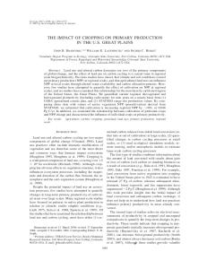
Title Implantation Relation of fetal membranes to wall of the uterus. Placental villi Intra-decidual sign Normal gestational sac Yolk sac Float test for chorionic villi Transvaginal ultrasound image showing the embryo (E), yolk sac (YS) and a subchorionic hematoma (SCH) Line diagram for classification of hematomas in and around placenta An empty gestation sac mimics a subchorionic collection Non-fusion and separation of chorion and amnion Intra-amniotic hemorrhage Prominent vessels mimicking a subchorionic hematoma Clustered bar chart showing the miscarriage outcome in the subchorionic hematoma group versus no hematoma group. Clustered bar chart showing the association between the hematoma size and miscarriage.
vii
Page 4 5 6 8 8 9 11 12 14 17 18 18 19 29
30
List of Tables Item Table 1 Table 2
Table 3
Table 4
Table 5 Table 6 Table 7
Comment Early Pregnancy Discriminatory Findings Maternal age, body mass index and gestational age for cases presented with threatened miscarriage with subchorionic hematoma versus no hematoma. Maternal parity for cases presented with threatened miscarriage with subchorionic hematoma versus no hematoma. History of maternal previous miscarriages for cases presented with threatened miscarriage and subchorionic hematoma versus no hematoma. Placental site in cases with subchorionic hematoma versus no hematoma The outcome of the subchorionic hematoma group versus no hematoma. The association between the hematoma size and miscarriage.
viii
Page 9 25
26
27
27 28 30
Abstract Background Subchorionic hematomas are common ultrasonographic findings that may be associated with first trimester bleeding. First-trimester bleeding occurs in 16% to 25% of all pregnancies. Pre-existing medical conditions, autoimmune diseases, and immunological factors have been associated with subchorionic hematoma, but the etiology of this condition is still unknown. Objectives To varify the relations and effects of having an ultrasonographic evidence of subchorionic hematoma in women presenting with threatened miscarriage in the first trimester. Patients and Methods This observational prospective study was conducted at Slemani Maternity Teaching Hospital in Iraq from the beginning of February 2016 to the end of september 2016.The study sample consisted of 140 cases of threatened miscarriage, divided into two groups 65 cases with ultrasound evidence of subchorionic hematoma (cohort) and another 75 cases without any ultrasound evidence of subchorionic hematoma (control).Inclusion criteria include a viable, singleton intrauterine pregnancy, a history of regular menstrual cycle and a known LMP presented with threatened miscarriage in their first trimester (between 6 weeks and 13 weeks).Informed consents were obtained from all patients with detailed history and clinical examination including the patients’ age, BMI, gravity, parity and number of previous miscarriages were noted. Pregnancy was confirmed by serum pregnancy test and abdominal ultrasound was used to confirm intrauterine pregnancy, viability, measure the crown rump length to calculate the gestational age, determine the site of the placenta (anterior or posterior) and search for evidence of subchorionic hematoma. The size of the subchorionic hematoma were noted in all cases then compared with the size of the gestational sac during the examination and classified as small (<20% of the gestational sac), medium (20%–50% of the gestational sac), or large (>50% of the gestational sac). The patients were followed up regularly every week in the antenatal clinic and repeat ultrasound scans were done weekly. In patients with subchorionic hematoma, scans were repeated until resolution of hematoma. The ix
gestational age at first bleeding attack was noted. All the patients were instructed to report any further bleeding attack, passage of clots or pieces and come to the hospital immediately. The patients were followed up till 24 weeks of pregnancy and the outcomes were recorded. Results Pregnant ladies presented with subchorionic hematoma in their first trimester were statistically significant older (30.52 ± 3.878 years) than the control group (23.49 ± 3.241 years, p<0.05), having more BMI (31.43± 1.732kg/m2 vs. 28.69 ± 1.770 kg/m2, p<0.05) and presented at a statically significant earlier gestational age (7.85 ± 0.95weeks vs. 11.24 ± 0.92weeks, p<0.05). There were no statistically significant difference in the parity and history of previous miscarriages between the two groups. Similarly, there was no statistically significant difference between the two groups regarding the placental site. Miscarriage rate was statistically significant higher in the subchorionic hematoma group (35.4%) than the control group (12%), p <0.05. There was a statistically significant association between the hematoma size and miscarriage. The majority of cases with small hematoma (83%) continued beyond 24 weeks versus (11.9%) and (4.8%) for medium and large size hematoma respectively. Conclusions Subchorionic hematoma in the first trimester of pregnancy increases the risk of spontaneous miscarriage, the size of the hematoma also have a significant effect on the outcome, the larger the hematoma the higher the rate of miscarriage. Key Words: threatened miscarriage, subchorionic hematoma
x
Chapter 1 Introduction
1
1.1 Overview Vaginal bleeding is a frequent complication of pregnancy during the first trimester, with an incidence of 16%-25%. Intrauterine bleeding without cervical dilatation and tenderness during the early pregnancy period is defined as threatened miscarriage. Generally, it is not associated with pain and excessive bleeding. These bleedings result in maternal anxiety and may be associated with fetal/maternal adverse outcomes.1 Intrauterine hemorrhages are commonly observed features on ultrasound examinations, especially among patients with clinically evident bleeding in early pregnancy, and the incidence has been reported to be 4%-22%. Subchorionic hematomas (SCHs) usually appear as hypoechoic or anechoic crescent-shaped areas on ultrasonography. Although the exact etiology is uncertain, they are believed to result from partial detachment of the chorionic membranes from the uterine wall. Uterine malformations, history of recurrent pregnancy loss, and infections are the possible predisposing factors.2 The clinical significance of SCH remains controversial. It is also not certain if these hemorrhages result in miscarriage. However, according to the results of a recent meta-analysis, the presence of SCH increases the risk of early or late pregnancy loss by 2-fold.3 It is suggested that the presence of SCH increases the risk of an adverse obstetric outcome, and fetal outcome is associated with the size of the hematoma, maternal age, and gestational age.4
2
1.2 Definitions5, 6 , 7 , 8 Spontaneous Miscarriage— spontaneous loss of a pregnancy prior to 24 weeks gestation. May be further described as: • Incomplete — occurs when some but not all of the products of conception have passed • Complete — the complete passage of all products of conception Threatened Miscarriage— bleeding before 24 weeks in the presence of an embryo with cardiac activity and closed cervix. • Inevitable — bleeding in the presence of a dilated cervix, indicating that passage of the conceptus is unavoidable • Missed — The fetus or embryo has been dead but no tissue has been passed. The cervix is closed. These patients often present with no growth in uterine size or no fetal heart beats being heard. • Septic — Incomplete miscarriage associated with ascending infection of the endometrium, parametruim, adnexa, or peritoneum Subchorionic Hemorrhage — Ultrasonographic finding of blood between the chorion and uterine wall, usually seen in the setting of vaginal bleeding. Recurrent Pregnancy Loss — more than three consecutive pregnancy losses. Amnion – amniotic cavity immediately surrounds the embryo; the amniotic membrane is derived from the epiblast and extraembryonic mesoderm. Chorion – the fetal component of the placenta, derived from trophoblast and extraembryonic mesoderm, contains fetal blood vessels. Chorion frondosum – region of chorion with villi whose association with the decidua basalis is the essential unit of the placenta. Chorion laeve – abembryonic region of chorion that is without villi, therefore, smooth (laeve). Decidua – is the functional secretory endometrium of pregnancy , it is totally shed after labor.
3
1.3 Physiology and Embryology During the luteal phase of the menstrual cycle, that is, following ovulation, some of the stromal cells of the endometrium enlarge and accumulate glycogen and lipid. These decidual cells disappear unless pregnancy occurs, and the corpus luteum is maintained, providing a source of progesterone. With pregnancy, continuing stimulation by progesterone and estrogen causes widespread decidualization, which is greatly amplified by the presence of the implanting blastocyst. The glands of the decidua function initially as a source of essential nutrients for the embryo until it establishes associations with the maternal blood supply. The decidua also contributes to the protection of the embryo from immunologic rejection and the uterine wall from excessive invasion by the embryo (Figure 1).8
Figure 1. Implantation. (a)The syncytiotrophoblast invades the uterine epithelium and by 8 days has expanded into the stroma. The blastocyst is nourished by uterine glands until an association is formed with the maternal vasculature. (b , c) By 9 days the embryo is completely implanted. Lacunae appear in the syncytiotrophoblast into which maternal blood will spill as the trophoblast invades maternal vascular sinusoids.8
4
The characteristics of the decidua (Figure 2) differ in various regions of the uterus defined by the implantation site. Immediately beneath the embryo the chorionic villi anchor into the region known as the decidua basalis. This forms part of the placenta. The thin layer of endometrium that surrounds the implanted embryo is the decidua capsularis. By 20 weeks this fuses with the endometrium lining the remainder of the uterus, the decidua parietalis, obliterating the uterine lumen.8 The placenta begins to develop upon the implantation of the blastocyst into the maternal endometrium. As the blastocyst differentiates, the placenta begins to form. There are four extra embryonic membranes that are part of the placenta including the yolk sac, the amnion, the allantois, and the chorion. 9 The fully developed placenta is a combination of a fetal portion, formed by the chorion and a maternal portion, formed by the decidua basalis. The embryonic trophoblast must invade the stroma of the uterine wall and form an association with the maternal bloodstream, but not invade more deeply into the uterine wall than the endometrium.8
Figure 2.Relation of fetal membranes to wall of the uterus. A. End of the second month. Note the yolk sac in the chorionic cavity between the amnion and chorion. At the abembryonic pole, villi have disappeared (chorion laeve). B. End of the third month. The amnion and chorion have fused, and the uterine cavity is obliterated by fusion of the chorion laeve and the decidua parietalis.8
5
Figure 3. Placental villi (a) Primary stem villus. (b) Secondary stem villus. (c) Tertiary stem villus 10
In the first trimester, placental growth is more rapid than that of the fetus. But by approximately 17 postmenstrual weeks, placental and fetal weights are approximately equal. By term, placental weight is approximately one sixth of fetal weight. At full term, the placenta is discoid with a diameter of 15 to 25 cm, is approximately 3 cm thick, and weighs about 500 to 600 g.10
6
1.4 Normal First Trimester Pregnancy Progress and Discriminatory Criteria Clinically, pregnancy is dated from the first day of the last normal menstrual period (LMP), an observable event, rather from the conception date. Conception is approximately two weeks later.11 The placenta produces human chorionic gonadotropin (β-hCG) after implantation. Implantation occurs at around 23 menstrual days, which is about eight days after conception. Commonly available urine pregnancy tests can detect the beta subunit of hCG at levels of 50 to 100 mIU/ml IRP (International Reference Preparation) and some can detect hCG at levels as low as 20 mIU/ml. Serum tests can detect hCG as low as 5 mIU/ml. It is therefore technically possible to detect pregnancy prior to a missed period, the majority of over the counter home pregnancy tests will not be positive until the first week after a missed menstrual period.11 Quantitative serum β-hCG levels should double every two to three days during weeks four to eight in normal early pregnancy. Falling levels, or those that plateau are strong evidence of impending poor outcome but do not distinguish between spontaneous miscarriage and ectopic pregnancy.12 The gestational sac first becomes visible sonographically during the fifth menstrual week as a 2 to 5 mm sonolucent area surrounded by an echogenic ring of chorionic villi. This early gestational sac is visible only using a high frequency transducer (5 MHz or greater) and the transvaginal scan route. A small sonolucent fluid collection, or pseudosac, may also be present in cases of ectopic pregnancy, so additional features of a normal gestational sac can be sought including the “intradecidual sign” indicating that the gestational sac is embedded in the endometrium and not within the endometrial stripe (Figure 4, Figure 5).13
7
Figure 4. Intra-decidual sign 13
Figure 5. Normal gestational sac, regularly round, located in the fundus and surrounded by an echogenic ring. 13 The yolk sac appears during transvaginal scanning during the sixth menstrual week and provides clear evidence of an intrauterine pregnancy. By the end of the sixth menstrual week, the “fetal pole” 2 mm to 8 mm in length with embryonic cardiac activity becomes visible during transvaginal scanning. All these 8
sonographic landmarks are visible with transabdominal scanning about one week later than with transvaginal scanning(Figure 6). 13
Figure 6. Yolk sac. 13 Discriminatory criteria based on clinical, β-hCG and sonographic findings are closely correlated and are displayed inTable 1. Ultrasonography is so helpful that, when it is readily available, many clinicians use it as a primary tool in the evaluating first trimester complications, leaving serum β-hCG and/or progesterone testing as a secondary tool to use only if sonographic findings are equivocal.14 Table 1. Early Pregnancy Discriminatory Findings.
9
Once the embryo is visible sonographically, first trimester menstrual age is calculated from crown-rump length. Between eight and 13 weeks, gestational age can be approximated using the simple formula: Menstrual age (weeks) = crown rump length (CRL) in centimeters (cm) plus 6.5 Or: Menstrual age (days) = crown-rump length in millimeters (mm) plus 42.15
1.5 Miscarriage: Pathophysiology, Clinical Course and Diagnosis One of the suggested mechanisms for threatened miscarriage is placental dysfunction, which can also cause several late complications, such as preeclampsia, preterm labor, preterm birth, placental abruption, placenta previa, intrauterine growth restriction, and perinatal mortality. Similarly, insufficient angiogenesis is associated with early pregnancy losses, and maternal serum AFP and β-hCG are suggested to be used as markers of angiogenesis in the first trimester. Together with these markers, chronic inflammation of the decidua might also be the underlying cause of early pregnancy bleedings.16 The cause of spontaneous miscarriage is rarely determined in clinical practice, but it is known that about half are due to major genetic abnormalities, usually trisomy, triploidy, or monosomy.17 Environmental factors linked to spontaneous miscarriage:
Uterine anomalies Leiomyomata Incompetent cervix Tobacco, alcohol or cocaine Progesterone deficiency due to luteal phase defect Irradiation Maternal diethylstilbestrol (DES) exposure Advanced maternal age Infections Occupational chemical exposure.
10
Spontaneous miscarriage may present clinically in several different ways. Most commonly, vaginal bleeding and cramping are present, but occasionally, regression of pregnancy symptoms or lack of Doppler-detected fetal heart tones by 10 to 12 weeks are the first clinical signs in the setting of anembryonic pregnancy or embryonic demise. Clinical examination should include palpation of the abdomen and pelvis. The size and position of the uterus, the location of any tenderness, the presence of rebound tenderness and the presence of masses should be noted. Adnexal tenderness and any masses should heighten concern for ectopic pregnancy, although a normal corpus luteum cyst may also be the cause. If the patient’s last menses was at least nine to ten weeks ago, consider listening for the fetal heart beat during the bimanual pelvic exam while elevating the uterus with the examining hand.18 The speculum examination will reveal non-uterine causes of bleeding, the degree of cervical dilation and, if present, tissue being passed. The quantity of blood in the vault and the source of bleeding (from the os vs. other sites) should be noted. If an intact gestational sac, an embryo, or the characteristic fronds of chorionic villi are seen, miscarriage is proven and ectopic pregnancy is virtually ruled out except in the rare case of simultaneous intrauterine and ectopic pregnancy (heterotopic pregnancy). To look for chorionic villi, rinse and float the tissue with saline. Low magnification, backlighting and “teasing” the tissue may help. Passed tissue should be submitted for pathological examination which is definitive in questionable cases. Figure 7.18
Figure 7. Float test for chorionic villi. 18
11
Both the gestational sac and the embryo if present should each grow at the rate of approximately 1 mm/day, thus allowing for appreciable interval change in that time period. 19 When ultrasound reveals a heartbeat in a patient who is bleeding, the probability of miscarriage is between 2.1 percent for women under 35 years of age to 16.1 percent in women over 35 years of age.20
1.5.1 Subchorionic Hemorrhage Intrauterine or subchorionic hematoma is defined as a collection of fluid in the uterine cavity, and it is believed to result from subchorionic bleeding caused by a partial detachment of the trophoblast from the uterine wall. This condition can be diagnosed only by ultrasonography. Mantoni and Pedersen first described its sonographic patterns. On ultrasound examination, it appears as an anechoic area that has a falciform shape, and it is usually observed behind or below the gestational sac, separating the chorion from the inner wall of the uterus. Small echogenic structures can be found in such areas, and they are believed to be blood clots.21 In subchorionic hemorrhage, a gestational sac and embryo will be present but sonography will reveal a hematoma between the chorion and uterine wall. When subchorionic hemorrhage is seen by ultrasound, the likelihood for miscarriage averages about ten percent, even when a heartbeat is detected, and varies by maternal age, size of the hematoma and gestational age.22
Figure 8. Transvaginal ultrasound image showing the embryo (E), yolk sac (YS) and a subchorionic hematoma (SCH)22 12
Therefore the patient should be warned to expect bleeding. The quantity of bleeding only predicts pregnancy loss when it is heavy. A prospective analysis of 4,510 women who were followed in the early first trimester showed that 1,204 (27 percent) had some bleeding or spotting. There was no increase in risk of miscarriage in early pregnancy when there was spotting or light bleeding. However risk of miscarriage increased significantly in the eight percent of women who reported heavy bleeding. This was the only group to have an increased risk of miscarriage (Adjusted OR 2.84, 95% CI 1.82-4.43).23 The underlying mechanism of how SCH causes adverse pregnancy outcomes is still controversial. One of the possible mechanisms is the premature perfusion of the intervillous space, as occurs with subchorionic hemorrhage, before the development of placental adaptations to cope with oxidative stress. In normal pregnancy, there is a burst of oxidative stress as the maternal circulation is established, which may serve a physiologic role in stimulating placental differentiation and regulate cell function. 24 Along with the rise in oxygen tension, there is a parallel rise in the expression of antioxidant enzymes within the placental tissues. A delicate balance is, therefore, achieved between the production of free radicals and protective antioxidant activity. A change in the equilibrium, resulting in an increase in free radical formation, such as a premature influx of oxygenated blood with hematoma formation, the presence of substances that form free radicals, or a reduction in local antioxidant levels may well result in an impairment in placentation and subsequent pregnancy complications including miscarriage.25 Another possible mechanism might be the underlying cause of the subchorionic bleeding and secondary mechanical effects of the hematoma. Shallow trophoblast invasion and impaired angiogenesis with resultant friable blood vessels may predispose one to subchorionic hemorrhage, as well as adverse outcomes. The presence of a hematoma, especially in a retroplacental location, may create an area of weakness, where further separation of the placenta from the uterine wall may occur, resulting in placental abruption.3The presence of an SCH and detachment of the gestational sac from the endometrium may result in miscarriage. However, if the gestational sac survives, reattachment to the
13
endometrial wall might be enough for further progression of the pregnancy without any other adverse effects.24 Bleeding in the first trimester with or without the presence of a hematoma may be associated with a chronic inflammatory reaction in the decidua resulting in persistent myometrial activity and expulsion of the pregnancy. It is known that in about two thirds of early pregnancy failures, there is defective placentation, which is mainly characterized by a thinner and fragmented trophoblast shell and reduced cytotrophoblast invasion of the lumen at the tips of the spiral arteries. 26
1.5.2 Differential Diagnosis of SCH: 27
twin gestational sac uterine leiomyoma (fibroid) focal myometrial contraction chorioamniotic separation prominent retroplacental veins retroplacental hematoma sub-amniotic hematoma (Figure 9)
Figure 9. Line diagram for classification of hematomas in and around placenta. P: Placenta, Dark grey color: Hematoma, Grey line: Amnion, Black line: Chorion A: Retroplacental bleeding is found behind placenta. B: Subchorionic bleeding dissects chorion and endometrium. When such bleeding involves the margin of placenta, it is called marginal SCH. C: Sub-amniotic hemorrhage is contained within the amnion and chorion and thus extends anteriorly to placenta but is limited by reflection of amnion on placental insertion site of umbilical cord. Sub-amniotic bleeding is rare.27
14
1.5.3 Implications of Subchorionic Hemorrhage Subchorionic hemorrhage (subchorionic hematoma) is the most common sonographic abnormality in the presence of a live embryo. Vaginal bleeding affects 25% of all women during the first half of pregnancy and is a common reason for first-trimester ultrasonography. Sonographic visualization of a subchorionic hematoma is important in a symptomatic woman because pregnant women with a demonstrable hematoma have a prognosis worse than women without a hematoma. However, small, asymptomatic subchorionic hematomas do not worsen the patient's prognosis.23 In women whose sonogram shows a subchorionic hematoma, the outcome of the fetus depends on the size of the hematoma, the mother's age, and the fetus's gestational age. Rates of miscarriage increase with advancing maternal age and increasing size of hematoma. Late first- or second-trimester bleeding also worsens the prognosis. The presence of sonographically detected subchorionic hemorrhage (subchorionic hematoma) increases the risk of miscarriage, stillbirth, abruptio placentae, and preterm labor. The subchorionic hemorrhage (subchorionic hematoma) collects between the uterine wall and the chorionic membrane and may leak through the cervical canal. Later in the first trimester and early second trimester, the subchorionic hematoma may partially strip the developing placenta away from its attachment site. Therefore, the prognosis of patients with this type of hematoma is worse than the prognosis of patients with hematoma early in first trimester. The subchorionic hematoma often regresses, especially if it is small or moderate in size. Large hematomas, which strip at least 30-40% of placenta away from endometrium, may enlarge further, compressing the gestational sac and leading to premature rupture of membranes with consequent spontaneous miscarriage.28
1.5.4 Role of radiology in the diagnosis of SCH I. Ultrasonography Ultrasonography is the imaging modality of choice for subchorionic hemorrhage (subchorionic hematoma) because it can be performed rapidly at the patient's bedside and because it has no known risk, as with radiation. The sensitivity of sonography is low and varies between 2% and 20%, as blood may
15
pass vaginally and not collect in the subchorionic space. Hematomas may also appear iso-echoic relative to the placenta.29 Acute subchorionic hemorrhages (subchorionic hematomas) vary in echogenicity and are seen between the chorion and the uterine wall on sonograms. Iso-echoic hematomas may be missed on initial sonograms, or they may be recognized as heterogeneous and thickened placentas.30 Color Doppler sonography may help in distinguishing the avascular hematoma from the highly vascular placenta. Follow-up sonography may also help in resolving hematomas. Hematomas may resolve over 1-2 weeks. During this time, they may be seen as complex fluid collections with mixed echogenicity. In addition, sonographic findings also confirm fetal viability and can help in differentiating and diagnosing other conditions associated with miscarriage in the first trimester, such as ectopic pregnancy, blighted ovum, and twin gestation. Follow-up ultrasonography should be performed as clinically indicated.31 Ultrasonography lacks high sensitivity for small bleeds. However, it is the most useful modality in a pregnant patient with vaginal bleeding.32 The size of the hematoma can be classified as small, medium, or large according to the size relative to the gestational sac. A SCH can be considered large if it is greater than 50% of the size of the gestation sac, medium if it is 20-50%, and small if it is less than 20%. Large hematomas by size (>30-50%) and volume (>50 mL) worsen the patient's prognosis.33 Another option is to measure the volume of the hematoma. Volumes are estimated by measuring the anteroposterior, longitudinal, and transverse diameters and multiplying these values by 0.523 (the ellipsoid formula). Because the hematoma may wrap around the gestational sac, the ellipsoid formula would overestimate the size. 33 Uterine fibroids or focal myometrial contractions (Figure 10) can cause the placenta to appear thickened, or they may look like subchorionic or retroplacental hematomas. Hematomas are avascular on color Doppler scanning. Fibroids have a characteristic hypoechoic appearance with or without calcifications and typical peripheral blood flow in color Doppler images. Color flow is seen in contracted 16
myometrium, and transient myometrial contractions usually resolve within 30 minutes of scanning.33
Figure 10. Sagittal endovaginal scan of a dichorionic-diamniotic twin pregnancy in the 13th week of gestation. An empty gestation sac mimics a subchorionic collection (red arrow). Also note the transient uterine contraction (yellow arrow).33
Chorioamniotic separation has an anechoic appearance and usually resolves by 16 weeks (Figure 11 ). It can be distinguished from an anechoic hematoma by finding elevation of the membrane in chorioamniotic separation that extends over the fetal surface of the placenta and that terminates at the origin of the umbilical cord. The amniotic membrane is also thinner than the chorionic membrane.33
17
Figure 11. Sagittal endovaginal scan of the uterus in 9th week of gestation demonstrates non-fusion and separation of chorion and amnion.33 Primary intra-amniotic hemorrhage can occur with a large subchorionic hematoma, with trauma, or with an invasive procedure such as chorionic villous sampling or amniocentesis. Floating echoes (Figure 12) or echogenic clots may be seen in the amniotic cavity.34
Figure 12. Transverse endovaginal scan demonstrating intra-amniotic hemorrha ge as fine echoes dispersed in the amniotic fluid.34 Hematomas of the umbilical cord are rare and may result from amniocentesis and sampling of blood from the umbilical cord. These hematomas are seen as echogenic masses limited to the cord itself.34 Prominent retroplacental and myometrial vessels should be distinguished from heterogeneous bleeding. The vessels appear as coily, anechoic structures and demonstrate flow on color Doppler sonography (Figure 13); these features differentiate them from avascular hematomas.35 18
Figure 13. Sagittal gray-scale endovaginal scan (top) of the uterus demonstrates a hypoechoic area adjacent to the lower edge of the placenta; this finding suggests a small subchorionic hematoma. However, color Doppler scan (bottom) at the same level confirms that this finding represents prominent vessels mimicking a subchorionic hematoma.35
19
II. Computed Tomography CT scanning is relatively contraindicated during pregnancy because of the risk of radiation to the fetus. Pregnant patients may undergo CT for reasons such as an evaluation of trauma or acute abdomen. Scans may show an incidental or injury-related hyper-attenuating subchorionic hemorrhage (subchorionic hematoma).The sensitivity of CT may be high compared with that of sonography. However, because of the risk of radiation with CT, no large comparisons of the 2 modalities have been reported. Normal chorioamniotic separation should not be confused with placental abruption.36
III. Magnetic Resonance Imaging MRI is not routinely performed to detect subchorionic hemorrhage (subchorionic hematoma); a more common indication is the detection of fetal anomalies. MRI may incidentally show a subchorionic hematoma and help in characterizing and determining the acuity of the hematomas by showing changes in signal intensity produced by various blood products. T1-weighted spin-echo and gradient-echo images are particularly useful in evaluating the hemorrhage.37
20
Chapter 2 Patients and Methods
21
2.1 Patients and Methods This is an observational prospective comparative study was conducted at Slemani Maternity Teaching Hospital – Slemani/Iraq during the period from the beginning of February 2016 to the end of September 2016. The study sample consisted of 140 cases of threatened miscarriage, the sample then was subdivided into two groups depending on ultrasound finding of SCH, 65 cases with ultrasound evidence of subchorionic hematoma (the cohort group) and 75 cases without any ultrasound evidence of subchorionic hematoma (the control group). The study protocol was approved by the scientific committee of University of Slemani and the Department of Obstetrics and Gynecology in Slemani Maternity Teaching Hospital as well as the ethical committee. Inclusion criteria: Pregnant ladies with a viable, singleton intrauterine pregnancy, a history of regular menstrual cycle and a known LMP presented with threatened miscarriage in their first trimester between 6 weeks and 13 weeks were included. Exclusion criteria: grand multiparity , multiple pregnancy ,non-viable or pregnancy of unknown location, fetal abnormalities diagnosed by ultrasound, recurrent miscarriage, uterine fibroids, polyps, uterine malformations , previous elective termination of pregnancy, previous cesarean section(s) and chronic medical illness (i.e., chronic hypertension, diabetes mellitus, anemia or using anticoagulants) were excluded. Informed consents were obtained from all patients after explaining to them the aim of the study followed by detailed history and clinical examination including patients’ age, BMI, gravity, parity and number of previous miscarriages were recorded. Pregnancy was confirmed by serum pregnancy test. Abdominal ultrasound was used to confirm intrauterine pregnancy, viability, measure the crown rump length (CRL) to calculate the gestational age, determine the site of the placenta (anterior or posterior) and search for evidence of subchorionic hematoma. The size of the gestational sac was recorded in all cases. 22
The size of the hematoma was then compared with the size of the gestational sac during the examination and classified as small (<20% of the gestational sac), medium (20%–50% of the gestational sac), or large (>50% of the gestational sac). The amount of vaginal bleeding was noted. If simple spotting, it was considered as light. If similar to patient’s menstrual bleeding it was considered as moderate if more, it was considered as heavy. Patients with threatened miscarriage were managed with bed rest until the bleeding stopped, folic acid supplementation, and were given micronized progesterone (Uterogestan®) 200 mg bid. The patients were followed up regularly every week in the antenatal clinic and repeat ultrasound scans were done weekly. In patients with subchorionic hematoma, scans were repeated until resolution of hematoma. The gestational age at first bleeding attack was noted. All the patients were instructed to report any further bleeding attack, passage of clots or pieces and come to the hospital immediately. The patients were followed up till 24 weeks and the outcomes were recorded.
2.2 Statistics The SPSS 22 software for Windows (Statistical Packages for Social Sciencesversion 22) was used for statistical analysis. Categorical data were compared using the Chi square (χ2) test or Fisher’s exact test, where appropriate. Continuous variables between groups were compared using the Student t -test or Mann – Whitney U test where appropriate. A value of p< 0.05 was considered significant.
2.3 Aim of the Study To verify the relations and effects of having an ultrasonograhic evidence of subchorionic hematoma in women presenting with threatened miscarriage in the first trimester.
23
Chapter 3 Results
24
Results In this observational prospective study 140 cases of threatened miscarriage which were included and divided into two groups 65 cases with ultrasound evidence of subchorionic hematoma (the cohort group) and another 75 cases without any ultrasound evidence of subchorionic hematoma (the control group). Table 2 shows a statistically significant difference in the mean maternal age for the cases presented with threatened miscarriage and subchorionic hematoma (30.52 ± 3.878 years) versus (23.49 ± 3.241 years) in the cases without a hematoma, p<0.05 BMI was significantly more in the cases presented with subchorionic hematoma (31.43± 1.732kg/m2) versus (28.69 ± 1.770 kg/m2) in the cases without a hematoma, p<0.05 The gestational age was significantly earlier for cases presented with subchorionic hematoma (7.85 ± 0.95weeks) versus (11.24 ± 0.92weeks) in the no hematoma group, p<0.05
Table 2 Maternal age, body mass index and gestational age for cases in both groups. Maternal Characteristics
Age (years)
2
BMI (kg/m )
GA (weeks)
Subchorionic Hematoma n=65 Mean ± SD
No Hematoma n=75 Mean ± SD
p value
30.52 ± 3.878
< 0.05 (<0.000) 23.49 ± 3.241 (5.8055E-22) *
31.43± 1.732
< 0.05 (<0.000) 28.69 ± 1.770 (4.5827E-16)*
7.85 ± 0.95
11.24 ± 0.92
< 0.05 (<0.000) (1.9601E-45)*
*Significant difference using Students-t-test for two independent means at 0.05 level.
25
Table 3 shows that in cases with subchorionic hematoma primi parity was 41.5% versus 45.3% in the control group. Para 1-2 was 20% of the subchorionic hematoma cases versus 30.7% of the control group. While para 3-4 was 38.5% in the subchorionic hematoma versus 24% in the control group. With a p value = 0.134 which indicates that there was no statistically significant difference in the parity of the pregnant ladies presented with threatened miscarriage whether associated with US evidence of SCH or not.
Table 3 Maternal parity for cases presented with threatened miscarriage with subchorionic hematoma versus no hematoma. Maternal Parity
Parity
Total
Subchorionic Hematoma
No Hematoma
primi
N 27
% 41.5%
N 34
% 45.3%
1-2
13
20%
23
30.7%
3-4
25
38.5%
18
24%
65
100%
75
100%
p value
0.134
*Significant difference between proportions using Pearson Chi-square test at 0.05 level.
Table 4shows that in the subchorionic hematoma group there was no history of miscarriage in 78.5% of cases versus 78.7% of cases in the control group. While 12.3% of cases in the subchorionic hematoma group had a one previous miscarriage versus 14.7% in the control group. 9.2% of cases in the subchorionic hematoma group had previous 2 miscarriages versus 6.7% in the control group, with a p value = 0.805 which indicates that there is no statistically significant difference regarding the history of previous miscarriages between the two groups.
26
Table 4 History of maternal previous miscarriages for cases presented with threatened miscarriage and subchorionic hematoma versus no hematoma. Pervious Miscarriages
Subchorionic Hematoma
No Hematoma
0
N 51
% 78.5%
N 59
% 78.7%
1
8
12.3%
11
14.7%
2
6
9.2%
5
6.7%
65
100%
75
100%
Total
p value
0.805
*Significant difference between proportions using Pearson Chi-square test at 0.05 level.
Table 5shows that the placenta was anterior in 41.5% of the subchorionic hematoma group versus 44%in the no hematoma group, and it was posterior in 58.5%in the subchorionic hematoma group versus 56%in the no hematoma group with a p = 0.769 which indicates that there is no statistically significant difference in the placental site between the two groups. Table 5 Placental site in cases with subchorionic hematoma versus no hematoma
Anterior
Subchorionic Hematoma N % 27 41.5%
No Hematoma N % 33 44%
Posterior
38
58.5%
42
56%
65
100%
75
100%
Placental Site
Total
p value
0.769
*Significant difference between proportions using Pearson Chi-square test at 0.05 level.
Table 6and Figure 14 show that there is a statistically significant difference in the miscarriage rate between the two groups, the miscarriage rate was significantly higher in the subchorionic hematoma group (35.4%) versus (12%) in the no hematoma group with a p <0.05. While (64.6%) of the ladies presented with threatened miscarriage and subchorionic hematoma continued the pregnancy versus (88%) in the no hematoma group. 27
The total number of cases in the hematoma group who ended by miscarriage was 23 cases, 7 cases ended by fetal demise, 4 ended by incomplete miscarriage and 12 cases ended by complete miscarriage. While all the 9 cases who miscarried in the control group ended as incomplete miscarriage. Table 6 the outcome of the subchorionic hematoma group versus no hematoma. Subchorionic Hematoma N % 23 35.4%
No Hematoma N % 9 12%
No miscarriage
42
64.6%
66
88%
Total
65
100%
75
100%
Outcome Miscarriage
p value
0.001*
*Significant difference between proportions using Pearson Chi-square test at 0.05 level.
28
Figure 14 Clustered bar chart showing the miscarriage outcome in the subchorionic hematoma group versus no hematoma group.
Table 7 shows that there is a statistically significant association between the hematoma size and miscarriage rate. 13% of cases with small subchorionic hematoma (less than 20% gestational sac size) underwent miscarriage versus 83.3% continued beyond the 24 weeks. 34.8% of cases with medium sized subchorionic hematoma (20 – 49% gestational sac size) underwent miscarriage versus 11.9%. And 52.2% of large subchorionic hematoma cases (greater than or equal to 50% gestational sac size) underwent miscarriage versus 4.8%, with a p <0.05.
29
Table 7 the association between the hematoma size and miscarriage. Miscarriage
No miscarriage
Hematoma Size Small
N 3
% 13%
N 35
% 83.3%
n 38
% 58.5%
Medium Large
8 12
34.8% 52.2%
5 2
11.9% 4.8%
13 14
20% 21.5%
23
100%
42
100%
65
100%
Total
Total
p value
< 0.05 (<0.000) (4.5827E16)*
*Significant difference between proportions using Pearson Chi-square test at 0.05 level.
Figure 15. Clustered bar chart showing the association between the hematoma size and miscarriage. 30
Chapter 4 Discussion
31
Discussion The reported incidence of intrauterine hematoma has a wide range, between 0.5% and 22%38, mainly associated with vaginal bleeding. The discrepancy in these rates might be related to different patient populations, study design, range of gestational ages, and lack of a standard definition. Moreover, the different approaches to ultrasound scanning, i.e., transabdominal or transvaginal, may be a factor in this epidemiological issue. The clinical significance of this sonographic finding remains controversial, and observational studies focusing on this topic reported conflicting results.39In this study maternal demographic and obstetric characteristics were compared between the pregnant ladies presented with threatened miscarriage between the 6th and 13th completed weeks of pregnancy who had a SCH confirmed by US versus those without SCH. In the current study pregnant ladies in the SCH group were statistically significant older than the control group. This finding is consistent with what was observed by Okan et al (2008) 40 who found that the incidence of SCH increased with increasing maternal age. Similarly Bennett et al. 41 observed that the spontaneous abortion rate was approximately twice as high for women aged 35 years or older as for younger women (13.8% and 7.3%, respectively). Johns et al (2003) 42 reported that there is no significant statistical difference in maternal age between the hematoma group (32.3 years) and control group (32.3 years) , Leite et al (2006)43also reported that the maternal age did not show a significant difference between the hematoma group ( 32 ± 1.66 years) and control group ( 30.77 ± 1.27 years) ,(p= 0.4688 ) and reported that maternal age is considered an independent risk factor for adverse outcome because there is a strong correlation with chromosomal and structural fetal anomaly .Giobbe et al (2001)44 , Nagy et al (2003)45,Maso et al.(2005) 2and S. Agrawal et al. (2014)46reported a similar finding. There was a statistically significant difference in the body mass index between the two groups in this study. The BMI in SCH group was significantly more than the control group. On the contrary S. Agrawal et al. (2014)44didn’t find any statistically significant difference in the BMI between the two groups. 32
In this study there was no statistically significant difference regarding parity and previous miscarriage history between the two study groups. Johns et al.(2003)42,Şükür et al. (2014)47and S. Agrawal et al. (2014)44found that there is no statistical difference in parity and previous miscarriages between the SCH group and the control group, a finding which is consistent with the results of the current study. In the current study the mean gestational age at diagnosis is significantly lower in the subchorionic hematoma group than those in the no hematoma group with a p value <0.05.This is consistent with what was observed by Leite et al. (2006)43who found that an early gestational age at the diagnosis may be considered a risk factor for poor outcome (p = 0.0227). Bennett et al. 41 found a gestational age of 8 weeks or less was predictive of an increased percent of spontaneous abortion. Maso et al. (2005) 2reported that the risk of spontaneous abortion is nearly 15 times greater for cases diagnosed before 9 weeks of gestation than for those observed after this period. This observation can be helpful to the clinician in the risk assessment of the pregnancy complicated by subchorionic hematoma, bearing in mind that a diagnosis made before 9 weeks gives a 20% likelihood of miscarriage, whereas this possibility is far more remote (less than 2%) when the hematoma is diagnosed after this cutoff gestational age. Gupta R et al.(2007) 48 and Okan et al.(2008)40 showed that at the end of the first trimester and early second trimester, hematoma can disturb and displace the subchorionic placental growth from attached place, so that the prognosis is worse than the hematoma that occurs early in the first trimester. On the other hand Yavuz et al. (2014)47found that there was no statistical significant difference in the gestational age at diagnosis between the hematoma group ( 9.3± 2.8 weeks) and control group ( 10.2 ± 3.3 weeks) , p = 0.085. Ben-Haroush et al. 49assessed 2556 pregnant patients who were admitted with vaginal bleeding during the first 20 gestational weeks. The incidence of SCH was 9%. They reported that gestational age at diagnosis, size of SCH, and duration of bleeding did not affect the pregnancy outcome. They also reported significantly decreased miscarriage rates with bed rest. Similarly Saurbrei and Pham 50 did not 33
find any statistical significant effect of gestational age at diagnosis of SCH on pregnancy outcome. In the current study there was a statistically significant association between the hematoma size and miscarriage rate. Ball et al. 29 found that large volumes, defined as a degree larger than the 2/3 of the gestational sac circumference elevated by the hematoma, increased 2.4fold the risk of spontaneous abortion, similarly Esen Çağsar et al (2000) 51 found that the presence of the subchorionic hematoma and largeness of its volume significantly increase the miscarriage rate, both findings are consistent with what was observed in the current study. In addition Leite et al (2006) 43 suggested that the presence of a very large first-trimester hematoma is associated with a 46% risk of spontaneous miscarriage. Bennett et al.41 conducted a retrospective study of 516 patients with first-trimester vaginal bleeding, a live fetus, and a subchorionic hematoma. The overall spontaneous rate was 9.3%. This figure almost doubled when the separation was large 18.8% compared to 7.7% for small hematoma and 9.2% for moderate ones. On the contrary Maso et al. (2005) 2and Uluğ et al. (2006) 52 reported that there is no statistical significant relationship between the prognosis and presence or size of the SCH. Similarly Nagy et al. 45and Pedersen et al.53 found no association between the size of the hematoma and adverse outcome. While Donogol et al. (2011) 54and Giobbe et al. (2001)44found that when hematoma is small and asymptomatic it may not be of clinical significance. The explanation for these discrepancies on this variable (SCH size) might be addressed by considering the physio pathological mechanism of formation of the subchorionic hematoma. In fact, the size might be the final result of 2 processes: the amount of subchorionic bleeding and the amount of the bleeding through the cervix. Therefore, the size of the hematoma may not represent a reliable estimation of the overall severity of the process, which could ultimately be calculated only by knowing the total amount of blood collected in the uterus, reabsorbed, and lost through the cervix. On this basis, therefore, it might be postulated that the presence and location of a hematoma, as a sign of the impaired placentation, rather than its volume, is important for pregnancy outcome. 29 34
In the current study there was no statistical significant difference in the placental site between the SCH group and the control group, the placenta was anterior in 41.5% of the subchorionic hematoma group versus 44% in the control group, and it was posterior in58.5%in the subchorionic hematoma group versus 56% in the control group with a p =0.769. This observation is consistent with what was observed by Donogol et al (2011) 54 who reported that there is no statistically significant association between the SCH and placental site compared to control group. Similarly, Nagy et al. (2003)45 compared 187 patients who had SCH with 6488 controls, and they found increased miscarriage rates in the presence of SCH. However, they failed to show an association between the size of the SCH, location of the placenta and ongoing pregnancy outcome. In the current study there was a statistically significant difference in the miscarriage rate between the two groups, the miscarriage rate was significantly higher in the subchorionic hematoma group (25.3%) versus (11.6%) in the nonhematoma group with a p <0.05. This is consistent with what was observed by Ozkaya et al (2011) 55 they reported that the presence of subchorionic hematoma is associated with increased risk of spontaneous abortion and Nagy et al (2003)45who showed that the rate of spontaneous abortion was ( 18.7 % ) which is two times higher than in those without hematoma ( p= 0.02 ). Ketut et al (2011)56and Okan et al (2008)40 showed that the risk of miscarriage in those with threatened miscarriage and had subchorionic hematoma was 3 times higher than those with threatened miscarriage and without subchorionic hematoma, which is consistent with this study. Bennett et al.41 also reported that first-trimester threatened miscarriage with a subchorionic hematoma on ultrasound has been associated with a higher incidence of miscarriage. Similarly Ball et al. 29evaluated 238 patients with ultrasonographically detected SCH in a retrospective case control study and reported a significant association between SCH and miscarriage. On the contrary Pedersen and Mantoni53 who followed up 342 pregnancies with vaginal bleeding between 9 to 20 gestational weeks, in which 18% had SCH found no association between the presence of SCH and miscarriage. Similarly 35
Johns et al. (2003) 42 reported that first-trimester vaginal bleedings with the presence of SCH had no effect on the prognosis.
Conclusions The risk of having a subchorionic hematoma increases with increased maternal age, maternal BMI, and presentation at earlier gestational age. Subchorionic hematoma in the first trimester of pregnancy increases the risk of spontaneous miscarriage, the size of the hematoma also have a major effect on the outcome, the larger the hematoma the higher the rate of miscarriage.
Recommendations Preconception counselling and patient education about pregnancy complications and commitment to regular antenatal care attendance to prevent, detect and manage early pregnancy complications including threatened miscarriage and subchorionic hematoma. Dedicated early pregnancy unit is needed to deal with women presented with early pregnancy bleeding. Ultrasound examination should provide details about the presence or absence of SCH and its size for proper patient counseling.
36
References 1 . Saraswat L, Bhattacharya S, Maheshwari A, Bhattacharya S. Maternal and perinatal outcome in women with threatened miscarriage in the first trimester: A systematic review. BJOG 2010; 117:245-57. 2 . Maso G, D’Ottavio G, De Seta F, Sartore A, Piccoli M, Mandruzzato G. First-trimester intrauterine hematoma and outcome of pregnancy. Obstet Gynecol 2005; 105: 339-44. 3 . Tuuli MG, Norman SM, Odibo AO, Macones GA, Cahill AG. Perinatal outcomes in women with subchorionic hematoma: a systematic review and meta-analysis. Obstet Gynecol. 2011 May. 117(5):1205-12 4 . Abu-Yousef MM, Bleider JJ, Williamson RA, Weiner CP. Subchorionic hemorrhage: Sonographic diagnosis and clinical significance. Am J Roentgenol 1987; 149: 737-40. 5 . Creinin MD, Schwartz JL, Guido RS, Pymar HC. Early pregnancy failure--current management concepts. Obstet Gynecol Surv. 2001 Feb;56(2):105-13. 6 . Chen BA, Creinin MD. Contemporary management of early pregnancy failure. Clin Obstet Gynecol. 2007 Mar;50(1):67-88. 7 .Philip N Baker, Louise C Kenny. Obstetrics by Ten Teachers-19th ed. London: Hodder Arnold,2011. ISBN - 13 978 0 340 983 539 8 . Langman’s Medical Embryology, 8th ed., TW Sadler, Lippincott Williams & Wilkins, Philadelphia (2000). 9 . Baergen, R.N., Early Villous Stage: Day 13 to 28 Post Conception. Pathology of the Human Placenta, 2005, 7; p: 73. 10 .Larsen, WI, Human Embryology, 3rd ed., Churchill Livingstone, New York (2001) 11 . Cole LA, Sutton-Riley JM, Khanlian SA, Borkovskaya M, Rayburn BB, Rayburn WF. Sensitivity of over-thecounter pregnancy tests: comparison of utility and marketing messages. J Am Pharm Assoc. 2005 Sep-Oct;45(5):608-15. 12 . Barnhart KT. Ectopic pregnancy. NEJM 2009;361:379-87 13 . Laing FC, Frates MC. Ultrasound evaluation dring the first trimester of pregnancy. In: Callen PW ed. Ultrasonography in Obstetrics and Gynecology 4th ed. 2000 W.B. Saunders Co. Philadelphia 14 .Paspulati RM, Bhatt S, Nour S. Sonographic evaluation of first-trimester bleeding. Radiol Clin North Am. 2004 Mar;42(2):297-314.
37
15 . Whitworth M, Bricker L, Neilson JP, Dowswell T. Ultrasound for fetal assessment in early pregnancy. Cochrane Database of Systematic Reviews 2010, Issue 4. Art. No.: CD007058. DOI: 10.1002/14651858. CD007058.pub2 (Level I) 16 . Kutluer G, Çiçek NM, Moraloğlu Ö, Ertargın P, Sarıkaya E, Artar İ, Erdem Ö. Low VEGF expression in conceptus material and maternal serum AFP and β-hCG levels as indicators of defective angiogenesis in first-trimester miscarriages. J Turk Ger Gynecol Assoc 2012; 13: 111-7. 17 . Goddign M, Leschot NJ. Genetic Aspects of Miscarriage. Bailliere’s Clinical Obstetrics and Gynaecology 2000;14(5):855-865, 18 .Neville F. Hacker, Joseph C. Gambone, Calvin J. Hobel. Hacker and Moore’s essentials of obstetrics and gynecology-5th ed.Philadelphia: Saunders Elsevier,2010.ISBN:978-1-4160-5940-0 19 . Chen BA, Creinin MD. Contemporary management of early pregnancy failure. Clin Obstet Gynecol. 2007 Mar;50(1):67-88. 20 . Smith KE, Buyalos RP. The profound impact of patient age on pregnancy outcome after early detection of fetal cardiac activity. Fertil Steril 2006;65:35-40. (Level III) 21 . Mantoni M, Pedersen JF. Intrauterine hematoma: an ultrasonic study of threatened abortion. Br J Obstet Gynaecol 1981;88:47–51. 22 . Bennett GL, Bromley B, Lieberman E, Benacerraf BR. Subchorionic Hemorrhage in FirstTrimester Pregnancies: Prediction of Pregnancy Outcome with Sonography. Radiology 1996;200:803-806 23 . Hasan R, Baird DD, Herring AH, et al. Association between first-trimester vaginal bleeding and miscarriage. Obstet Gynecol 2009;114:860-7. (Level II) 24 . Jauniaux E, Watson AL, Hempstock J, Bao YP, Skepper JN, Burton GJ. Onset of maternal arterial blood flow and placental oxidative stres. A possible factor in human early pregnancy failure. Am J Pathol 2000; 157: 2111-22. 25 . Watson AL, Palmer ME, Jauniaux ER, Burton GJ. Variations in expression of copper/zinc superoxide dismutase in villous trophoblast of the human placenta with gestational age. Placenta 2007;18:295–9. 26 . Hustin J, Jauniaux E, Schaaps JP. Histological study of the materno-embryonic interface in spontaneous abortion. Placenta 2000;11:477–86. 27 . Avneesh Chhabra (Author), Eugene C Lin (Chief Editor). Subchorionic haemorrhage: Overview. Available at: http://emedicine.medscape.com/article/404971- overview. 28 . Yamada T, Atsuki Y, Wakasaya A, Kobayashi M, Hirano Y, Ohwada M. Characteristics of patients with subchorionic hematomas in the second trimester. J Obstet Gynaecol Res. 2012 Jan. 38(1):180-4. 38
29 . Ball RH, Ade CM, Schoenborn JA, Crane JP. The clinical significance of ultransonographically detected subchorionic hemorrhages. Am J Obstet Gynecol. 2006 Mar. 174(3):996-1002. 30 . Trop I, Levine D. Hemorrhage during pregnancy: sonography and MR imaging. AJR Am J Roentgenol. 2001 Mar. 176(3):607-15. 31 . Nyberg DA, Cyr DR, Mack LA, et al. Sonographic spectrum of placental abruption. AJR Am J Roentgenol. 1987 Jan. 148(1):161-4. 32 .Dighe M, Cuevas C, Moshiri M, Dubinsky T, Dogra VS. Sonography in first trimester bleeding. J Clin Ultrasound. 2008 Jul-Aug. 36(6):352-66. 33 . Mazzariol FS, Roberts J, Oh SK, Ricci Z, Koenigsberg M, Stein MW. Pearls and pitfalls in firsttrimester obstetric sonography. Clin Imaging. 2015 Mar-Apr. 39 (2):176-85. 34 . Akhlaghpoor S, Tomasian A. Safety of chorionic villus sampling in the presence of asymptomatic subchorionic hematoma. Fetal Diagn Ther. 2007. 22(5):394-400. 35 . Podrasky AE, Javitt MC, Glanc P, Dubinsky T, Harisinghani MG, Harris RD, et al. ACR appropriateness Criteria® second and third trimester bleeding. Ultrasound Q. 2013 Dec. 29 (4):293-301. 36 . Deutchman M, Tubay AT, Turok D. First trimester bleeding. Am Fam Physician. 2009 Jun 1. 79(11):985-92. 37 . Gupta R, Sharma R, Jain T, Vashisht S. Antenatal MRI diagnosis of massive subchorionic hematoma: a case report. Fetal Diagn Ther. 2007. 22(6):405-8. 38 . Seki H, Kuromaki K, Takeda S, Kinoshita K. Persistent subchorionic hematoma with clinical symptoms until delivery. Int J Gynaecol Obstet 2010;63:123– 8. 39 . Pearlstone MM, Baxi LV. Subchorionic hematoma: a review. Obstet Gynecol Surv 2003;48:65– 8. 40 . Okan O, Mekin S, Hakan K. Serum malondialdehyde,Erythrocyte Glutation Peroxidase, and Erythrocyte Superoxide Dismutase Levels in Woman With Early Threatened Abortion Accompanied by Vaginal Hematoma. Med Sci Monit, 2008; 1:47-51 41 .Bennett GL, Bromley B, Lieberman E, Benacerraf BR.Subchorionic hemorrhage in first trimester pregnancies:prediction of pregnancy outcome with sonography. Radiology 1996;200:803– 6. 42 . .Johns J, Hyett J, Jauniaux E. Obstetric outcome after threatened miscarriage with and without a hematoma on ultrasound. Obstetric gynecol,2003;22(10):466-465 43 . Leite J, Ross P, Rossi AC, Jeanty P. Prognosis of very large first trimester hematomas. J Ultrasound Med. 2006 ; 25(11):1441-5.
39
44 . Giobbe M, Fazzio M, Boni T. Current role of bed-rest in threatened abortion. Minerva Gynecol, 2001; 53:337-40 45 . Nagy S, Bush M, Stone J, Lapinski RL, Gardo S. Clinical significance of subchorionic and retroplacental hematomas detected in the first trimester of pregnancy. Obstet Gynecol 2003; 102:94–100. 46 . Swati Agrawal, Susheela Khoiwal, Kumar Jayant, Rajendra Agarwal. Predicting adverse maternal and perinatal outcome after threatened miscarriage. Open Journal of Obstetrics and Gynecology, 2014, 4, 1-7 47 . Yavuz Emre Şükür , Göksu Göç , Osman Köse , Gökhan Açmaz , Batuhan Özmen , Cem Somer Atabekoğlu , Acar Koç , Feride Söylemez .The effects of subchorionic hematoma on pregnancy outcome in patients with threatened abortion. J Turk Ger Gynecol Assoc 2014; 15: 239-42 48 . Gupta R, Sharma R, Jain T, Vashisht S. Antenatal MRI diagnosis of massive subchorionic hematoma: a case report. Fetal Diagnostic and Therapy, 2007; 22(6):405-8 49 . Ben-Haroush A, Yogev Y, Mashiach R, Meizner I. Pregnancy outcome of threatened abortion with subchorionic hematoma: possible benefit of bed-rest? IMAJ 2003; 5: 422-4 50 . Saurbrei EE, Pham DH. Placental abruption and subchorionic hemorrhage in the first half of pregnancy: US appearance and clinical outcome. Radiology 2006;160(1):109-12. 51 . Esen Çağsar, Significance of Subchorionic Hematomas in Patients with Threatened Abortion, Maternity and Teaching Hospital, Department of Perinatology, yenisehirIZMIR ,2000;9:44- 49. 52 . Uluğ U, Jozwiak EA, Tosun S, Bahçeci M. Preterm delivery risk among pregnancies with history of first trimester vaginal bleeding and intrauterin hematoma. Zeynep Kamil Tıp Bülteni 2006; 37: 47-51 . 53 . Pedersen JF, Mantoni M. Prevalence and significance of subchorionic hemorrhage in threatened abortion: a sonographic study. AJR Am J Roentgenol. 1990 Mar;154(3):535-7. 54 . Dongol A, Mool S, Tiwari P. Outcome of Pregnancy Complicated by Threatened Abortion. Kathmandu Univ Med J. 2011;33:41- 44. 55 . Özkaya E, Altay M, GelisenO .Significance of subchorionic haemorrhage and pregnancy outcome in threatened miscarriage to predict miscarriage, pre-term labour and intrauterine growth restriction. J ObstetGynaecol. 2011;31: 210 – 212 56 . Ketut R.D. Wijayanti, Anak Agung N.J. Kusuma , Subchorionic Hematoma on Threatened Abortion as Risk Factors Occurrence of Spontaneous Abortion, Department of Obstetrics and Gynecology Sanglah General Hospital Denpasar, Jln. Pulau Bali no.1 Sanglah, 2011;35-4:170-72.
40