Monitoring of tissue thermal modification with a bundle-based full-field speckle analyzer Dmitry A. Zimnyakov, Alexander P. Sviridov, Liana V. Kuznetsova, Stepan A. Baranov, and Natali Yu. Ignatieva
Speckle-contrast monitoring of laser-mediated tissue modification is examined for the specific case of delivery of speckle-modulated light from the tissue to detector (CCD camera) with a fiber-optic element (bundle). The influence of the transfer properties of a bundle-based optical system on the decorrelation rate of detected dynamic speckles is analyzed. Compared with the widely used method on the base of speckle-contrast analysis in the image plane, the considered technique is characterized by a more pronounced correlation between variations of the contrast of time-averaged speckle patterns and changes in the temperature of the modified tissue. The possibility of characterization of the modification kinetics (in particular, by the evaluation of the characteristic activation energy) using the developed speckle technique is demonstrated. © 2006 Optical Society of America OCIS codes: 030.6140, 170.6930.
1. Introduction
Recent advances in optical biomedical technologies are particularly related to the development and use of the effective speckle methods of tissue diagnostics. Because these methods are applied for the study of nonstationary scattering media of biological nature, they are successfully used for characterization of the examined medium. Among other speckle techniques, the full-field method pioneered by Fercher and Briers1 and Briers and Webster2,3 and known as laser speckle-contrast analysis (LASCA) is widely used now for the monitoring of blood perfusion4 – 8 because of the relative simplicity of instrumentation and data processing. Also, the LASCA technique provides the statistical robustness of diagnosis due to the simultaneous processing of a great number of statistically independent time series of speckle intensity fluctuations in the various detection points. Because of the relatively short time intervals necessary for the accumulation of the raw data (blurred speckle patD. A. Zimnyakov (
[email protected]) and L. V. Kuznetsova are with the Department of Optics, Saratov State University, 155 Moscowskaya Street, Saratov 410012, Russia. A. P. Sviridov, S. A. Baranov, and N. Yu. Ignatieva are with the Institute of Laser and Information Technologies of the Russian Academy of Sciences, Troitsk, Moscow Region 142190, Russia. Received 15 August 2005; revised 28 January 2006; accepted 10 February 2006; posted 16 February 2006 (Doc. ID 64147). 0003-6935/06/184480-11$15.00/0 © 2006 Optical Society of America 4480
APPLIED OPTICS 兾 Vol. 45, No. 18 兾 20 June 2006
terns), this method can be applied for the analysis of high-rate processes in living tissues, which are induced by changes in the physiological state or by external factors. In particular, the high potential of the full-field speckle technique was demonstrated in the case of monitoring laser-mediated modification of cartilage.9 The contrast of speckle-modulated images of the treated zone of tissue, which are partially blurred due to the exposure time comparable with the speckle decorrelation time, appears to be an adequately sensitive indicator of characteristic changes in the cartilage structure at various stages of modification. Application of fiber-optic units (for instance, a fiber-optic bundle) in a full-field speckle instrument can be considered as a further step from the benchtop studies toward the practical use of speckle monitoring of tissues. In particular, because of its mechanical flexibility, such a bundle-based speckle monitor can be combined with a fiber-optics laser system for thermal treatment of tissues and used as a feedback control system of treatment. In addition, the incorporation of a multiple-channel fiber-optic unit in the speckle-based instrument can provide an additional potential for the processing of dynamic speckle patterns due to specific filtering, sampling, and phasemodulating properties of such a unit. In comparison with classical fiber-optic Doppler systems, which are applied to monitor the blood perfusion in tissues (see, e.g., Ref. 10), the bundle-based full-field speckle instrument has the additional advantage of multi-
speckle analysis of detected optical signals. This advantage is beneficial in the case of a dynamic scattering probe of nonergodic scattering systems characterized by the time-varying scatter dynamics (for instance, such nonergodic behavior is typical for thermally modified cartilage and is manifested in timevarying nonuniform distributions of the statistical characteristics of image-modulating speckles across the probed area9). Application of the multispeckle principle to analysis of the speckle correlation decay (i.e., by evaluation of the exposure-dependent speckle contrast related to the ensemble-averaged integrand of the normalized autocorrelation function of speckle intensity fluctuations) can diminish the negative effect of nonergodicity of analyzed speckles. In particular, the potential of multispeckle diffusing light spectroscopy for the study of nonergodic scattering systems (such as, e.g., the aged soft glassy materials) was discussed in detail in Ref. 11. In this sense, the method of full-field speckle monitoring with the use of a fiber-optic bundle and CCD array in the detection unit, which is discussed in this work, is in some points similar to the two-cell technique applied for diffusing-wave spectroscopy of nonergodic scattering systems12–14 (the probed nonergodic and nonstationary medium acts like the first cell, and the detection unit with a fiber-optic bundle is similar to the second cell in the two-cell scheme). Note that, from the viewpoint of nonergodicity suppression, the application of a fiber-optic bundle in the speckle instrument instead of a multimode optical fiber (as applied in classical fiber-optic Doppler monitors10) has a principal significance because of the necessity to collect and mix the optical signals from a great number of statistically independent coherence areas (speckles) across the nonergodic speckle-modulated image of a probed region. Note that the important problem arising in this case is the noiselike disturbance of speckle dynamics, which can appear because of unexpected movements of the bundle. This problem can be particularly avoided by providing mechanical stability of the speckle instrument during the time required to capture a set of speckle-modulated images. The goal of this work is the study of peculiarities of the speckle-contrast analysis with the use of a bundle-based full-field instrument applied to monitor the thermal modification of collagenous tissue (cartilage). 2. Speckle Correlation Transformation in the Bundle-based Full-field Analyzer
Figure 1 displays the scheme of the considered fullfield speckle analyzer with a fiber-optic bundle as a light-delivering unit and speckle detection in the diffraction zone. A probed object is illuminated by a collimated laser beam. The area of interest on the object surface is imaged by lens L1 on the input tip of the bundle. The dynamic speckle pattern is captured with a CCD camera without a lens, which is placed behind the output tip of the bundle at the distance
Fig. 1. Multicascade transformation of the optical field by the bundle-based full-field analyzer. 1, object; L1, image-transferring component; 2, fiber-optic bundle; 3, detector; x ⫽ 共x, y兲, object ⫽ 共X, Y兲, image plane (input plane of the bundle); x plane; X ⫽ 共x, y兲, output plane of the bundle; x2 ⫽ 共x2, y2兲 , detector plane.
Z ⬎⬎ d2兾 (d is the diameter of the single fiber in the bundle, is the wavelength of light). For these conditions, the transformation of the scattered light in the optical system is the result of (i) spatial filtration of the optical field in the object plane by the imagetransferring component L1, (ii) spatial sampling and random-phase modulation of propagating light by the fiber-optic bundle, and (iii) diffraction in free space between the output tip of the fiber and the CCD sensor. Such sequential transformation of the speckle field causes changes in the correlation properties of detected light with respect to that of light leaving the object. The problem of correlation transformation can be addressed to the general problem of coherence transfer in optical systems.15 We consider the effect of light transfer in the optical system (Fig. 1) on the spatial–temporal correlations of detected light in the framework of the scalar approach. Despite the limitations of the scalar model in the case of depolarization of incident radiation by the probed medium and the fiber-optic bundle, it will allow us to find basic relationships between the transfer properties of the optical system and the correlation decay parameters of speckles in the detection plane. In addition, the results can be applied for the case of polarizationsensitive speckle diagnostics (when incident light is linearly polarized and speckle statistics are analyzed using the polarization discrimination of detected light, see, e.g., Ref. 16). Considering the transformation of the speckle-modulated optical field by the L1 component, we can write (see, for instance, Refs. 17 and 18) 1, X 2, t1, t2兲 ⫽ 具E共X 1, t1兲E*共X 2, t2兲典 G1共X ⫽
冕冕
具E共x1, t1兲E*共x2, t2兲 典
1, x1兲K*共X 2, x2兲dx1dx2 . ⫻ K共X (1) 1, X 2, t1, t2兲 is the correlation function of Here G1共X 20 June 2006 兾 Vol. 45, No. 18 兾 APPLIED OPTICS
4481
the field fluctuations in the image plane, E共x , t兲 is the , x 兲 is the field amplitude in the object plane, and K共X point-spread function of the image-transferring component under coherent illumination; we assume the statistical homogeneity and ergodicity of spatial fluctuations of E共x , t兲. In the case of Gaussian statistics of , t兲其, Im兵E共X , t兲其, and 具E共x , t兲典 ⫽ 0, the Siegert Re兵E共X formula is valid and the normalized temporal correlation functions of the intensity and field fluctuations are related to each in an arbitrarily chosen point X 19 other as g2共兲 ⫽
, t兲I共X , t ⫹ 兲典 具I共X ⫽ 1 ⫹ ⱍg1共兲ⱍ2, , t兲典2 具I共X
(2)
, t兲E*共X , t ⫹ 兲典兾 where g1共兲 ⫽ G1共兲兾G1共0兲 ⫽ 具E共X 2 具 E共X, t兲 典. In the case of a conventional full-field speckle technique, the time averaging of speckle patterns in the image plane is used and the contrast of the averaged speckle pattern is introduced for characterization of the speckle decorrelation due to scatter dynamics20:
ⱍ
ⱍ
VT˜ ⫽
再冕 1 ˜ T
˜ T
0
关g2共兲 ⫺ 1兴d
冎
0.5
,
(3)
˜ is the exposure time used to capture speckwhere T les. Usually, the fixed value of ˜ T is set comparable with the speckle decorrelation time, and the local estimates of VT˜ reflect the relative changes in scatter dynamics across the analyzed region (the blood perfusion imaging7,8). Thus, analysis of Eqs. (1)–(3) for the given detection conditions allows us to characterize the influence of the transfer properties of the image-transferring component 共L1兲 on the decorrelation rate of dynamic speckles in the image plane. It is obvious that for the , x 兲 ⬀ ␦共X ⫺ x 兲兴 this influence is ideal conditions 关K共X absent. Conversely, with the low-quality imaging component, the role of correlation transformation becomes significant. In the extreme case of ␦-correlated speckles in the object plane 共x 兲, the spatial–temporal speckle dynamics in the image plane is controlled by the transfer properties of a speckle-imaging system.17,18,21 The important feature is the strong effect of the type of speckle dynamics in the object plane on the 兲 for speckle decorrelation rate in the image plane 共X the given transfer properties of L1. Considering the extreme cases of speckle dynamics in the object plane (the pure translation or the pure boiling), we obtain in the latter case the relatively weak dependence of the speckle decorrelation time c,i in the image plane, which is determined by a 1兾e decay of g2共兲 ⫺ 1, on the FWHM value ⌬⍀ of the square of the point-spread , x兲|2. For boiling speckles, function modulus |KL1共X the decrease in the c,i plane with respect to that in the object plane 共c,o兲 is controlled not only by the 4482
APPLIED OPTICS 兾 Vol. 45, No. 18 兾 20 June 2006
Fig. 2. Theoretical dependencies of VT˜ in the image plane on ˜ T兾c,o for the case of a soft aperture of ’ L1. Solid curve, ⌬⍀ ⬍⬍ r0; dashed curve, ⌬⍀ ⫽ r0; dotted curve, ⌬⍀ ⫽ r0. Inset: dependencies of the characteristic exposure time on ⌬⍀ ⫽ r0 . Solid curve, uniform distribution of d; dashed curve, quadratic model; dotted curve, linear model.
properties of the imaging system, but also by the peculiarities of 具E共x1, t1兲E*共x2, t2兲典 decay with the increasing |x1 ⫺ x2| and |t1 ⫺ t2|. The rigid and detailed consideration of the spatial–temporal correlation properties of multiply scattered optical fields in the object plane is beyond the framework of this paper, but we can choose a reasonable model of the decay in G1共x1, x2, t1, t2兲 for further analysis of the influence of correlation transfer in the considered full-field speckle instrument on the exposuredependent speckle contrast in the image plane. Figure 2 displays the calculated dependencies of VT˜ 兲 plane, which for partially blurred speckles in the 共X are imaged by L1 with the soft apodized aperture (the , x兲|2), on the normalized Gaussian function |KL1共X ˜兾 . Different curves correspond to exposure time T c,o different relationships between ⌬⍀ and the average speckle size ro in the object plane (the high-resolution image-transferring element with ⌬⍀兾ro ⬍⬍ 1, the medium-resolution element with ⌬⍀兾ro ⫽ 1, and the low-resolution element with ⌬⍀兾ro ⬎⬎ 1). The Gaussian second-order statistics of the spatial fluctuations and the Lorentzian spectrum of the temporal fluctuations of speckles in the object plane were assumed in the framework of the considered scatter model. Also, the linearly decreasing decorrelation time d of G1共x1, x2, 兲 was assumed with the increasing ⌬ ⫽ |x1 ⫺ x2|: d ⬇ c,o兵1 ⫺ 共⌬兾2.12r0兲其. The value of d for ⌬ ⫽ 0 was taken equal to c,o and equal to 0 for ⌬ exceeding the cutoff value 2.12ro, which corresponds to ⬇exp共⫺4.5兲 decay of the Gaussian correlation function G1共x1, x2, 0兲 ⫽ exp兵⫺共⌬兾r0兲2其. This model gives c,o close to c,o in the case of the narrow pointspread function of the image-transferring element. With the increasing ⌬⍀, the value of c,i monotonically decreases and approaches the value character-
istic for the linear model of d decay. The inset in Fig. 2 shows the dependencies of the characteristic exposure time (which corresponds to a twofold decay in VT˜ with respect to VT˜⫽0) normalized by c,o on the ratio ⌬⍀兾r0 for apodized L1 and various models of d decay: the constant value of d across the coherence area, the linear decay of d, and the quadratic decay d ⬇ c,o兵1 ⫺ 共⌬兾2.12r0兲2其. Consideration of the simulation data obtained in the framework of the above-mentioned models shows that the extreme value of the decay factor c,o兾c,i at large ⌬⍀兾ro, which is estimated from the specklecontrast analysis, strongly depends on the characteristic rate 共⌬d兾r0兲 of a decrease in d for ⌬ varying from 0 to r0. Thus, c,o兾c,i ⫽ 1 for ⌬d兾r0 ⫽ 0, c,o兾c,i ⬇ 1.32 for ⌬d兾r0 ⬇ 0.78c,o兾r0 (the quadratic model), and c,o兾c,i ⬇ 1.86 for ⌬d兾r0 ⬇ 0.53c, o兾r0 (the linear model). A similar analysis provided for the case of L1, with a circular pupil and uniform transmittance has shown the insignificant deviations of the extreme values of c,o兾c,i from that obtained for the case of an apodized aperture. Multicascade transformation of a speckle-modulated optical field by imagetransferring elements with ⌬⍀兾ro ⬎⬎ 1 will cause the gradual decrease in the c,o兾c,i decay factor. The situation dramatically changes if the speckle translation occurs in the object plane. In this case, the , x兲|2 leads to an increasing FWHM value of |KL1共X increasing c,i. With image-modulating speckles much larger in comparison with speckles in the object plane (i.e., for ⌬⍀兾ro ⬎⬎ 1), the correlation time c,i is defined by ⌬⍀兾t,i, where t,i is the velocity of the speckle translation in the image plane. Note that a similar effect of the transfer properties of the optical system on the decorrelation of translating speckles was discussed in Ref. 18. Further transformation of the speckle-modulated optical field in the considered optical system is controlled by the spatial sampling in regular or stochastic fashion (depending on the fiber-optic bundle that is used), the random-phase modulation of light propagating in separate fibers, and the propagation in free space between the output tip of the bundle and the detector. We present the complex amplitude in the arbitrarily chosen detection point 共x2, y2兲 as E共x2, y2, t兲 ⬀
再
⫻ exp
j Z
N
I共x2, y2, t兲 ⬀ ⱍE共x2, y2, t兲ⱍ2 ⬀ 兺 ⱍEi共t兲ⱍ2 i⫽1
⫹ 兺 Ei共t兲Ei⬘共t兲cos兵关i共t兲 ⫺ i⬘共t兲兴 ⫹ 关⌽i ⫺ ⌽i⬘兴其, i⫽i⬘
(5) where i共t兲 is the fluctuating component of phase related to speckle fluctuations at the input tip, and ⌽i is the stationary random component determined by the transfer properties of the ith fiber and free space between the bundle and the detector. In the case of deep phase modulation 共⌽i⫽1, . . ., N ⬎⬎ 2兲 and for large values of N, the outgoing light induces fully developed speckle patterns with the circular Gaussian statistics and the zero mean value of the complex amplitude. In this case, after some simple calculations, the temporal correlation function of the intensity fluctuations in the arbitrarily chosen detection point is obtained as G2共兲 ⫽ 具I共x2, y2, t兲I共x2, y2, t ⫹ 兲典 ⫽
具兺 N
冎
关共x2 ⫺ xi兲2 ⫹ 共y2 ⫺ yi兲2兴 , (4)
where N is the number of fibers in the fiber-optic bundle; Ei共t兲, i共t兲 are the amplitude and phase of the fluctuating field at the output tip of the ith fiber caused by the time-varying speckle-modulated optical field at the input tip of the given fiber; is is the phase delay related to the optical length of the ith fiber; and xi and yi characterize the position of the ith fiber in the plane. Here we assume formation of
典
N
兺 Ii共t兲Ii⬘共t ⫹ 兲 ,
i⫽1 i⬘⫽1
(6)
where Ii共t兲 is the intensity related to the contribution from the ith fiber in the detected signal. Ensemble averaging, which was carried out under the conditions of ergodicity and spatial homogeneity of the ensemble of pointlike sources in the plane, leads to G2共兲 ⬇ NG2,s共兲 ⫹
N
兺i Ei共t兲exp兵j关i共t兲 ⫹ is兴其
speckles in a far diffraction zone as a result of the superposition of contributions from the ensemble of randomly fluctuating pointlike sources. The instantaneous complex amplitude Ei共t兲exp兵ji共t兲其 of the ith effective pointlike source is taken in consideration by assuming in the generalized form the effects related to the modal structure of optical fields propagating in each fiber and the antenna properties of fibers excited by speckle-modulated light at the input tip of the bundle. This model is valid under the condition Z ⬎⬎ d2兾. The time-varying intensity in the detection point can be expressed as
kmax⫽N⫺1
兺
k⫽1
G2,k共兲P共k兲,
(7)
N where G2,s共兲 ⫽ 共1兾N兲兺i⫽1 具Ii共t兲Ii共t ⫹ 兲典 and the second cross-correlation term is caused by temporal correlations between optical signals sampled from different regions of the same speckle in the input plane. G2,k共兲 are the cross-correlation functions of Ii共t兲 and Ii⬘共t兲 with k ⫽ |i ⫺ i⬘|, and P(k) are the weight factors satisfying the following normalization kmax⫽N⫺1 condition: 兺k⫽1 P共k兲 ⫽ N共N ⫺ 1兲. The influence of the cross-correlation term on G2共兲 rises up with an increase of the average speckle size in the input plane above the distance between the neighboring sampling zones determined by the antenna properties of the fibers. Thus, the effect of light propagation in the
20 June 2006 兾 Vol. 45, No. 18 兾 APPLIED OPTICS
4483
bundle and free space on decorrelation of detected speckles is related to the averaging of d in the input plane over the discrete groups of partially correlated optical signals sampled from the same coherence area in the input plane. Therefore the two-stage transformation of boiling speckles (by the image-transferring element and by the bundle in combination with free space) will be accompanied by a sequential decrease in the speckle decorrelation time with respect to that in the object plane. 3. Experimental Technique and Results
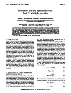
The full-field speckle analyzer with the bundle-based light delivery unit [Fig. 3(a)] was applied to monitor laser-mediated modification of cartilage. The laser beam emitted by a single-mode He–Ne laser (1) 共632.8 nm linear polarization, 5 mW output power) was expanded by a 50⫻ telescopic system (2) and fell on the surface of a sample (3) of ex vivo bovine nasal septum cartilage. The samples under study were taken from freshly slaughtered animals and prepared as square pieces with 25 mm ⫻ 25 mm lateral dimensions and 1.5 mm thickness. The diffuse reflected light was collected by the imaging lens (4) (focal length of 16 mm and diameter of 6 mm), and the speckle-modulated image of the area of interest on the sample surface was formed on the input tip of a fiber-optic bundle (5). The bundle with the hexagonal packing of fibers (the fiber diameter is 25 m and the separation between the fibers in a row is ⬇3 m) was 1.2 m in length. Light from the output tip was detected by a CCD sensor (6) (Sony ICX415 chip-based unit, 25 frames兾s frame rate, 10 bit analog-to-digital (conversion) coaxially placed at a distance of 300 mm from the output tip. In the experiments, the capture of subframes (450 pixels ⫻ 450 pixels) at the frame rate of 25 frames兾s was used. This allowed us to set the constant value of exposure time equal to 20 ms with the value of the CCD readout time approximately equal to 22 ms. To characterize the optical system of the bundlebased speckle monitor, we estimated the average speckle size in the image plane of lens 4 and in the detector plane. A Teflon plate 共10 mm thick) was placed in the object plane as a source of backscattered speckle-modulated light. The pixel size of the CCD array is 8.3 m ⫻ 8.3 m, and the average speckle size in the detection plane was evaluated as ⬇65 m. Thus, the average coherence area in the detection plane covered approximately 50 CCD pixels. Also, the quantification of the intermediate speckle pattern was carried out by positioning the CCD module without a lens instead of the input tip of the fiber-optic bundle. In this case the average speckle size appeared comparable with the pixel size of the CCD array, which resulted in distortions of detected intensity distributions (due to the effect of integration of speckle intensity fluctuations over the area of each pixel). Statistical analysis of the captured speckle pattern allowed us to estimate the correlation radius of intensity fluctuations in the image plane of lens 4 as ⬇11 m. Thus, we can conclude that the average 4484
APPLIED OPTICS 兾 Vol. 45, No. 18 兾 20 June 2006
Fig. 3. (a) Scheme of the experimental setup with the bundlebased full-field analyzer. 1, He–Ne laser; 2, telescopic system; 3, object; 4, image-transferring element; 5, fiber-optic bundle; 6, CCD detector; 7, erbium laser; 8, light-delivering fiber; 9, thermograph. (b) Scheme of the experimental setup with the LASCA modality. 1, He–Ne laser; 2, telescopic system; 3, object; 4, CCD camera; 5, erbium laser; 6, light-delivering fiber; 7, thermograph, uppergraph; the temperature distributions across the surface of a treatment zone at various stages of modification: 1, tissue heating; 2, quasi-stationary phase of modification.
speckle size in the image plane did not exceed ⬇22 m and was comparable with the diameter of the fibers in the fiber-optic bundle. Thermal treatment of the samples was carried out by radiation of an erbium fiber-optic laser (7)
共 ⫽ 1.56 m, adjustable output power up to 5 W) arranged with multimode quartz fiber (8) 共0.6 mm in diameter) as a radiation-delivering unit. The diameter of the treatment zone on the sample surface was approximately equal to 15 mm. The wavelength of 1.56 m provides the bulk absorption of laser radiation by tissue.9 In our experiments the recording of sequences of speckle-modulated images with the use of the CCD camera was started synchronously with the acquisition of the temperature of tissue surface with the use of the thermograph, whereas the laser treatment of tissue was started manually after the beginning of image recording. Such experimental procedures resulted in the random delay (typically, ⬃2–4 s) of the alterations in speckle contrast and tissue temperature with respect to the initial moment of each experiment. This feature is not important for further data analysis because of synchronization of the temperature and contrast time series and application of delay-independent procedures of data processing (in particular, estimates of the contrast decrement). For comparison, a similar experiment was carried out with the conventional LASCA modality [Fig. 3(b)] arranged with the CCD sensor of the same type and high-quality imaging lens (LMZ13A5M). The magnification of the tissue surface image at the CCD sensor was ⬇0.4⫻. Similar to the bundle-based speckle instrument, the average speckle size in the detector plane was estimated for the LASCA modality. In this case the focal length and the aperture of the CCD camera objective [Fig. 3(b)] equal to 75 and 5 mm were chosen to provide the optimal detection conditions (the sufficient brightness of speckle patterns and the appropriate speckle size; see Ref. 9 for more details). The speckle size estimated using the Teflon plate was equal to ⬇15 m. An experiment with the conventional LASCA modality was carried out only with the single value of IR laser power 共4.5 W兲 to demonstrate the higher efficiency of the bundle-based speckle instrument for cartilage modification monitoring. Simultaneously with the capture of dynamic speckle patterns, the spatial–temporal distributions of the tissue surface temperature were monitored with the use of a thermograph (IRTIS-200, manufactured in Russia). Typical profiles of the instantaneous temperature, which correspond to different stages of cartilage modification, are illustrated by the upper graph in Fig. 3(b). Analysis of speckle dynamics in the course of modification has shown the pure boiling type of speckle motions in the detector plane (without any noticeable translations). Characterization of the dynamics type can be carried out using the analysis of the normalized spatial–temporal correlation function of speckle intensity fluctuations in the detector plane: g2共⌬x2, ⌬y2, 兲 ⫽ 具关I共x2 ⫹ ⌬x2, y2 ⫹ ⌬y2, t ⫹ 兲 ⫺ 具I典兴 ⫻ 关I共x2, y2, t兲 ⫺ 具I典兴典兾关具I2典 ⫺ 具I典2兴.
With the zero time lag ⫽ 0, the 1兾e decay of g2共⌬x2, ⌬y2, 0兲 characterizes the average speckle size 具rs典 ⬇ 2冑共⌬x2兲2 ⫹ 共⌬y2兲g2⫽1兾e2. The presence of a noticeable translational component in speckle dynamics should manifest itself in the appearance of a visible correlation peak for the given sufficiently nonzero value of 共 ⬎ c兲. This peak should be shifted in the coordinate plane 共⌬x2, ⌬y2兲 to the position 共x2, y2兲, where x2y2 are the x2 and y2 components of the speckle translation velocity. Figure 4(a) displays the normalized temporal correlation functions g2共兲 of the detected optical signal at various stages of modification; spatial correlations of captured speckle patterns are illustrated in Fig. 4(b) for the cases of the same frame 关g2共⌬x2, ⌬y2, 0兲兴 and two frames 关g2共⌬x2, ⌬y2, 兲兴 captured with the time delay slightly exceeding the speckle decorrelation time in the detector plane [Fig. 4(c)]. Note the absence of any correlation peak in the latter case. This indicates the absence of any remarkable translational dynamics of speckles in the detector plane. Also, the direct observation of the sequences of captured speckle patterns showed only the boiling of speckles at the stages of tissue heating and modification. The boiling type of speckle dynamics is obviously caused by the absence of macroscopic regular motions of scattering sites in the thermally treated tissue. Because of the backscattering geometry and contributions of multiply scattered diffuse components, the detected light is characterized by the significantly more broadband intensity fluctuations in comparison with the case of transillumination monitoring of thermally treated ex vivo cartilage.9 The remarkable blur of speckle patterns occurs at the frame rate of 25 frames兾s (with the above-mentioned value of the exposure time equal to 20 ms). Consequently the value of VT˜ was estimated for each frame in the recorded sequence without any additional time averaging (as it was carried out in Ref. 9). Figure 5 displays the dependencies of the temperature in the central region of the treatment zone and the normalized speckle contrast VT˜共t兲兾VT˜共0兲 on the time lapse t in the course of the treatment procedure provided with the different values of the output power of IR radiation. The appearance of some artifacts on the experimental dependencies plotted in Fig. 5(b) [in particular, the minimal value of contrast at 4.8 W appears later than the minimal values of VT˜共t兲兾VT˜共0兲 at 4.2 and 4.5 W] is related to the arbitrary shift of the origin for each data sequence because of the abovementioned random delay in the start of laser treatment. As mentioned above, this circumstance is insufficient for further data analysis. Similar to previously reported results on the transillumination speckle monitoring of cartilage modification,9 the looplike behavior of VT˜共t兲兾VT˜共0兲 occurs with the changes in tissue temperature. This peculiarity is related to the observed nonmonotonic behavior of the contrast: The abrupt decrease in ˜ V at the stage of tissue heating from room temperature to ⬇65 °C changes to a slight increase at the stage of 20 June 2006 兾 Vol. 45, No. 18 兾 APPLIED OPTICS
4485
Fig. 4. (a) Normalized temporal correlation functions of speckle intensity fluctuations in the detector plane. 1, tissue heating (the temperature in the central region of the treatment zone is ⬃50 °C); 2, the quasi-stationary phase of tissue modification (the temperature is ⬃68 °C). (b) and (c) Normalized spatial correlation functions of dynamic speckle patterns in the detector plane: (b) the normalized spatial autocorrelation function, (c) the cross-correlation function for two frames with the time delay ⌬ approximately equal to 2.5 c,detector. The scale for c graphs. (b) and (c) are the same.
4486
APPLIED OPTICS 兾 Vol. 45, No. 18 兾 20 June 2006
Fig. 5. (a) Dependencies of the tissue temperature in the central region of the treatment zone on the time lapse. Inset displays the looplike behavior of V˜T共T兲 (the output power of the erbium laser is 4.5 W; I, tissue heating; II, modification; III, thermal relaxation). (b) The dependencies of VT˜ on the time lapse (bundle-based analyzer). (c) The same as in (b) but for the LASCA modality (IR laser output power is 4.5 W; low-amplitude fluctuations related to nonergodic behavior of the speckle-modulated image are marked by arrows).
Fig. 6. Values of ln共⌬V˜T兲 obtained with the bundle-based analyzer (1–3) and the LASCA modality (4) and plotted against the inverse absolute temperature in the central region of the treatment zone. The output power of the erbium laser is 1, 4.5 W; 2, 4.2 W; 3, 4.8 W; 4, 4.5 W.
modification [Fig. 5(b)]. This results in the larger value of ˜ V at the final phase of modification (when the treatment is terminated) in comparison with the initial phase (when T approaches ⬇65 °C) and, correspondingly, in the looplike form of ˜ V共T兲 dependence. This looplike feature is displayed by the inset in Fig. 5(a). Figure 5(c) shows the dependence VT˜共t兲兾VT˜共0兲 obtained with the LASCA modality. In calculation of the speckle contrast from raw images obtained with the LASCA unit, the area of the analyzed zone of speckle-modulated images was 0.7 mm ⫻ 0.7 mm, which corresponded to the analyzed area of the central part of the treatment zone with a size of 1.75 mm ⫻ 1.75 mm. An area of same size was analyzed with the bundle-based modality. 4. Discussion of Results
Considering the contrast decrement ⌬VT˜共t兲 ⫽ VT˜共t兲 ˜兲, we can assume the first-order ap⫺VT˜共t ⫹ T proximation of ⌬VT˜ for the small values of ˜ ˜兲兾dT ˜兴兾2冑g 共T ˜兲 ⫺ 1其T ˜ ⬇ K共T ˜ 兾 兲 T兾c: ⌬VT˜ ⬇ 兵关dg2共T 2 c ˜ and, respectively, c ⬀ 1兾⌬VT. The factor K is controlled by the asymptotic behavior of the temporal correlation function of intensity in the object plane and the transfer properties of the bundle-based analyzer as well. The static character of captured speckle patterns at room temperature and an abrupt increase in the contrast decrement with the increasing tissue temperature allow us to consider the decay in c at the first stage of tissue modification as a thermally activated process with a certain value of the activation energy. Figure 6 displays the values of ln共⌬VT˜兲 estimated for the pairs of sequential frames and plotted against the instantaneous inverse absolute temperature in the central part of the treatment zone. This plot corresponds to data obtained with the conventional
LASCA modality (with the single value of output power of an IR laser) and the bundle-based speckle analyzer. For the relatively narrow interval of T, the plotted data can be approximated with reasonable accuracy by the linear dependencies of ln共⌬VT˜兲 on T⫺1, which are also shown in Fig. 6. This allows us to assume the Boltzmann-like dependence of the inverse speckle decorrelation time on the absolute temperature of modified tissue: c,o⫺1 ⬇ K1 exp共⫺Ea兾RT兲, where the parameter K1 depends on various factors such as the frequency factor of the basic thermally activated process with the activation energy of Ea and the transfer properties of the speckle-imaging system. Note that both data sets are characterized by the close values of Ea 共61 ⫾ 4.5 kJ兾mol for data obtained with the LASCA modality and 64 ⫾ 6 kJ兾mol for the bundle-based speckle analyzer, respectively) but significantly differ in the values of the normalization parameter 共K1,LASCA兾K1,bundle ⬇ 2.9兲. This difference is obviously caused by a difference in the transfer properties of the conventional LASCA system and the bundlebased speckle analyzer. In the latter case the influence of the transfer properties of the light-delivering channel is more expressed because of the cascade transformation of speckle patterns and the lower quality of the image-transferring component in comparison with the examined LASCA modality. The above-considered linear and quadratic models of d decay predict the extreme value of the ratio c,o兾c,detector in the case of a sequential two-stage transformation of boiling speckle patterns and varies from ⬇1.74 (the quadratic model) to ⬇3.24 (the linear model). Thus, under the semiqualitative assumptions used in theoretical analysis and without knowledge of correlation properties of dynamic speckles in the object plane, the experimentally obtained ratio K1,LASCA兾K1,bundle ⬇ 2.9 reasonably agrees with the above-presented values. Let us consider some probable mechanisms of the laser-induced cartilage modification, which cause dynamic light scattering in the treated tissue. One mechanism can be related to the temperaturedependent changes in the rate of scatter diffusion in the treated volume. For many condensed media, the dependence of the diffusion coefficient on the temperature is described as D ⫽ D0 exp共⫺E0兾RT兲, where E0 is the activation energy of transition of the scattering particle from one stable position to another one. Among other factors, the contrast decrement is determined by the average displacement of scatters L ˜⬀ over the observation time ˜ T [i.e., ⌬VT˜ ⬀ L ⬀ 冑DT exp共⫺E0兾2RT兲]. Thus, this consideration leads to the Boltzmann dependence of the contrast decrement on the characteristic temperature of tissue. Another probable mechanism is related to the thermally induced alterations in tissue structure, which are accompanied by the generation of new effective scattering sites in the treated volume. Assuming the monomolecular process of thermally induced transformation of one of the basic components of cartilage 20 June 2006 兾 Vol. 45, No. 18 兾 APPLIED OPTICS
4487
and the insignificant changes in its concentration during the observation time ˜ T, we can obtain the contrast decrement proportional to the temperature.
.
˜ is ˜ (where N dependent transformation rate ⌬VT˜ ⬀ N the rate of transformation of scattering centers in the probed volume; see Appendix A). With these assump. ˜ will be controlled by the factor tions, the value of N exp共⫺Ea兾RT兲. Thus, both mechanisms lead to the Boltzmann-like dependence of ⌬VT˜ on T. The obtained effective activation energy can correspond to the thermally induced transfer of tissue liquid, which causes the changes in local configurations of scattering sites, or to the low-energy modification of the collagen matrix. Note that the thermal denaturation of collagen itself can be excluded from the considered mechanisms, which causes the pronounced structural changes at the initial phase of modification. Typically, the expressed denaturation of collagen in cartilage occurs at temperatures above ⬃70 °C.22 However, the thermally induced dynamics of the collagen matrix can be related to the partial denaturation of proteoglycan aggregates (PGAs)23 or to the conformational transitions in glycosaminoglycans as one of the PGA components. Note that each disaccharide unit in the glycosaminoglycan macromolecule is stabilized by three hydrogen bonds24 with a total energy of ⬃60–75 kJ兾mol,25 which is close to the found value of the effective activation energy. The comparison of VT˜共t兲 dependencies obtained with the LASCA modality and the bundle-based analyzer allows us to emphasize the expressed nonmonotonic behavior of the exposure-dependent speckle contrast in the first case. The stationary phase of thermal modification, which corresponds to the plateaulike region of the T(t) curve, and the phase of thermal relaxation (after switching off the erbium laser) are featured by the remarkable oscillations in VT˜共t兲 [Fig. 5(c)]. Such nonmonotonic behavior is related to macroscopically heterogeneous nonergodic spatial distributions of modulating speckles in the image plane, which are associated with inhomogeneities of the cartilage structure. Similar statistical inhomogeneities of image-modulating speckles varying in the course of tissue modification were observed in the case of transillumination monitoring.9 On the contrary, dynamic speckle patterns obtained with the use of the bundle-based modality exhibit significant suppression of the contrast oscillations due to the specific conditions of speckle formation (randomphase modulation of partial optical signals sampled by the bundle and their mixing in the diffraction zone; these features result in the formation of ergodic dynamic speckle patterns in the detector plane). This gives us the opportunity for more detailed analysis of peculiarities in behavior of the exposure-dependent speckle contrast at the various stages of thermal treatment. In particular, the existence of inverse peaks on VT˜共t兲 dependencies [marked by arrows in Fig. 5(b)] and dramatic changes in the contrast behavior at the quasi-stationary phase of modification 4488
APPLIED OPTICS 兾 Vol. 45, No. 18 兾 20 June 2006
(the trend to increase with the increasing tissue temperature) can be emphasized. The latter peculiarity is obviously manifested for data obtained with the higher values of treatment power [curve in Fig. 5(b), which corresponds to P ⫽ 4.8W] and is correlated with previously reported looplike behavior of VT˜共t兲 in the transillumination mode.9 The presumable interpretation of such behavior is related to the change in a predominating mechanism of dynamic light scattering in the thermally treated tissue at temperatures above 70 °C (from generation of new scattering centers due to partial denaturation of the basic components of cartilage structure toward migration of accumulated denaturation products in the treatment zone). Note that in the case of too high a value of treatment power [curve in Fig. 5(a), which corresponds to P ⫽ 4.8 W], the heat consumption in the treated tissue is insufficient, and overheating of the tissue takes place [see region in Fig. 5(a), which is marked by arrow]. This situation is characterized by the remarkably lower values of VT˜ at the stage of thermal modification itself [the time interval from ⬇20 to ⬇40 s, Fig. 5(b)] in comparison with the smoother regimes of the tissue treatment (curves responding to P ⫽ 4.2 W and 4.5 W, respectively). Thus, we can summarize that the analysis of the exposure-dependent speckle contrast in the backscattering mode with the developed bundle-based analyzer is the appropriate tool for the monitoring of the laser-mediated cartilage modification. The specific conditions of speckle pattern formation in the developed full-field speckle unit make it possible to obtain the statistically reliable estimates of the instantaneous values of a diagnostical parameter (the exposure-dependent contrast) by processing the small fragments of speckled images (subframes). In turn, this will provide a sufficient decrease in the data processing time as a further step toward the design of a real-time speckle monitor, which is appropriate for clinical conditions. 5. Conclusions
We have examined the potential of speckle-contrast monitoring of laser-mediated cartilage modification by means of the developed full-field speckle analyzer, which is featured by application of the fiber-optic bundle as the light-transferring unit and by analysis of time-integrated dynamic speckles in the diffraction zone. These features provide the suppression of lowfrequency noiselike fluctuations of the exposuredependent speckle contrast, which are typical in the case of application of the conventional LASCA technique (the results of the experiment were for backscattered light detection and an erbium laser output of 4.5 W, the results of our previous study were for the transillumination mode with various levels of IR laser output power9). These fluctuations are caused by a sufficient nonergodic character of dynamic speckle-modulated images of the surface of modified tissue. The additional transformation of the specklemodulated optical field by the detection unit on the base of a fiber-optic bundle leads to the formation of
an ergodic dynamic speckle pattern in the detector plane due to mixing on each detector pixel of the optical signals from a large number of speckles across the nonergodic speckle-modulated image. Some analogies can be drawn between the discussed technique and the two-cell technique.11–14 Because of the partial suppression of these noiselike fluctuations, a more detailed picture of the thermally induced alterations in cartilage structure can be obtained with the use of the developed speckle analyzer. It was found that the changes in the specklecontrast decrement due to an increase in the tissue temperature up to ⬇65 °C can be approximated by the Boltzmann-like law with an activation energy ⬇共6.1–6.4兲 ⫻ 104 J兾mol, which is close to the energy of conformational transitions in glycosaminoglycan macromolecules. Above 65 °C, the treatment of the tissue is accompanied by dramatic alterations in thermally induced speckle dynamics due to the change in the dynamic scattering mechanism. Appendix A
With the discrete scattering model (see, e.g., Refs. 26 and 27), the correlation function of the scattered field fluctuations, which are induced by alterations in the ensemble of static scattering sites due to the appearance and disappearance of a small part of them, can be presented as
ⱍG1共兲ⱍ ⫽
冏冓冋兺 ˜ N
i⫽1
⫹
˜ N⬘()
兺
k⫽1
Ei exp共ji兲
册冋 兺 册 冔冏
Ek⬘ exp共jk⬘兲
˜ ˜ N⫺N⬘() i⫽1
Ei exp共ji兲
*
,
(A1)
where the complex amplitude of the multiply scattered field in the observation point is considered as the sum of statistically independent partial contributions scattered many times in the probed volume and ˜ ⬘共兲 is the number of contributions that are changed N due to the disappearance of part of old scattering centers and the appearance of new ones (the asterisk denotes the complex conjugation). With the quasi˜ adiabatic approach (variations in the total number N of partial contributions and in the average value of Ei over the time interval can be neglected), the normalized temporal correlation function of the scattered field fluctuations can be written as
ⱍg1共兲ⱍ ⫽ ⱍG1共兲ⱍ兾具I典 ⫽ 1 ⫺ ˜共兲,
(A2)
˜⬘共兲兾N ˜ is the relative weight of the where ˜ 共兲 ⫽ N modified partial contributions, which can be obtained in analytical or numerical form on the basis of the analysis of the path-length statistics of multiple scattered light in the probed volume. In the case of large ˜ and deep phase modulation of the probe coherent N light by the scattering system, 2 ⬇ 1 ⫺ 2 ˜共兲→0. 关g2共兲 ⫺ 1兴→0 ⫽ ⱍg1共兲ⱍ→0
(A3)
The first cumulant dg 共兲兾d→0 ⫽ ⫺2d˜共兲兾d ⬃ c⫺1 is . 2 ˜ of thermally induced transforrelated to the rate N mation of scattering centers in the probed volume. This work was supported by grants from the Russian Foundation for Basic Research (04-02-16533, 0402-97203, and 04-02-16743). References 1. A. F. Fercher and J. D. Briers, “Flow visualization by means of single-exposure speckle photography,” Opt. Commun. 37, 326 – 329 (1981). 2. J. D. Briers and S. Webster, “Quasi-real time digital version of single-exposure speckle photography for full-field monitoring of velocity or flow fields,” Opt. Commun. 116, 36 – 42 (1995). 3. J. D. Briers and S. Webster, “Laser speckle contrast analysis (LASCA): a non-scanning, full-field technique for monitoring capillary blood flow,” J. Biomed. Opt. 1, 174 –179 (1996). 4. J. D. Briers and A. F. Fercher, “Retinal blood-flow visualization by means of laser speckle photography,” Invest. Ophthalmol. Visual Sci. 22, 255–259 (1982). 5. G. Richards and J. D. Briers, “Capillary blood flow monitoring using laser speckle contrast analysis (LASCA): improving the dynamic range,” in Proc. SPIE 2981, 160 –171 (1997). 6. A. F. Fercher, M. Peukert, and E. Roth, “Visualization and measurement of retinal blood flow by means of laser speckle photography,” Opt. Eng. 25, 731–735 (1986). 7. J. D. Briers, G. Richards, and X. W. He, “Capillary blood flow monitoring using laser speckle contrast analysis (LASCA),” J. Biomed. Opt. 4, 164 –175 (1999). 8. A. K. Dunn, H. Bolay, M. A. Moskowitz, and D. A. Boas, “Dynamic imaging of cerebral blood flow using laser speckle,” J. Cereb. Blood Flow Metab. 21, 195–201 (2001). 9. D. A. Zimnyakov, D. N. Agafonov, A. P. Sviridov, A. I. Omel’chenko, L. V. Kuznetsova, and V. N. Bagratashvili, “Speckle-contrast monitoring of tissue thermal modification,” Appl. Opt. 41, 5989 –5996 (2002). 10. G. E. Nilsson, E. G. Salerud, N. O. T. Stromberg, and K. Wardell, “Laser Doppler perfusion monitoring and imaging,” in Biomedical Photonics Handbook, T. Vo-Dinh, ed. (CRC Press, 2003), pp. 15-1–15-24. 11. V. Viasnoff, F. Lequeux, and D. J. Pine, “Multispeckle diffusing-wave spectroscopy: a tool to study slow relaxation and time-dependent dynamics,” Rev. Sci. Instrum. 73, 2336 – 2344 (2002). 12. S. E. Skipetrov and R. Maynard, “Dynamic multiple scattering of light in multilayer turbid media,” Phys. Lett. A 217, 181– 185 (1996). 13. S. Romer, F. Scheffold, and P. Schurtenberger, “Sol-gel transition of concentrated colloidal suspensions,” Phys. Rev. Lett. 85, 4980 – 4983 (2000). 14. F. Scheffold, S. E. Skipetrov, S. Romer, and P. Schurtenberger, “Diffusing-wave spectroscopy of non-ergodic media,” Phys. Rev. E 63, 061404 (2001). 15. L. Mandel and E. Wolf, Optical Coherence and Quantum Optics (Cambridge U. Press, 1995). 16. D. A. Zimnyakov, J. T. Oh, Yu. P. Sinichkin, V. A. Trifonov, and E. V. Gurianov, “Polarization-sensitive speckle spectroscopy of scattering media beyond the diffusion limit,” J. Opt. Soc. Am. A 21, 59 –70 (2004). 17. T. Yoshimura, “Statistical properties of dynamic speckles,” J. Opt. Soc. Am. A 3, 1032–1054 (1986). 18. T. Yoshimura, K. Nakagawa, and N. Wakabayashi, “Rotational and boiling motion of speckles in a two-lens imaging system,” J. Opt. Soc. Am. A 3, 1018 –1022 (1986). 19. H. Z. Cummins and E. R. Pike, eds., Photon Correlation and 20 June 2006 兾 Vol. 45, No. 18 兾 APPLIED OPTICS
4489
20.
21. 22.
23.
Light-Beating Spectroscopy, NATO Advanced Study Institute Series B: Physics (Plenum, Press, 1974). D. A. Zimnyakov, J. D. Briers, and V. V. Tuchin, “Speckle technologies for monitoring and imaging of tissues and tissuelike phantoms,” in Handbook of Optical Medical Diagnostics, V. V. Tuchin, ed. (SPIE Press, 2002), pp. 987–1036. N. Takai, T. Iwai, and T. Asakura, “Correlation distance of dynamic speckles,” Appl. Opt. 22, 170 –177 (1983). N. Yu. Ignatieva, V. V. Lunin, S. V. Averkiev, A. F. Maiorova, V. N. Bagratashvili, and E. N. Sobol, “DSC investigation of connective tissues treated by IR-laser radiation,” Thermochim. Acta 422, 43– 48 (2004). A. M. Jamieson, J. Blackwell, H. Reihanian, H. Ohno, R. Gupta, D. A. Carrino, A. I. Caplan, L. H. Tang, and L. C. Rosenberg, “Thermal and solvent stability of proteoglycan ag-
4490
APPLIED OPTICS 兾 Vol. 45, No. 18 兾 20 June 2006
24.
25. 26.
27.
gregates by quasielastic laser light-scattering,” Carbohydr. Res. 160, 329 –341 (1987). J. E. Scott, “Secondary and tertiary structures of hyaluronan in aqueous solution. Some biological consequences,” Science of Hyaluronan Today, V. C. Hascall and M. Yanagishita, eds., http://www.glycoforum.gr.jp/science/hyaluronan/hyaluronanE. html (1998). G. C. Pimentel and A. L. McClennan, The Hydrogen Bond (Freeman, 1960), p. 189. G. Maret and P. E. Wolf, “Multiple light scattering from disordered media. The effect of Brownian motions of scatterers,” Z. Phys. B 65, 409 – 413 (1987). F. C. MacKintosh and S. John, “Diffusing-wave spectroscopy and multiple scattering of light in correlated random media,” Phys. Rev. B 40, 2382–2406 (1989).