Osteoporos Int (2011) 22:1581–1591 DOI 10.1007/s00198-010-1371-6
ORIGINAL ARTICLE
Evidence for an additional effect of whole-body vibration above resistive exercise alone in preventing bone loss during prolonged bed rest D. L. Belavý & G. Beller & G. Armbrecht & F. H. Perschel & R. Fitzner & O. Bock & H. Börst & C. Degner & U. Gast & D. Felsenberg
Received: 2 March 2010 / Accepted: 2 August 2010 / Published online: 3 September 2010 # International Osteoporosis Foundation and National Osteoporosis Foundation 2010
Abstract Summary The addition of whole-body vibration to highload resistive exercise may provide a better stimulus for the reduction of bone loss during prolonged bed rest (spaceflight simulation) than high-load resistive exercise alone. Introduction Prior work suggests that the addition of whole-body vibration to high-load resistive exercise (RVE) may be more effective in preventing bone loss in spaceflight and its simulation (bed rest) than resistive exercise alone (RE), though this hypothesis has not been tested in humans. Methods Twenty-four male subjects as part of the 2nd Berlin Bed Rest Study performed RVE (n=7), RE (n=8) or no exercise (control, n=9) during 60-day head-down tilt bed rest. Whole-body, spine and total hip dual X-ray absorptiometry (DXA) measurements as well as peripheral quantitative computed tomography measurements of the tibia were conducted during bed rest and up to 90 days afterwards. Electronic supplementary material The online version of this article (doi:10.1007/s00198-010-1371-6) contains supplementary material, which is available to authorized users. D. L. Belavý (*) : G. Beller : G. Armbrecht : O. Bock : H. Börst : C. Degner : U. Gast : D. Felsenberg Center for Muscle and Bone Research, Charité Universitätsmedizin Berlin, Hindenburgdamm 30, 12203 Berlin, Germany e-mail:
[email protected] F. H. Perschel : R. Fitzner Zentralinstitut für Laboratoriumsmedizin und Pathobiochemie, Charité Universitätsmedizin Berlin, Hindenburgdamm 30, 12203 Berlin, Germany
Results A better retention of bone mass in RVE than RE was seen at the tibial diaphysis and proximal femur (p≤ 0.024). Compared to control, RVE retained bone mass at the distal tibia and DXA leg sub-region (p≤0.020), but with no significant difference to RE (p≥0.10). RE impacted significantly (p=0.038) on DXA leg sub-region bone mass only. Calf muscle size was impacted similarly by both RVE and RE. On lumbar spine DXA, whole-body DXA and calcium excretion measures, few differences between the groups were observed. Conclusions Whilst further countermeasure optimisation is required, the results provide evidence that (1) combining whole-body vibration and high-load resistance exercise may be more efficient than high-load resistive exercise alone in preventing bone loss at some skeletal sites during and after prolonged bed rest and (2) the effects of exercise during bed rest impact upon bone recovery up to 3 months afterwards. Keywords Countermeasures . Inactivity . Microgravity . Training . Weightlessness
Introduction Developing optimal exercise modalities to maintain or increase bone formation is an important priority not only for the management of osteoporosis but also for longduration manned spaceflight. With a view to long-duration spaceflights to Mars, a priority of space agencies worldwide is the development of appropriate countermeasures against bone loss. Prior work suggests that up to a loss of 1.5% per month of bone occurs during prolonged exposure to weightlessness, depending on body region considered [1].
1582
A greater loss of bone occurs in the regions of the skeleton involved in postural activities (e.g. spine, pelvis and legs) [1–3]. Evidence [4, 5] from bed rest studies, a methodology commonly used to simulate the effects of spaceflight [6], suggests that exercise countermeasures that include highload resistive exercise for the lower limbs and spine would be more effective in reducing bone loss in spaceflight than low load/low impact endurance exercise [1, 7, 8]. This suggestion corresponds to findings from animal studies [9, 10] that loading of bone with larger forces (such as during resistance exercise), leading to greater bone strain [11], generates a greater stimulus for osteogenesis. Evidence also suggests, however, that loading applied to bone at higher strain rates [12–14] or at higher loading frequencies [15] can also provide an osteogenic stimulus. As it is difficult for humans to conduct voluntary movement at high rates, such higher loading rates can be applied externally via mechanical vibration devices [16–18]. Importantly, however, the addition of low-magnitude, high-frequency vibration to slower, high-magnitude loading (such that experienced during voluntary resistance exercise) can have a greater effect on bone formation than either vibration alone or the high-magnitude strain loading alone [19, 20]. This leaves open the possibility that high-load resistance exercise, combined with higher frequency vibration (RVE) may present a more potent stimulus for preventing bone loss in spaceflight or prolonged bed rest than high-load resistance exercise alone. Prior work in the 1st Berlin Bed Rest Study [5, 21] has shown that RVE can reduce bone loss during prolonged bed rest. Whilst animal studies may strongly suggest that this countermeasure would be more effective than high-load resistance exercise alone (RE), evidence in support of this hypothesis in humans is lacking. In the current work, we wished to address this hypothesis by examining RVE in comparison to RE and no countermeasure (CTR) on changes in bone in the spine and lower quadrant during 60-day bed rest in the 2nd Berlin Bed Rest Study.
Osteoporos Int (2011) 22:1581–1591
computed tomography >120 mg/ml). Subjects attended the facility for 9 days of baseline data collection (BDC-9 to BDC1), underwent 60 days of 6° head-down tilt bed rest (HDT), remained in the facility for 7 days after re-ambulation (R+1 to R+7) and returned to the facility 14, 30 and 90 days after re-ambulation (R+14, R+30, R+90) for follow-up testing. The study was approved by the ethical committee of the Charité Universitätsmedizin Berlin, and the radiological examinations were approved by the Bundesamt für Strahlenschutz. All subjects gave their informed written consent prior to participation in the study. Sample size estimates based upon data from an earlier bed rest study [5, 23] suggested that for the comparison between RVE and control, six subjects were necessary per group and that for the comparison between RVE and RE, nine subjects per group were required. Based on psychological advice, subjects were paired into “room-mates” prior to the beginning of the study. In the evening 2 days before the beginning of bed rest (BDC-2) after all subjects completed countermeasure exercise familiarisation and maximal force testing, the room-mate pairs were randomised to one of three groups: one that performed resistive exercises with whole-body vibration during bed rest (RVE), one that performed resistive exercise only (RE) and one that performed no exercise and served as a control group (CTR). One subject who was randomised to the RVE group could not perform the countermeasure programme due to exercise induced headache but remained as a CTR subject. Another subject (RE group) withdrew from the study at HDT30 for medical reasons [22]. Thus, nine subjects comprised the CTR group, eight in the RE group (until HDT30; n=7 beyond this) and seven in the RVE group. The baseline characteristics of these subjects are given in Table 1. Two subjects (one RE, one CTR) did not attend follow-up examinations at R+90. Countermeasure exercise during bed rest Details of the countermeasure programme have been published elsewhere [22]. However, countermeasure exer-
Materials and methods Table 1 Baseline beginning of bed rest subject characteristics
Study characteristics Details of the study protocol have been published elsewhere [22]; however, in brief, 24 medically and psychologically healthy males participated in the 2nd Berlin Bed Rest Study which was conducted at the Charité Campus Benjamin Franklin in Berlin, Germany, by the Centre for Muscle and Bone Research. The subjects had no history of bone or joint disease and were not osteopenic or osteoporotic (i.e. lumbar spine and hip dual X-ray absorptiometry (DXA) more than −1.5 SD and lumbar spine trabecular bone density on quantitative
Group
Number
Age (years)
Weight (kg)
Height (cm)
CTR RE RVE
9 8 7
33.1 (7.8) 31.1 (5.1) 32.2 (10.4)
80.6 (5.2) 75.0 (12.8) 81.5 (6.2)
181.3 (6.0) 179.3 (7.7) 179.6 (5.8)
Values for age, weight and height are mean (SD). There were no differences between groups for any of these variables (F all<1.2, p> 0.33). One subject (RE group) dropped out after the 30th day of bed rest for medical reasons unrelated to the investigations reported here CTR inactive control group, RE resistive exercise-only group, RVE resistive exercise with whole-body vibration group
Osteoporos Int (2011) 22:1581–1591
cise was performed 3 days a week with one session of exercise performed on each day. Prior to bed rest, familiarisation sessions and then maximal force testing were performed to determine initial force level settings at the beginning of bed rest. The RVE group performed the same exercise programme as the RE group except with the addition of whole-body vibration applied at the feet (Fig. 1). After a warm-up exercise (bilateral squats at 50% of maximal force, eight repetitions), the following exercises were performed: &
&
Bilateral squats were begun at 75% of pre-bed rest maximum. Exercises were performed until exhaustion and force was progressed over the course of the study. In the RVE group, the vibration frequency was progressed from 20 to 24 Hz (amplitude 3.5–4 mm) from HDT1 to HDT5 and then maintained at this level for the rest of the study. Single leg heel raises were performed on the left and right legs from maximal plantarflexion to maximal dorsiflexion against a force equivalent to approximately 1.3 times their HDT1 body weight. The exercise was performed until exhaustion. Vibration frequency was set
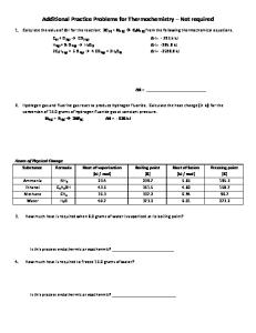
Fig. 1 Countermeasure exercise. Both the resistance exercise-only and resistance exercise with whole-body vibration groups performed their exercises on the specially designed Galileo Space exercise device. Subjects were positioned in head-down tilt on a movable platform with shoulder pads and hand grips preventing downward movement and permitting application of force via the platform. A pneumatic system generated the force, applied through the moveable platform, against which the subject needed to resist and move (via the shoulder pads and hand grips). The feet were positioned on either side of a platform which was set to vibrate in the RVE group. Subjects were given visual feedback of their actual and target position in the exercise via a monitor placed in the subjects’ field of view. As the force output was dictated by the exercise device, the feedback focussed on ensuring the subjects performing the exercise in the desired range of motion and at the desired speed. During the heel raises, the sport scientist monitored the range of movement and encouraged the subject to go to the end of range in each direction. Here the subject is performing a back extension exercise
1583
&
&
to 26 Hz (amplitude 3.5–4 mm) in the RVE group. The exercise was progressed as appropriate. Double leg heel raises were performed in the same manner as for single leg heel raises except that the resistive force was set to approximately 1.8 times body weight. The exercise was performed until exhaustion and progressed as appropriate. Vibration frequency was set to 26 Hz (amplitude 3.5–4 mm) in the RVE group. Back and toe raise with their feet positioned on the platform, subjects extended their hips and lumbar spine, dorsiflexed their ankles and maintained their knee at full extension. Subjects were required to maintain this position for 60 s; 1.5 times body weight was applied during the exercise. Vibration frequency was set to 16 Hz (amplitude 3.5–4 mm) in the RVE group. The exercise was not progressed.
The total duration of loading was thus 5 to 6 min per exercise session, with the entire training protocol requiring approximately 23 min per session including rest periods and changes in position for each exercise. A vibration amplitude of 3.5–4 mm was chosen as it was the maximum possible on the device: Higher vibration amplitude results in greater forces being imposed on the subject during exercise. Whole-body vibration frequencies for the squat exercise were chosen based on our prior experience that previously untrained people can more easily gain mastery of the manoeuvre with 20 Hz vibration. Progression of vibration frequency during squats was done to increase exercise intensity and depended upon the ability of the subject. A maximum vibration frequency of 26 Hz was chosen due to physiological time limitations of the muscle stretch reflex arc. At frequencies higher than 26 Hz, we assumed there to be inadequate time for muscle fibre contraction and relaxation. For the heel-raise exercises, 26 Hz vibration, as our experience, shows that subjects can typically master this (simpler) exercise already with higher vibration frequencies. For the back-raise exercise, a lower vibration frequency (16 Hz) was chosen in order to better target type I (slow twitch) muscle fibres, which are typically activated with lower frequency motor unit potentials Peripheral quantitative computed tomography An XCT 2000 (Stratec Medizintechnik, Pforzheim, Germany) was used to obtain peripheral quantitative computed tomography (pQCT) scans from the left leg as described previously [24, 25]. Scout views were generated in the frontal plane to identify the tibiotalar cleft to position the reference line. Sectional images were then obtained from the tibia at 4% (distal epiphysis) and 66% (diaphysis) of its length.
1584
Measurements were performed prior to bed rest (BDC; double measurement), during bed rest on HDT16, HDT30, HDT46, HDT59 and after bed rest on R+3, R+14, R+30 and R+90. As changes in body posture and fluid shifts can influence muscle cross-sectional area (CSA) measurements, subjects were positioned in the horizontal position (no headdown tilt) for at least 2 h prior to scanning. The integrated XCT 2000 software (version 6.20A) was used to analyse the pQCT images. The total bone mineral content (BMC) of the tibia was determined using a detection threshold of 180 mg/cm3 (contour mode 1, peel mode 1) for the epiphysis, and cortical BMC of the tibial diaphysis was assessed with a threshold of 710 mg/cm3 (cortical mode 1). Cortical BMC, and not total BMC, was assessed at the tibial diaphysis (66% position) as we expected intracortical remodelling which can be better assessed with a cortical bone measure. Also, in contrast to the distal tibia, at the 66% position, there is very little trabecular bone and a cortical bone measure is more appropriate. Gross anatomical muscle CSA was obtained from the scans taken at 66% of the tibia length (diaphysis) and was calculated as total bone area (detection threshold of 280 mg/cm3; contour mode 1, peel mode 1, filter 2: F03) subtracted from the combined muscle and bone area (detection threshold of 45 mg/cm3; contour mode 3, peel mode 1, filter 2: F03F05). Dual X-ray absorptiometry BMC (in grams) of the lumbar spine (L1–L4 in anteroposterior projection), the proximal femur (total) and whole body were measured using DXA with a Lunar iDXA (General Electric Company, Waukesha, WI, USA). Scans were performed according to the standard iDXA protocol 3 days prior to the start of bed rest (BDC-3; double measurement of the proximal femur and lumbar spine), on head-down tilt day HDT2 (whole body only), HDT30, HDT59 and on post-bed rest recovery (R+) days R+3, R+ 14, R+30 and R+90. Whole-body BMC data excluded the head sub-region. BMC in the arms, trunk and legs subregions was derived from the whole-body scan. All scanning and analyses were performed by the same operator to ensure consistency and followed standard quality control procedures. Similar to pQCT measurements, subjects remained recumbent in the horizontal position for at least 2 h prior to DXA scans.
Osteoporos Int (2011) 22:1581–1591
course of the day into larger collecting flasks by the nursing staff. Every 24 h (at 7 a.m.), the subject was requested again to void his urinary bladder, and the total volume over the 24-h period was measured. Aliquots were taken from each daily collection and stored at −80°C. To avoid precipitation of calcium salts, samples were acidified using HCl. Calcium levels in each sample were measured by atomic absorption spectrophotometry (Unicam 9200 PX, Philips, Kassel, Germany), and the total volume of calcium excreted in each 24-h period from BDC-2 until 5 days after bed rest (R+5) was calculated. Data processing and statistical analyses Where double baseline measurements were performed, these data were averaged prior to further analysis. Linear mixed-effects models [26] were used to assess changes with respect to subject group (RVE, RE and CTR), study date (with each day from the BDC phase and beyond factorised) and a group × study date interaction. As bone loss due to bed rest and the impact of countermeasures can be expected to continue beyond the end of bed rest [5], separate analyses of bed rest and recovery phases were not performed. Subject age, height and weight were included as linear covariates. Where necessary, allowances for heterogeneity of variance according to study date and/or subject group were applied. Random effects for each subject were permitted. An α of 0.05 was taken for statistical significance. Where significant effects were seen on analysis of variance (ANOVA), further linear mixedeffects models then considered differences between pairs of subject groups (i.e. RVE vs. RE, RVE vs. CTR and RE vs. CTR) using a similar modelling approach. Furthermore, subsequent analyses determined which study days differed from baseline (BDC) as well as differences between groups on a given study date. As multiple measurement sessions were undertaken on the same subjects, a Bonferroni adjustment was not performed, rather we looked for consistent significant differences across time points. Where a significant group × study date interaction was seen, analyses were also repeated using data expressed as percentage change compared to baseline (to ensure that subtle differences in baseline data did not influence the outcome). All analyses were performed in the “R” statistical environment (version 2.10.1, www.r-project.org).
Urine collection and analyses
Results
To assess changes in the amount of calcium excreted by the body, daily urine collections were performed. Urine was collected from 7 a.m. 2 days prior to the beginning of bed rest (BDC-2). Urine bottles were emptied regularly over the
At baseline, no differences were apparent between groups for any of the outcome measures (F all≤2.2, p all ≥0.13; Table 2). The main outcomes of the current study do not change if the subject who was unable to perform exercise
Osteoporos Int (2011) 22:1581–1591 Table 2 Baseline values of outcome measures
1585 Parameter
Subject group CTR
All values are mean (SEM). No differences between groups were apparent at baseline (F all≤2.2, p all ≥0.13) CTR inactive control group, RE resistive exercise-only group, RVE resistive exercise with whole-body vibration group, BMC bone mineral content, CSA cross-sectional area, pQCT measurement via peripheral quantitative computed tomography, DXA measurement via dual X-ray absorptiometry
Distal tibial epiphysis total BMC (pQCT; g/cm) Tibial diaphysis cortical BMC (pQCT; g/cm) Muscle CSA tibial diaphysis (pQCT; cm2) Lumbar spine BMC L1–L4 (DXA; g) Proximal femur (total region) BMC (DXA; g) Total body BMC (DXA; g) Arms BMC (DXA; g) Trunk BMC (DXA; g) Legs BMC (DXA; g) 24-h urinary calcium output (mmol/day)
but remained as part of the CTR group is excluded from analyses (see online supplementary material).
417.8 397.9 8,363 76.6 38.3 3,085 441 874 1,213 4.1
RE (15.1) (18.2) (276) (3.2) (2.2) (86) (15) (33) (46) (0.7)
443.2 424.5 8,281 77.9 40.8 3,197 467 933 1,247 3.9
RVE (16.5) (20.1) (301) (3.5) (2.5) (95) (16) (37) (51) (0.7)
374.6 382.9 7,938 74.3 35.9 2,892 418 840 1,133 3.8
(17.1) (20.8) (329) (3.6) (2.6) (98) (17) (39) (53) (0.8)
muscle CSA (p≤0.005). Differences between the RE and CTR groups did not reach significance for pQCT BMC measurements.
Peripheral quantitative computed tomography Dual X-ray absorptiometry The countermeasures influenced impact of bed rest and recovery on total BMC of the distal tibial epiphysis, cortical BMC of the tibial diaphysis and muscle cross-sectional area at the level of the tibial diaphysis (group × study date: F all ≥2.2, p all≤0.0065). During bed rest, BMC at the distal tibia decreased in all groups, but this change was more pronounced in the CTR and RE groups than in the RVE group (Fig. 2). Reductions in distal tibia BMC continued after re-ambulation, and the differences between the RVE and the RE/CTR groups became significant at 14 days after bed rest and beyond. A similar pattern was seen for BMC of the tibial diaphysis (Fig. 2), though the RVE group showed no loss of bone at this region, with significant increases seen during bed rest. The RE and CTR groups, in contrast, both showed decreases in BMC of the tibial diaphysis, and the difference of these groups to the RVE group reached statistical significance during and after bed rest (Fig. 2 and online supplementary material). Muscle CSA at the tibial diaphysis decreased during bed rest in the CTR group more so than in the RVE and RE groups (Fig. 3), but with no differences between the RVE and RE groups (p all >0.27). Repeating these analyses using percentage change in BMC and muscle CSA compared to baseline yielded similar results (see online supplementary material) indicating that any subtle (non-significant) differences between in baseline data (Table 2) did not influence the changes over the course of bed rest. Further ANOVAs with two-group comparisons over the course of the study (see online supplementary material) showed differences (p= 0.007) between the RVE and RE groups for tibial diaphysis BMC, with differences between RVE and CTR over the course of the study for distal tibia, tibial diaphysis and
Loss of BMC at the proximal femur seen in the CTR group was attenuated in the RVE group, but not in the RE group (group × study date: F=2.5, p=0.0066; study date: F=14.9, p<0.0001; Fig. 4). The RVE group showed stable levels of proximal femur BMC during bed rest and after bed rest, BMC at the proximal femur increased beyond pre-bed rest levels and this reached significance at R+90 (Fig. 4). The RE group in contrast showed significant losses of BMC during and after bed rest and the differences between the RE and RVE groups reached statistical significance at R+ 15 and R+90. Repeating analysis of these data using the data expressed as percentage change compared to baseline yielded similar results from ANOVA, with further twogroup ANOVAs showing the differences in behaviour between RVE vs. RE and RVE vs. CTR being significant (p≤0.024) but not for RE vs. CTR (see online supplementary material). The countermeasures did not, however, influence changes in lumbar spine BMC (group × study date: F= 0.8, p=0.69) although this parameter did change during bed rest and recovery (study date: F=10.7, p<0.0001). When data from all groups were combined (Table 3), the loss of lumbar spine BMC reached significance at R+3, but only 11 days later (at R+14) lumbar spine BMC had increased by 1.7% to be significantly greater than before bed rest and this persisted at R+30. Changes in whole-body BMC differed between groups (group × study date: F=1.8, p=0.045), with the RE group having shown no significant decreases in whole-body BMC during or after bed rest (ESM Table 1), but with decreases seen in the CTR and RVE groups. Further, two-group
1586
Osteoporos Int (2011) 22:1581–1591
Fig. 2 Percentage changes in tibia bone mineral content from pQCT. Top image: distal tibia, total bone mineral content. Bottom image: tibial diaphysis, cortical bone mineral content. Values are mean (SEM) percentage change compared to baseline. CTR inactive control group, RE resistive exerciseonly group, RVE resistive exercise with whole-body vibration group. Significance of difference to baseline within each group indicated by a p<0.05, b p<0.01 and c p<0.001. HDT day of head-down tilt bed rest, R+ day of recovery. Superscripts d, e and f indicate, respectively, significant differences in change compared to baseline in CTR vs. RE, RVE vs. RE, RVE vs. CTR comparisons. See online supplementary material for further details on comparison between groups from ANOVA
ANOVAs did not suggest a strong effect for differences between groups, however (online supplementary material). Consideration of whole-body data in sub-regions revealed both the RVE and RE groups lost less leg BMC than the CTR group (group × study date: F=2.6, p=0.0039; though only the RVE group lost significantly less leg BMC than the CTR group; ESM Table 1. See online supplementary material for results from two-group ANOVAs). At the trunk and arms, although ANOVA did not suggest a significant difference in response in the different subject groups (group × study date: F both≤1.0, p both ≥0.44), the RVE showed marginal reductions in arm and trunk BMC whereas the RE and CTR showed either no change or an increase in BMC. Urinary calcium excretion Daily urinary calcium excretion increased during bed rest (study date: F=7.2, p<0.0001). After bed rest, calcium
excretion returned to baseline levels either by day 2 (RVE and RE) or day 3 (CTR group; Fig. 5). Whilst evidence existed from ANOVA suggesting a different response between the groups on different days (group × study date: F=1.6, p<0.0001; such as on HDT2 and HDT4, data not shown), conducting an analysis of the average calcium output for each 6-day period of bed rest indicated no significant difference between the groups in changes during bed rest (F=1.2, p=0.26; Fig. 5).
Discussion The current study found evidence that high-load resistive exercise with whole-body vibration was more effective in impeding bone loss at the tibial diaphysis and proximal femur during prolonged bed rest than high-load resistive exercise alone. Whilst these differences were apparent during bed rest (and significant at the tibial diaphysis
Osteoporos Int (2011) 22:1581–1591
1587
Fig. 3 Percentage changes in muscle cross-sectional area at the tibial diaphysis from pQCT. Values are mean (SEM) percentage change compared to baseline. CTR inactive control group, RE resistive exercise-only group, RVE resistive exercise with whole-body vibration group. Significance of difference to baseline within each group indicated
by a p<0.05, b p<0.01 and c p<0.001. HDT day of head-down tilt bed rest, R+ day of recovery. Superscripts d, e and f indicate, respectively, significant differences in change compared to baseline in CTR vs. RE, RVE vs. RE, RVE vs. CTR comparisons. See online supplementary material for further details on comparison between groups from ANOVA
during bed rest), these differences became more significant (statistically) as bone loss continued up to 15 days after bed rest and with subsequent recovery beyond this time point. Compared to the control group, the RVE group showed less loss of bone mass at the distal tibia and for the leg subregion of DXA whole-body scanning, though the differences compared to the RE group on these parameters were non-significant. A significant impact of resistive exercise alone, compared to control, on bone was seen only for the leg sub-region of DXA whole-body scanning. Both the RE
and RVE groups showed less muscle CSA loss at the calf than the CTR group, but there was no difference between the two training groups. There were few differences between groups for changes in lumbar spine bone mass, whole-body bone mass or for urinary calcium excretion. These results of this study provide evidence that the addition of whole-body vibration to high-load resistive exercise can provide a more effective stimulus for preventing bone loss at some skeletal sites during bed rest than high-load resistive exercise alone. This suggestion is in line
Fig. 4 Percentage changes in proximal femur (total region) bone mineral content from DXA. Values are mean (SEM) percentage change compared to baseline. CTR inactive control group, RE resistive exercise-only group, RVE resistive exercise with whole-body vibration group. Significance of difference to baseline within each group indicated by a p<0.05, b p<0.01
and c p<0.001. HDT day of head-down tilt bed rest, R+ day of recovery. Superscripts d, e and f indicate, respectively, significant differences in change compared to baseline in CTR vs. RE, RVE vs. RE, RVE vs. CTR comparisons. See online supplementary material for further details on comparison between groups from ANOVA
1588
Osteoporos Int (2011) 22:1581–1591
Table 3 Percentage change in bone mineral content of the lumbar spine, whole-body and leg, trunk and arm sub-regions Subject group
Study date HDT30
HDT59
R+3
R+14
Lumbar spine BMC L1-L4 (DXA; %) ANOVA group × study date: p=0.69 CTR +0.25 (0.84) −0.87 (0.87) −1.30 (0.77) RE +0.35 (0.56) +0.01 (0.53) −0.02 (0.84) RVE −0.50 (1.04) −0.75 (0.83) −1.13 (0.99) Combined +0.20 (0.42) −0.29 (0.38) −0.87 (0.44)* Total body BMC (DXA; %) ANOVA group × study date: p=0.045 CTR −0.01 (0.29) −0.64 (0.29)* −0.44 (0.25) RE −0.18 (0.42) +0.58 (0.51)d −0.12 (0.26) RVE −0.58 (0.34) −0.38 (0.42) −0.92 (0.34)** Legs BMC (DXA; %) ANOVA group × study date: p=0.0039 CTR −0.80 (0.39)* −2.51 (0.48)*** −2.35 (0.43)*** RE −1.34 (0.97) −0.69 (0.97) −1.33 (0.88) RVE −1.02 (0.76) −1.05 (0.52)* f −0.72 (0.34)* f Trunk BMC (DXA; %) ANOVA group × study date: p=0.44 CTR +0.92 (0.96) +0.69 (0.89) +1.86 RE +0.10 (0.56) +1.68 (0.97) +1.41 RVE +0.61 (1.88) −0.08 (1.25) −1.43 Arms BMC (DXA; %) ANOVA group × study date: p=0.87 CTR +0.63 (0.79) +0.95 (0.84) +0.25 RE +1.77 (0.89)* +1.08 (1.03) −0.22 RVE −0.97 (1.31) −0.35 (1.00) −1.19
+0.52 +1.34 +0.24 +0.83
R+30
(0.85) (0.59)* (0.83) (0.39)*
+1.77 +1.95 −0.19 +1.60
R+90
(0.87)* (0.53)*** (0.98) (0.42)***
+0.42 +0.55 −0.23 +0.42
(0.89) (0.76) (1.04) (0.47)
−0.86 (0.25)*** −0.05 (0.17)d −1.23 (0.37)** e
−0.63 (0.21)** −0.39 (0.35) −0.80 (0.38)*
−0.41 (0.19)* −0.36 (0.40) −0.33 (0.40)
−2.87 (0.38)*** −0.87 (0.50)d
−2.49 (0.40)*** −1.64 (0.32)***
−1.27 (0.47)** −1.24 (0.35)***
−1.67 (0.55)** f
−0.88 (0.34)* f
−0.05 (0.22)ef
(0.77)* (1.13) (1.33)f
+1.87 (0.77)* +1.11 (0.73) −0.90 (1.37)
+1.29 (0.83) +1.39 (0.63)* −0.44 (1.44)
+0.80 (0.87) +0.72 (1.06) +0.14 (1.47)
(0.76) (0.99) (0.88)
−0.89 (0.86) +0.03 (0.74) −1.55 (1.02)
+0.04 (0.68) −0.88 (0.87) −2.03 (0.98)*
−0.63 (0.65) −0.13 (0.46) −1.65 (0.92)
Values are mean (SEM) percentage change compared to baseline. “d”, “e” and “f” indicate, respectively, significant differences in change compared to baseline in CTR vs. RE, RVE vs. RE, RVE vs. CTR comparisons. See online supplementary material for further details on comparison between groups from ANOVA CTR inactive control group, RE resistive exercise-only group, RVE resistive exercise with whole-body vibration group, HDT day of head-down tilt bed rest, R+ day of recovery *p<0.05; **p<0.01; ***p<0.001 and indicate significance of difference to baseline within each group
with evidence from animal models [19, 20] that the addition of a low amplitude vibration stimulus to largemagnitude bone strain generates greater bone formation than large-magnitude bone strain alone. The additional effect of vibration may be via increasing the rate (or frequency) of bone strain, such that there is a lower minimum bone strain threshold required for stimulating bone [27], and/or via increasing the magnitude of applied bone strain [19] than would be possible as part of resistive exercise alone. Although the duration (5 to 6 min per exercise day) and frequency (3 days per week) of loading were small, the findings in the RVE group are in line with the suggestion that when a loading stimulus is of sufficient magnitude (high-impact/large-magnitude strain/ high rates of strain), it is not necessary to conduct a large number of loading cycles per day to impact upon bone metabolism [28–30]. Nonetheless, further work needs to be conducted to further delineate other aspects of vibration loading, such as the optimal loading frequency
[15, 31, 32], duration of loading and number of loading sessions (per day or week). There have been some studies in ambulatory subjects examining the effect of whole-body vibration on bone. Some works [33, 34] have provided evidence that wholebody vibration alone can increase bone mass or density. There is also evidence [35] that whole-body vibration during weight-bearing activities may be better than highload resistive exercise targeting similar muscle groups. In another study [36], however, the addition of whole-body vibration to a programme involving high-impact exercise, balance training and strengthening exercises did not provide additional benefit on increases in bone mineral density at the lumbar spine. With the exception of the current study, we have not identified any other work which has examined the addition of whole-body vibration to the same resistance exercise protocol. Hence, the current study provides evidence that high-load resistive exercise with whole-body vibration may be a useful protocol for improving
Osteoporos Int (2011) 22:1581–1591
1589
Fig. 5 Percentage change in urine calcium output over the course of bed rest and recovery. For ease of data presentation, data from bed rest have been averaged into 6-day phases. X-axis gives day of bed rest. Values are mean (SEM) percentage change in calcium output compared to baseline (average of BDC-2 and BDC-1 values). Changes of 30% or more typically reach statistical significance (p<0.05).
Vertical line separates bed rest and recovery phases. Calcium output returns to pre-bed rest levels by the end of the 2nd post-bed rest day in the RVE and RE groups (p>0.58) but first on third day after bed rest in the CTR group (p=0.028 on day 2 post-bed rest). No significant differences between groups
bone health. However, as the subjects of the current study were relatively young and physically fit, caution should be applied in using the current protocol in the training of osteopenic, elderly or in sedentary individuals. Furthermore, whilst findings from animal studies [19, 20] suggest that highload resistive exercise with whole-body vibration would be more effective in improving bone mass than whole-body vibration alone, this issue has not yet been evaluated in humans. Although high-load resistance exercise can be effective for increasing bone mass in an ambulatory population [37, 38] or for preventing bone loss during bed rest [4], the current study showed little benefit of high-load resistance exercise alone on bone. In comparison to prior work [4], the current RE protocol was performed at similar loading levels but consisted of fewer exercises which were repeated in three sets, and not progressed from two to six sets as in this prior study. Overall, this may suggest that the number of loading cycles (per day or per week) of the loading levels applied in the current study as part of RE were not sufficient to prevent loss of bone. Similarly, there was limited impact of either exercise protocols on urinary calcium excretion, whereas in our prior work [21], in which 11 exercise sessions were performed per week (rather than three in the current work), the countermeasure exercise completely prevented any significant increase in urinary calcium excretion. Aside from the number of loading cycles, it is also possible that the rate of loading (8 s, or 0.125 Hz, for each squat repetition, calf raises performed at rates from 0.4 to 0.7 Hz and static lumbar extension exercises; [22]) may not have been wholly
sufficient to stimulate bone, with animal studies [39] finding that slow rates of application (0.2 Hz and less) of a given load did not impact on bone formation despite showing an effect when the same load was applied at faster rates (0.5 Hz or greater). Overall, the findings suggest that the high-load resistance exercise alone, as performed in the current study, provided only a limited stimulus for preventing bone loss during prolonged bed rest. In considering the findings in the inactive control group, although a loss of 1.2% of proximal femur BMC and 1.7% of distal tibia BMC after 60-day bed rest may not at first glance seem like a large effect size, it should be considered that the BMD loss at the proximal femur in elderly women is approximately 0.7–1.0% per year [40, 41] and approximately 2–4% per year at the distal tibia in post-menopausal women [42]. The rate of bone loss during bed rest is therefore roughly five to seven times faster than that seen as part of age-related bone loss in more elderly populations. Also, in comparison to spaceflight, the loss of bone in bed rest is similar with losses per month of approximately 1–1.5% being seen, depending on body region [1]. Based on these data, it is reasonable to hypothesise that given a long enough time span (such as during long-term spaceflight), the losses in bone could become clinically relevant. It is worthy to highlight the finding that loss of bone did not stop as soon as bed rest was ceased. The current study and also prior work [5, 25] showed that bone loss continues for up to 14 days after bed rest. The turnaround in bone metabolism (reduction in bone resorption, increase in bone formation) has been shown to begin within approximately a
1590
week after bed rest [21]. Nonetheless, these available data suggest that bone strength may be further compromised in the ambulatory period immediately after bed rest (or spaceflight) when individuals are returning to their normal activities. The findings of the current study also stress that the impact of countermeasure exercise during bed rest, including the additional benefit of whole-body vibration above resistive exercise alone, continues to play a role in bone recovery after bed rest long after training has ceased. Furthermore, the effects of bed rest on bone are regional, with load-bearing body regions (e.g. hip, tibia) typically losing more bone during bed rest than non-load-bearing regions (e.g. arms). Consequently, the exercise programme was designed to target these regional differences. The RVE programme was successful in preventing losses of bone at the tibial diaphysis and proximal femur, but less successful at the distal tibia. Prior data have shown that it can be difficult to reduce bone loss at the distal tibia during bed rest [25], and greater effects at more proximal body regions (such as the hip) could be due to the impact of summation between resonant vibration frequencies [17]. There was also no significant impact of the countermeasures on spinal bone mass changes during bed rest. The countermeasures did not perfectly maintain bone (or muscle CSA) at all body regions, and it should be noted that our intent was also to investigate how great an effect could be attained with a short duration, relatively infrequent (3 days per week, 5 to 6 min of actual loading per countermeasure session), intervention, rather than attempting to design the “perfect” countermeasure. Further countermeasure optimisation and investigation of other exercise principles (such as exercise frequency, duration, loading levels) needs to be performed. Furthermore, although a consistent pattern of differences between the groups was seen over the course of the study for a number of variables from the lower limb, these between-group differences were not always statistically significant at every measurement time point during and/or after bed rest. This could be in part due to measurement error coupled with the relatively low subject numbers in each group. Regardless, it is a strength of the current study that “despite” the low subject numbers, significant effects are seen. It is worthy to point out that as the one subject who was unable to train in the RVE group remained as a control subject, this means that the current study could be considered a “quasi-randomised study”. As presented in the results, removing this one subject from analyses does not change the outcomes of the analyses. Hence, to ensure consistency of with other publications from the 2nd Berlin Bed Rest Study, this subject was retained in the analyses presented here. Finally, the current work did not examine whole-body vibration without any additional load. Whilst we would argue such a “vibration only” countermeasure would be of limited value in maintaining the musculature
Osteoporos Int (2011) 22:1581–1591
during prolonged bed rest (compare [43] and [44]), it is still possible that this kind of countermeasure may impact upon bone. In conclusion, the results provide evidence that combined whole-body vibration and high-load resistance exercise during bed rest are more effective exercise regimes than resistance exercise alone for some but not all skeletal sites. Whilst resistance exercise alone was less effective, there was still evidence of an impact in the leg sub-region of whole-body DXA scanning. In conjunction with findings from other studies, the results suggest that the high-load resistive exercise-alone protocol may have been performed at too slow movement rates or with too few repetitions to impact bone loss during bed rest. Thirdly, the effects of countermeasure exercise during bed rest were observed to impact upon bone recovery up to 3 months after bed rest was ceased. Overall, whilst further countermeasure optimisation and investigation of other exercise principles (such as exercise “dose”) is required, the results provide evidence that whole-body vibration can increase the efficiency of exercise in preventing bone loss at some skeletal sites during and after prolonged bed rest. Acknowledgments Inge Armbrecht, Martina Kratzsch, Erika May and Frank Touby are thanked for their assistance with the radiological measures. The 2nd Berlin Bed Rest Study (BBR2-2) was supported by grant 14431/02/NL/SH2 from the European Space Agency and grant 50WB0720 from the German Aerospace Center (DLR). The 2nd Berlin Bed Rest Study was also sponsored by Novotec Medical, Charité Universitätsmedizin Berlin, Siemens, Osteomedical Group, Wyeth Pharma, Servier Deutschland, P&G, Kubivent, Seca, Astra-Zeneka and General Electric. Daniel L. Belavý was supported by a post-doctoral fellowship from the Alexander von Humboldt Foundation. Conflicts of interest Dieter Felsenberg acts as a consultant to the European Space Agency and Novotec Medical for the exploitation of this study’s results. All other authors have no conflicts of interest.
References 1. Le Blanc A, Schneider V, Shackelford L et al (2000) Bone mineral and lean tissue loss after long duration space flight. J Musculoskelet Neuronal Interact 1:157–160 2. Le Blanc AD, Schneider VS, Evans HJ et al (1990) Bone mineral loss and recovery after 17 weeks of bed rest. J Bone Miner Res 5:843–850 3. Lang T, LeBlanc A, Evans H et al (2004) Cortical and trabecular bone mineral loss from the spine and hip in long-duration spaceflight. J Bone Miner Res 19:1006–1012 4. Shackelford LC, LeBlanc AD, Driscoll TB et al (2004) Resistance exercise as a countermeasure to disuse-induced bone loss. J Appl Physiol 97:119–129 5. Rittweger J, Beller G, Armbrecht G et al (2010) Prevention of bone loss during 56 days of strict bed rest by side-alternating resistive vibration exercise. Bone 46:137–147 6. Nicogossian AE, Dietlein LF (1982) Microgravity simulation and analogues. In: Nicogossian AE (ed) Space physiology and medicine. Lea & Febiger, Philadelphia, pp 240–248
Osteoporos Int (2011) 22:1581–1591 7. Oganov VS, Grigoriev AI, Voronin LI et al (1992) Bone mineral density in cosmonauts after 4.5–6 month long flights aboard orbital station MIR. Aerosp Environ Med 26:20–24 8. Le Blanc A, Lin C, Shackelford L et al (2000) Muscle volume, MRI relaxation times (T2), and body composition after spaceflight. J Appl Physiol 89:2158–2164 9. Rubin CT, Lanyon LE (1985) Regulation of bone mass by mechanical strain magnitude. Calcif Tissue Int 37:411–417 10. Srinivasan S, Ausk BJ, Poliachik SL et al (2007) Rest-inserted loading rapidly amplifies the response of bone to small increases in strain and load cycles. J Appl Physiol 102:1945–1952 11. Turner CH, Forwood MR, Rho JY et al (1994) Mechanical loading thresholds for lamellar and woven bone formation. J Bone Miner Res 9:87–97 12. Mosley JR, Lanyon LE (1998) Strain rate as a controlling influence on adaptive modeling in response to dynamic loading of the ulna in growing male rats. Bone 23:313–318 13. O’Connor JA, Lanyon LE, MacFie H (1982) The influence of strain rate on adaptive bone remodelling. J Biomech 15:767–781 14. Turner CH, Owan I, Takano Y (1995) Mechanotransduction in bone: role of strain rate. Am J Physiol 269:E438–E442 15. Rubin C, Turner AS, Bain S et al (2001) Anabolism. Low mechanical signals strengthen long bones. Nature 412:603–604 16. Pel JJ, Bagheri J, van Dam LM et al (2009) Platform accelerations of three different whole-body vibration devices and the transmission of vertical vibrations to the lower limbs. Med Eng Phys 31:937–944 17. Rubin C, Pope M, Fritton JC et al (2003) Transmissibility of 15hertz to 35-hertz vibrations to the human hip and lumbar spine: determining the physiologic feasibility of delivering low-level anabolic mechanical stimuli to skeletal regions at greatest risk of fracture because of osteoporosis. Spine 28:2621–2627 18. Kiiski J, Heinonen A, Jarvinen TL et al (2008) Transmission of vertical whole body vibration to the human body. J Bone Miner Res 23:1318–1325 19. Castillo AB, Alam I, Tanaka SM et al (2006) Low-amplitude, broad-frequency vibration effects on cortical bone formation in mice. Bone 39:1087–1096 20. Tanaka SM, Alam IM, Turner CH (2003) Stochastic resonance in osteogenic response to mechanical loading. FASEB J 17:313–314 21. Armbrecht G, Belavý DL, Gast G et al (2010) Resistive vibration exercise attenuates bone and muscle atrophy in 56 days of bed rest: biochemical markers of bone metabolism. Osteoporos Int 21:597–607 22. Belavý DL, Bock O, Börst H et al (2010) The 2nd Berlin BedRest Study: protocol and implementation. J Musculoskelet Neuronal Interact (in press) 23. Rittweger J, Belavy DL, Hunek P et al (2006) Highly demanding resistive vibration exercise program is tolerated during 56 days of strict bed-rest. Int J Sport Med 27:553–559 24. Rittweger J, Beller G, Ehrig J et al (2000) Bone-muscle strength indices for the human lower leg. Bone 27:319–326 25. Rittweger J, Frost HM, Schiessl H et al (2005) Muscle atrophy and bone loss after 90 days bed rest and the effects of flywheel resistive exercise and pamidronate: results from the LTBR study. Bone 36:1019–1029 26. Pinheiro JC, Bates DM (2000) Mixed-effects models in S and SPLUS. Springer, Berlin
1591 27. Hsieh YF, Turner CH (2001) Effects of loading frequency on mechanically induced bone formation. J Bone Miner Res 16:918–924 28. Umemura Y, Ishiko T, Yamauchi T et al (1997) Five jumps per day increase bone mass and breaking force in rats. J Bone Miner Res 12:1480–1485 29. Konieczynski DD, Truty MJ, Biewener AA (1998) Evaluation of a bone’s in vivo 24-hour loading history for physical exercise compared with background loading. J Orthop Res 16:29–37 30. Forwood MR, Turner CH (1994) The response of rat tibiae to incremental bouts of mechanical loading: a quantum concept for bone formation. Bone 15:603–609 31. Warden SJ, Turner CH (2004) Mechanotransduction in the cortical bone is most efficient at loading frequencies of 5–10 Hz. Bone 34:261–270 32. Zhang P, Tanaka SM, Jiang H et al (2006) Diaphyseal bone formation in murine tibiae in response to knee loading. J Appl Physiol 100:1452–1459 33. Ward K, Alsop C, Caulton J et al (2004) Low magnitude mechanical loading is osteogenic in children with disabling conditions. J Bone Miner Res 19:360–369 34. Rubin C, Recker R, Cullen D et al (2004) Prevention of postmenopausal bone loss by a low-magnitude, high-frequency mechanical stimuli: a clinical trial assessing compliance, efficacy, and safety. J Bone Miner Res 19:343–351 35. Verschueren SM, Roelants M, Delecluse C et al (2004) Effect of 6-month whole body vibration training on hip density, muscle strength, and postural control in postmenopausal women: a randomized controlled pilot study. J Bone Miner Res 19:352–359 36. von Stengel S, Kemmler W, Engelke K et al (2009) Effects of whole body vibration on bone mineral density and falls: results of the randomized controlled ELVIS study with postmenopausal women. Osteoporos Int. doi:10.1007/s00198-00010-01215-00194 37. Liu CJ, Latham NK (2009) Progressive resistance strength training for improving physical function in older adults. Cochrane Database Syst Rev (3):CD002759 38. Martyn-St James M, Carroll S (2006) Progressive high-intensity resistance training and bone mineral density changes among premenopausal women: evidence of discordant site-specific skeletal effects. Sports Med 36:683–704 39. Turner CH, Forwood MR, Otter MW (1994) Mechanotransduction in bone: do bone cells act as sensors of fluid flow? FASEB J 8:875–878 40. Greenspan SL, Maitland LA, Myers ER et al (1994) Femoral bone loss progresses with age: a longitudinal study in women over age 65. J Bone Miner Res 9:1959–1965 41. Reid IR, Ames RW, Evans MC et al (1993) Effect of calcium supplementation on bone loss in postmenopausal women. N Engl J Med 328:460–464 42. Ito M, Nakamura T, Tsurusaki K et al (1999) Effects of menopause on age-dependent bone loss in the axial and appendicular skeletons in healthy Japanese women. Osteoporos Int 10:377–383 43. Zange J, Mester J, Heer M et al (2008) 20-Hz whole body vibration training fails to counteract the decrease in leg muscle volume caused by 14 days of 6 degrees head down tilt bed rest. Eur J Appl Physiol 105:271–277 44. Belavý DL, Miokovic T, Armbrecht G et al (2009) Resistive vibration exercise reduces lower limb muscle atrophy during 56day bed-rest. J Musculoskelet Neuronal Interact 9:225–235
Online Supplementary Material Table 1: p-values from ANOVA models incorporating all three groups.
Parameter
pQCT total BMC 4% pQCT cortical BMC 66% pQCT Muscle CSA 66% DXA - total hip BMC DXA - lumbar spine BMC DXA whole-body BMC DXA Arm BMC DXA Leg BMC DXA Trunk BMC
Absolute values (group×study-date) All subjects
No S4P
0.007 0.000 0.000 0.007 0.678 0.045 0.870 0.004 0.443
0.009 0.000 0.000 0.018 0.719 0.077 0.908 0.007 0.392
Percentage change data (versus baseline) Group
group×studydate
Group
All subjects 0.393 0.106 0.000 0.163 0.678 0.042 0.262 0.016 0.242
0.014 0.001 0.000 0.007 0.761 0.235 0.874 0.043 0.490
group×studydate No S4P
0.446 0.109 0.002 0.173 0.660 0.044 0.300 0.032 0.204
0.031 0.001 0.000 0.023 0.840 0.273 0.933 0.041 0.471
P-values less than 0.05 have been bolded. Percentage change data refers to models where the data were expressed as percentage change compared to baseline value (to control for subtle, but not significant, differences between groups at baseline). Note that for percentage change data, a significant effect for the “group” factor or the “group×study-date” interaction implies a difference between the groups. “No S4P” implies models where the subject who was unable to perform exercise but remained as part of the CTR-group was excluded from analyses. BMC: bone mineral content; pQCT: data from peripheral quantitative computed tomography of the tibia (4% = distal tibia; 66% = tibial diaphysis); DXA: data from dual X-ray absorptiometry. The main outcomes of the study are not affected when percentage change data are considered, or when the subject-S4P is excluded from analyses.
Online Supplementary Material Table 2: p-values for the group×study-date interaction from 2-group ANOVAs (absolute data) Parameter
RVE vs RE
pQCT total BMC 4% pQCT cortical BMC 66% pQCT Muscle CSA 66% DXA - total hip BMC DXA - lumbar spine BMC DXA whole-body BMC DXA Arm BMC DXA Leg BMC DXA Trunk BMC
0.111 0.007 0.954 0.024 0.576 0.06 0.81 0.098 0.496
RVE vs CTR No S4P 0.005 0.0002 <.0001 0.002 0.348 0.498 0.708 0.02 0.147
0.032 0.0003 <.0001 0.005 0.443 0.529 0.744 0.045 0.078
RE vs CTR No S4P 0.054 0.155 <.0001 0.34 0.824 0.056 0.709 0.038 0.894
0.033 0.209 <.0001 0.482 0.799 0.136 0.793 0.02 0.915
Two group ANOVAs imply models with either RVE-RE, RVE-CTR or RE-CTR included “group” and “group×study-date”. P-values less than 0.05 have been bolded. “No S4P” implies models where the subject who was unable to perform exercise but remained as part of the CTR-group was excluded from analyses. BMC: bone mineral content; pQCT: data from peripheral quantitative computed tomography of the tibia (4% = distal tibia; 66% = tibial diaphysis); DXA: data from dual X-ray absorptiometry. These analyses indicated differences for: RVE vs. RE: tibial diaphysis BMC and proximal femur (total hip) BMC changes RVE vs. CTR: distal tibia BMC, tibial diaphysis BMC, muscle CSA, proximal femur (total hip) BMC and whole-body leg sub-region BMC changes RE vs. CTR: muscle CSA and whole-body leg sub-region BMC changes.
Online Supplementary Material Table 3: p-values from 2-group ANOVAs examining percentage change in each variable compared to its baseline value are detailed here. Parameter
RVE vs RE
RVE vs CTR
RE vs CTR
No S4P
No S4P
Group factor pQCT total BMC 4% pQCT cortical BMC 66% pQCT Muscle CSA 66% DXA - total hip BMC DXA - lumbar spine BMC DXA whole-body BMC DXA Arm BMC DXA Leg BMC DXA Trunk BMC
0.249 0.277 0.413 0.125 0.263 0.028 0.228 0.052 0.212
0.138 0.039 0.009 0.118 0.671 0.495 0.160 0.016 0.156
0.198 0.047 0.016 0.139 0.789 0.321 0.204 0.030 0.131
0.570 0.690 0.000 0.738 0.721 0.082 0.886 0.109 0.907
0.984 0.655 0.000 0.951 0.581 0.184 0.844 0.248 0.784
0.027 0.077 0.000 0.230 0.692 0.153 0.523 0.069 0.787
0.061 0.086 0.000 0.382 0.755 0.216 0.669 0.037 0.835
"group×study-date" interaction pQCT total BMC 4% pQCT cortical BMC 66% pQCT Muscle CSA 66% DXA - total hip BMC DXA - lumbar spine BMC DXA whole-body BMC DXA Arm BMC DXA Leg BMC DXA Trunk BMC
0.221 0.015 0.950 0.050 0.883 0.266 0.919 0.123 0.679
0.018 0.002 0.000 0.001 0.351 0.407 0.886 0.199 0.188
0.057 0.002 0.000 0.005 0.434 0.478 0.915 0.231 0.115
Is either group or group×study-date interaction p<0.05? pQCT total BMC 4% pQCT cortical BMC 66% pQCT Muscle CSA 66% DXA - total hip BMC DXA - lumbar spine BMC DXA whole-body BMC DXA Arm BMC DXA Leg BMC DXA Trunk BMC
p<.05 p<.05
p<.05 p<.05 p<.05 p<.05
p<.05 p<.05 p<.05 p<.05
p<.05
p<.05
p<.05
p<.05
p<.05 p<.05
P-values less than 0.05 have been bolded. Although initial analyses (see Table 1) did not suggest a difference between the groups for lumbar spine BMC, arm BMC or trunk BMC, these data have been included here for completeness. Since percentage change values were examined, if either the “group” factor or the “group×study-date” interaction were significant, then this implies a difference between the two groups – this is summarised in the bottom panel of the table. The reader will note that the results of analyses are similar to when absolute values are evaluated (Table 2).