Anal. Chem. 2003, 75, 6666-6672
Improving Fluorescent DNAzyme Biosensors by Combining Inter- and Intramolecular Quenchers Juewen Liu and Yi Lu*
Department of Chemistry, University of Illinois at UrbanasChampaign, Urbana, Illinois 61801 A previously reported DNAzyme-based biosensor for Pb2+ has shown high sensitivity and selectivity at 4 °C. In the system, the substrate and the enzyme strand of the DNAzyme are labeled with a fluorophore and a quencher, respectively. In the presence of Pb2+, the substrate strand is cleaved by the enzyme strand, and the release of the cleaved fragment results in significant fluorescence increase. However, the performance of the sensor decreases considerably if the temperature is raised to room temperature because of high background fluorescence. A careful analysis of the sensor system, including measurement of the melting curve and fluorescence resonance energy-transfer (FRET) study of the free substrate, suggests that a fraction of the fluorophore-labeled substrate strand is dissociated from the enzyme strand, resulting in elevated background fluorescence signals at room temperature. To overcome this problem, we designed a new sensor system by introducing both inter- and intramolecular quenchers. The design was aided by the FRET study that showed the dissociated substrate maintained a random coil conformation with an end-to-end distance of ∼39 Å, which is much shorter than that of the fully extended DNA. With this new design, the background fluorescence was significantly suppressed, with 660% increase of fluorescence intensity as compared to 60% increase for the previous design. This suppression of background fluorescence signals was achieved without losing selectivity of the sensor. The new design makes it possible to use the sensor for practical applications in a wide temperature range. The design principle presented here should be applicable to other nucleic acid-based biosensors to decrease background fluorescence. The periodic table of elements is dominated by metals, some of which are beneficial while others are harmful to human health. It is therefore important to identify and quantify metal ions in clinical, industrial, and environmental monitoring. Analytical techniques, such as atomic absorption spectrometry, inductively coupled plasma mass spectrometry, anodic stripping voltammetry, capillary electrophoresis, and X-ray fluorescence spectrometry, have been routinely used for metal ion analysis with high sensitivity.1 Another complementary approach to the above * Corresponding author. E-mail:
[email protected]. Tel: (217)-333-2619. Fax: (217)-333-2685. (1) Ewing, G. W. Analytical Instrumentation Handbook, 2 ed.; M. Dekker: New York, 1997.
6666 Analytical Chemistry, Vol. 75, No. 23, December 1, 2003
instrumental techniques is the design of sensors that can provide simple, on-site, real-time, and continuous monitoring of metal ions required for certain applications. Among different classes of metal sensors developed, those based on fluorescently labeled organic chelators,2,3 proteins,4,5 and peptides6-8 have received much attention. However, despite remarkable progress having been made in developing fluorescence-based sensors for metal ions such as Ca2+ 2,4 and Zn2+,5,6,8 designing and synthesizing sensitive and selective metal ion sensors remains a significant challenge. Perhaps the biggest challenge in fluorescence-based sensor research is the design and synthesis of sensing elements capable of metal binding with high affinity and specificity. Since our knowledge about the construction of metal-binding sites is limited, searching for sensors in a combinatorial way is of significant value. In this regard, in vitro selection of DNA/RNA from a library of 1014-1015 random DNA/RNA sequences offers considerable opportunity.9-13 In comparison to combinatorial searches of chemo- and peptidyl-based sensors, in vitro selection of DNA/ RNA is capable of sampling a larger pool of sequences, amplifying desired sequences by the polymerase chain reaction (PCR), and introducing mutations to improve performance by mutagenic PCR. For example, the in vitro selection method has been used to obtain DNA/RNA aptamers and aptazymes (or allosteric DNA/ RNAzymes) that are responsive to a broad range of analytes including metal ions, organic molecules, proteins, and intact viral particles.14-18 (2) Tsien, R. Y. In Fluorescenct Chemosensors for Ion and Molecule Recognition; Czarnik, A. W., Ed.; American Chemical Society: Washington, DC, 1993; Vol. 538, pp 130-146. (3) Czarnik, A. W. Chem. Biol. 1995, 2, 423-428. (4) Miyawaki, A.; Llopis, J.; Helm, R.; McCaffery, J. M.; Adams, J. A.; Ikura, M.; Tsien, R. Y. Nature 1997, 388, 882-887. (5) Thompson, R. B.; Maliwal, B. P.; Feliccia, V. L.; Fierke, C. A.; McCall, K. Anal. Chem. 1998, 70, 4717-4723. (6) Walkup, G. K.; Imperiali, B. J. Am. Chem. Soc. 1996, 118, 3053-3054. (7) Deo, S.; Godwin, H. A. J. Am. Chem. Soc. 2000, 122, 174-175. (8) Godwin, H. A.; Berg, J. M. J. Am. Chem. Soc. 1996, 118, 6514-6515. (9) Gold, L.; Polisky, B.; Uhlenbeck, O.; Yarus, M. Annu. Rev. Biochem. 1995, 64, 763-797. (10) Osborne, S. E.; Ellington, A. D. Chem. Rev. 1997, 97, 349-370. (11) Breaker, R. R. Chem. Rev. 1997, 97, 371-390. (12) Wilson, D. S.; Szostak, J. W. Annu. Rev. Biochem. 1999, 68, 611-647. (13) Joyce, G. F.; Orgel, L. E. In RNA World, 2nd ed.; Gesteland, R. F., Cech, T. R., Atkins, J. F., Eds.; Cold Spring Harbor Laboratory Press: Cold Spring Harbor, NY, 1999; Vol. 37, pp 49-77. (14) Jayasena, S. D. Clin. Chem. 1999, 45, 1628-1650. (15) Brody, E. N.; Gold, L. Rev. Mol. Biotechnol. 2000, 74, 5-13. (16) Hesselberth, J.; Robertson, M. P.; Jhaveri, S.; Ellington, A. D. Rev. Mol. Biotechnol. 2000, 74, 15-25. (17) Soukup, G. A.; Breaker, R. R. Curr. Opin. Struct. Biol. 2000, 10, 318-325. (18) Breaker, R. R. Curr. Opin. Biotechnol. 2002, 13, 31-39. 10.1021/ac034924r CCC: $25.00
© 2003 American Chemical Society Published on Web 11/01/2003
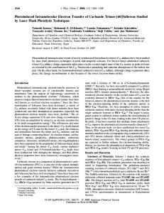
Figure 1. Comparison of the one- and two-quencher DNAzyme-based Pb2+ sensor design. (A1) The previous sensor design with one quencher. The primary and secondary structures of the DNAzyme are presented. The cleavage site (rA) is indicated in pink on the substrate strand. (A2) Schematic illustration of the two substrate populations at room temperature. One population is the substrate annealed with enzyme strands, and the other is free substrate. Since there is no quencher for the free substrate, high background fluorescence is observed. (A3) Roomtemperature steady-state fluorescence spectra of background fluorescence (solid line) and after complete cleavage of substrate (dashed line). The excitation wavelength is fixed at 560 nm. The signal increase is 60% over the background. Inset is a fluorescence image of the onequencher sensor sample without Pb2+ added (left) and with 6 µM Pb2+ added after 2 min (right). (B1) The new sensor design with the two quenchers. (B2) The free substrate fluorescence is partially quenched by the intramolecular quencher. (B3) The steady-state fluorescence spectra of background fluorescence (solid line), and fluorescence signal after complete cleavage of substrate (dashed line). The excitation wavelength is fixed at 473 nm. The signal increase is 660% over the background. Inset is a fluorescence image of the two-quencher sensor sample without Pb2+ added (left) and with 6 µM Pb2+ added after 2 min (right).
DNAzymes (also known as DNA enzymes, catalytic DNA, or deoxyribozymes elsewhere) are particularly attractive as a platform for designing biosensors. Long considered as a strictly genetic information storage material, DNA was shown in 1994 to carry out catalytic functions,19 and thus became the newest member of the enzyme family after proteins and RNA. Since then, DNAzymes have been shown to catalyze many of the same reactions as RNA or protein enzymes.20-22 In addition to being biocompatible, biodegradable, and amenable to in vitro selection, DNAzymes are relatively less expensive to produce and more stable to hydrolysis than RNA and protein enzymes. Unlike proteins, most DNAzymes can be denatured and renatured many times without losing binding ability or activity. DNA/RNAzymes that are highly specific for Pb2+,19,23,24 Cu2+,25-27 Zn2+,28 and Co2+ 29,30 have been obtained through in vitro selection. (19) Breaker, R. R.; Joyce, G. F. Chem. Biol. 1994, 1, 223-229. (20) Breaker, R. R. Nat. Biotechnol. 1997, 15, 427-431. (21) Sen, D.; Geyer, C. R. Curr. Opin. Chem. Biol. 1998, 2, 680-687. (22) Lu, Y. Chem. Eur. J. 2002, 8, 4588-4596. (23) Pan, T.; Uhlenbeck, O. C. Nature 1992, 358, 560-563. (24) Li, J.; Lu, Y. J. Am. Chem. Soc. 2000, 122, 10466-10467.
We are particularly interested in developing Pb2+ sensors, largely because of the adverse effects of Pb2+ exposure to human health, especially to children.31,32 Monitoring lead concentration in the environment on a regular basis is highly desirable. Although lead can be detected and quantified by a variety of instrumental methods,1 simple lead sensors7,24,33-35 may allow more practical on-site, real-time monitoring in household, environmental, and clinic applications. (25) Cuenoud, B.; Szostak, J. W. Nature 1995, 375, 611-614. (26) Carmi, N.; Shultz, L. A.; Breaker, R. R. Chem. Biol. 1996, 3, 1039-1046. (27) Wang, W.; Billen, L. P.; Li, Y. Chem. Biol. 2002, 9, 507-517. (28) Santoro, S. W.; Joyce, G. F.; Sakthivel, K.; Gramatikova, S.; Barbas, C. F., III. J. Am. Chem. Soc. 2000, 122, 2433-2439. (29) Bruesehoff, P., J.; Li, J.; Augustine, I. A. J.; Lu, Y. Comb. Chem. High Throughput Screening (CCHTS) 2002, 5, 327-335. (30) Mei, S. H. J.; Liu, Z.; Brennan, J. D.; Li, Y. J. Am. Chem. Soc. 2003, 125, 412-420. (31) Needleman, H. L. Human Lead Exposure; CRC Press: Boca Raton, FL, 1992. (32) Godwin, H. A. Curr. Opin. Chem. Biol. 2001, 5, 223-227. (33) Telting-Diaz, M.; Bakker, E. Anal. Chem. 2002, 74, 5251-5256. (34) Chen, C.-T.; Huang, W.-P. J. Am. Chem. Soc. 2002, 124, 6246-6247. (35) Blake, D. A.; Jones, R. M.; Blake, R. C.; Pavlov, A. R.; Darwish, I. A.; Yu, H. Biosens. Bioelectron. 2001, 16, 799-809.
Analytical Chemistry, Vol. 75, No. 23, December 1, 2003
6667
Table 1. Names and Sequences of DNA Used in the Papera
a
name
sequence
17DS 17DS-FD Rh-17DS 17DS-FR 17DDS Fl-17DDS Rh-17DDS 17E 17E-Dy
5′-ACTCACTATrAGGAAGAGATG-3′ FAM-5′-ACTCACTATrAGGAAGAGATG-3′-Dabcyl TMR-5′-ACTCACTATrAGGAAGAGATG-3′ FAM-5′-ACTCACTATrAGGAAGAGATG-3′-TMR 5′-ACTCACTATAGGAAGAGATG-3′ FAM-5′-ACTCACTATAGGAAGAGATG-3′ TMR-5′-ACTCACTATAGGAAGAGATG-3′ 5′-CATCTCTTCTCCGAGCCGGTCGAAATAGTGAGT-3′ 5′-CATCTCTTCTCCGAGCCGGTCGAAATAGTGAGT-3′-Dabcyl
The cleavage site of the substrate is shown in boldface type (rA).
After designing a strategy to obtain molecules such as DNAzymes that can recognize target metal ions with high affinity and specificity, the next challenge is how to convert molecular recognition into physically detectable signals.30,36-38 Toward this goal, we have communicated a novel strategy of converting a Pb2+dependent DNAzyme named the 8-17 motif,39-41 into a highly selective and sensitive fluorescence-based Pb2+ sensor.24 As shown in Figure 1A1, the sensor is composed of a fluorophore-labeled substrate strand (Rh-17DS) and a fluorescence quencher-labeled enzyme strand (17E-Dy). The substrate strand is 5′-end labeled with a 6-carboxytetramethylrhodamine (TMR). The enzyme strand is 3′-end labeled with a 4-(4′-dimethylaminophenylazo)benzoic acid (Dabcyl, a fluorescence quencher). The initial state of the sensor is when the substrate strand is annealed to the enzyme strand. The adjacent position of TMR and Dabcyl allows quenching of TMR fluorescence and gives low background fluorescence. The cleavage of the substrate in the presence of Pb2+ and the subsequent release of the cleaved product cause a significant increase in fluorescence intensity (Figure 1A2). Nearly 300% increase of signal upon complete cleavage has been achieved at 4 °C.24 The detection limit (10 nM) is 50-fold lower than the toxic lead level defined by the U.S. Center for Disease Control. The selectivity for Pb2+ over other divalent metal ions is over 5000fold for Mg2+ and Ca2+. For the closest competing metal ions, Zn2+ and Co2+, the selectivity is at least 80-fold higher. While the above DNAzyme-based biosensor is quite effective at 4 °C, its sensitivity suffers at room temperature because of elevated background signals. The fluorescence increase is only 60% upon cleavage (Figure 1A3) as compared to the 300% increase at 4 °C. The decreased sensitivity limits the practical application of the sensor. To improve the performance of the sensor at higher temperature, we carried out a detailed study of the system, including measuring the melting curve of the DNAzyme and determining the end-to-end distance of the free substrate strand. Based on the insight obtained from the study, we designed a new (36) Liu, Z.; Mei, S. H. J.; Brennan, J. D.; Li, Y. J. Am. Chem. Soc. 2003, 125, 7539-7545. (37) Jhaveri, S.; Rajendran, M.; Ellington, A. D. Nat. Biotechnol. 2000, 18, 12931297. (38) Fang, X.; Cao, Z.; Beck, T.; Tan, W. Anal. Chem. 2001, 73, 5752-5757. (39) Santoro, S. W.; Joyce, G. F. Proc. Natl. Acad. Sci. U.S.A. 1997, 94, 42624266. (40) Faulhammer, D.; Famulok, M. Angew. Chem., Int. Ed. Engl. 1996, 35, 28372841. (41) Li, J.; Zheng, W.; Kwon, A. H.; Lu, Y. Nucleic Acids Res. 2000, 28, 481488.
6668
Analytical Chemistry, Vol. 75, No. 23, December 1, 2003
general strategy that allows the DNAzyme sensor system to work over a wide temperature range without losing its sensitivity or selectivity. EXPERIMENTAL SECTION Fluorescently Labeled Oligonucleotides. The fluorescently labeled oligonucleotides were purchased from Integrated DNA Technology Inc. The sequences and modifications of the DNA molecules used in this work are summarized in Table 1. The cleavable substrate of the 8-17 DNAzyme (17DS) is a DNA/RNA chimer with a single RNA linkage. The all-DNA analogue of 17DS is named 17DDS, in which all the bases are DNA bases, so that no cleavage can occur. 17DS-FD is the 5′-end FAM (6-carboxyfluorescein) labeled and 3′-end Dabcyl-labeled 17DS (see Figure 1). Similarly, Rh-17DS is the 5′-end TMR-labeled 17DS. 17DS-FR is the 5′-end FAM-labeled and 3′-end TMR-labeled 17DS. Fl-17DDS is the 5′-end FAM-labeled 17DDS. The enzyme strand of the DNAzyme is named 17E. 17E-Dy is the 3′-end Dabcyl-labeled 17E. All DNA molecules were purified by HPLC or 20% polyacrylamide gel electrophoresis to ensure 100% labeling efficiency. Determination of the Melting Temperature. Two methods were used to determine the melting temperature of the 8-17 DNAzyme. In the fluorescence-based method, the substrate strand was Fl-17DDS. The use of an uncleavable substrate was to prevent cleavage in the process of measuring the melting curve. The enzyme strand used was 17E-Dy. Concentration of 50 nM Fl-17DDS and 17E-Dy were annealed in 50 mM Tris acetate buffer, pH 7.2, containing 50 mM NaCl, by heating to 90 °C for 5 min and then cooling slowly to 4 °C. The fluorescence of the sample was acquired on a SLM 8000S fluorometer operating in the photon counting mode. Upon melting, the substrate strand was dissociated from the enzyme strand, resulting in an increased fluorescence signal. The emission was monitored at 520 nm after excitation at 473 nm. The second method was based on the hyperchromic UV absorption of DNA. The temperature-dependent absorption at 260 nm of 2 µM annealed 17DDS and 17E without any fluorescent modification was monitored. In both methods, the sample was kept at target temperatures for at least 1 min after the temperature was reached to ensure that the sample was at the stated temperature. Determining the End-to-End Distance of the Free Substrate. The end-to-end distance was measured by fluorescence resonance energy transfer (FRET). A dual-labeled substrate 17DSFR was used. 17DS-FR contains two fluorophores, with the 5′end FAM as donor and the 3′-end TMR as acceptor. The (ratio)A
quantification was performed in the Fuji ImageGuage 3.12 program. A well containing pure water was used as the internal standard to minimize the difference between each scan. Pb2+ Detection in a Lake Michigan Water Sample. Lake Michigan water (Chicago, IL) was buffered at pH 7.2 with 50 mM Tris acetate buffer, and 50 mM NaCl. Pb(OAc)2 was added to make a series of water samples with Pb2+ concentration from 0.5 to 20 µM. A 95-µL aliquot of the leaded water sample was added to 5 µL of concentrated sensor solution, which contained 500 nM hybridized 17DS-FD and 17E-Dy in the same buffer to initiate the cleavage reaction for Pb2+ detection. Fluorescence intensity was collected after 2 min. Figure 2. FRET spectra of 17DS-FR (substrate 17DS labeled at the 5′-end with a FAM and at the 3′-end with a TMR). The solid line is the FRET spectrum of the free substrate. The dashed line is the FRET spectrum for the same substrate but annealed to a complementary strand DNA. Thus, the substrate is in the fully stretched state.
method was used to calculate the FRET efficiency.42 The (ratio)A is the ratio of the acceptor fluorescence from energy transfer and from direct excitation. From (ratio)A, FRET efficiency can be calculated, which can then be used to calculate the distance between the two fluorophores. The detailed methods for data fitting to acquire (ratio)A and the calculation of energy-transfer efficiency from (ratio)A have been described previously.42,43 Specifically, to obtain (ratio)A, 17DS-FR was excited at 490 nm, and the emission spectrum was recorded from 500 to 650 nm. With the 490-nm excitation, FAM was directly excited, and energy was transferred from FAM to TMR through FRET (Figure 2, solid line). A second excitation was performed by exciting the FRET acceptor (TMR) at 560 nm, and the emission spectrum was collected from 570 to 650 nm (spectrum not shown). In a separate sample, a FAM singly labeled substrate, Fl-17DDS was excited at 490 nm, and the emission spectrum from 500 to 650 nm was collected (spectrum not shown). This spectrum was used as a shape function for spectra fittings to eliminate the “red tail” emission from FAM at 570 nm, where TMR emits. From these spectra, the energy-transfer efficiency was calculated to be 0.81. The literature reported R0 value for the FAM/TMR pair is 50 Å. Thus, the distance of the two fluorophores can be calculated to be 39 Å from the equation E ) 1/(1 + (R/R0)6). Hybridization and Assay Procedures. Concentrations of 50 nM 17DS-FD and 50 nM 17E-Dy were annealed in 50 mM Tris acetate buffer, pH 7.2, in the presence of 50 mM NaCl by heating at 90 °C for 5 min and subsequently cooling to 4 °C slowly. The annealed sample was then taken to room temperature for subsequent assays. A 95-µL aliquot of the 50 nM hybridized DNA was loaded in each well of a 96-well plate. A 5-µL aliquot of 20× concentrated metal ions stock solution was then added into the DNA solution using an eight-channel pipet to initiate the cleavage reaction. Data Acquisition and Quantification. The fluorescence detection was performed on a multifunctional three-laser fluorescence image analysis system (model FLA-3000G, Fuji). The excitation laser wavelength was set at 473 nm, and the filter was set at 520 nm to monitor the fluorescence of FAM. Data (42) Clegg, R. M. Methods Enzymol. 1992, 211, 353-388. (43) Liu, J.; Lu, Y. J. Am. Chem. Soc. 2002, 124, 15208-15216.
RESULTS Melting Temperature of the DNAzyme. The bulged structure of the 8-17 DNAzyme (Figure 1A1) is different from the normal complementary double-stranded DNA. Therefore, the stability of the DNAzyme and thus its utility at higher temperatures may be affected by the bulged structure. To quantitatively determine the stability of the DNAzyme complex at different temperatures, the melting curve of the DNAzyme was measured. Two methods were used to determine the melting temperature (Tm) of the DNAzyme. In the method using fluorophore- and quencher-labeled DNAzyme, the fluorescence increase with respect to temperature was measured at 520 nm, the emission wavelength of FAM, and Tm was determined to be 34 °C. In the method based on the hyperchromic absorption of the fluorophoreand quencher-free DNAzyme at 260 nm, the DNA absorption increase with respect to temperature increase was measured at 260 nm. This increase is indicative of the conversion from doublestranded to single-stranded DNA. Tm was determined to be 35 °C by this method. (See Supporting Information for the melting curves.) From the above results, two conclusions can be reached. First, the nearly identical melting temperature determined by the above two methods suggests that the presence of the fluorophore and quencher exerts little effect on the stability of this DNAzyme. Labeling DNA with a fluorophore can affect the DNA melting temperature, depending on the identity of the fluorophore and its location on the DNA.44 The minimum effect of the fluorophore/ quencher on the stability of our DNAzyme sensor may be a result of the design strategy (shown in Figure 1A1) that separates the motif for molecular recognition from that for fluorescence detection and that places the fluorophore and the quencher at the end of the DNAzyme. Previous studies of the same DNAzyme24,45 also indicated that the Pb2+-dependnet activity was not influenced by the presence of a fluorophore and a quencher. Together these results strongly suggest that separating the molecular recognition motif from the signaling motif is an important feature for DNAzyme-based sensors; this feature makes it possible to design sensors without prior knowledge of three-dimensional structure of the system while maintaining metal-binding affinity and specificity of the selected DNAzymes. Second, the Tm of the DNAzyme (34 °C) is lower than that of a double-stranded DNA with the same sequence as the substrate strand 17DS but with perfect base (44) Marras, S. A. E.; Kramer, F. R.; Tyagi, S. Nucleic Acids Res. 2002, 30, e122/ 121-e122/128. (45) Brown, A. K.; Li, J.; Pavot, C. M. B.; Lu, Y. Biochemistry 2003, 42, 71527161.
Analytical Chemistry, Vol. 75, No. 23, December 1, 2003
6669
pairing and no bulging (Tm ) 54 °C). This result suggests that the bulged structure of the DNAzyme decreased the stability of the system significantly. With a Tm of 34 °C, the enzyme and the substrate strand of the DNAzyme may not be completely hybridized at room temperature or 37 °C, the physiologically relevant temperature. The incomplete hybridization means that the quencher may not quench the fluorophore completely, resulting in large background fluorescence. End-to-End Distance of the Free Substrate. Since incomplete hybridization at room temperature may result in a portion of free substrate that does not hybridize with the enzyme strand in solution, we investigated the conformation of the free substrate in solution in order to find ways to decrease background fluorescence of the free substrate. Based on the mfold program (available at http://www.bioinfo.rpi.edu/applications/mfold),46 the free substrate cannot form stable secondary structures. FRET was used to measure the average end-to-end distance of the unstructured free substrate. The substrate strand was labeled with a FAM at the 5′-end and with a TMR at the 3′-end. After excitation at 490 nm, the absorption wavelength of the FRET donor FAM, emission spectra were collected from 500 to 650 nm (Figure 2, solid line). The 520- and 570-nm peaks correspond to emission from FAM and TMR, respectively. Upon excitation at 490 nm, only FAM is directly excited; the emission from TMR at 570 nm results from energy transferred from FAM to TMR. The efficiency of energy transfer (EFRET) is dependent on the distance between the two fluorophores. If we know EFRET, the distance between the two fluorophores can be calculated. Using the method for distance calculation described in the Experimental Section and elsewhere,42,43 the average end-to-end distance was determined to be 39 Å for the free substrate. As a control experiment, the FRET spectrum of the substrate annealed to a complementary DNA strand was collected (Figure 2, dashed line). The end-to-end distance was calculated to be 71 Å, which is consistent with the length of a double-stranded B-form 20-mer DNA (68 Å). From Figure 2, it can also be observed that the fluorescence intensity of the FRET donor (FAM at 520 nm) is much weaker in the free state than in the fully base-paired double-helix form. Therefore, the shorter end-to-end distance makes it possible to decrease the background fluorescence from the free substrate by replacing the FRET acceptor TMR with a fluorescence quencher. New DNAzyme-Based Pb2+ Sensor Design. On the basis of the results described above, we conclude that the high background fluorescence of the previously designed sensor (Figure 1A1) at room temperature is due to the presence of free substrate, which maintains a random coil conformation. To decrease this contribution to the background fluorescence signal, a second quencher is attached to the 3′-end of the substrate to quench fluorescence from the free substrate. The new design is illustrated in Figure 1B1. Since the additional quencher is placed at the end of the substrate strand, its effect on the enzyme activity should be minimal. Thus, the new design has two quenchers, one intermolecular and one intramolecular. The advantage of the intermolecular quencher (the Dabcyl on the 3′-end of the enzyme strand) is that it gives almost 100% quenching efficiency; however, not all substrate strands are annealed to enzyme strands at room temperature. The distance between the intermolecular quencher
and the fluorophore is very short, allowing exciton interactions between the fluorophore and the quencher, resulting in nearly 100% quenching efficiency.47 The presence of exciton interactions is supported by the observation of significant changes in the absorption spectra of the fluorophore and quencher-labeled DNAzyme at 4 and 50 °C (see Supporting Information). The advantage of the intramolecular quencher (the Dabcyl on the 3′end of the substrate) is the 100% labeling efficiency; however, the quenching efficiency is not 100%. The quenching mechanism is through FRET and, therefore, depends on the fluorophore-toquencher distance. The combination of the inter- and intramolecular quenchers utilizes the strengths of both types of quenchers and minimizes the effects of their weaknesses. Although the origin of the intermolecular quenching described here is not based on FRET because of the extremely short distance between the fluorophore and the quencher,47 the intramolecular quenching in the free substrate is FRET-based. To obtain high FRET efficiency, or quenching efficiency, the emission spectrum of the fluorophore and the absorption spectrum of the quencher should have a large overlap.42 Because the spectral overlap between FAM and Dabcyl is much larger than those between TMR and Dabcyl, the fluorophore was changed from TMR to FAM. As shown in Figure 1B3, after the addition of a second quencher to the 3′-end of the substrate strand, 660% fluorescence increase was observed upon addition of Pb2+, as compared to the 60% increase of the previous design (Figure 1A3), resulting in a 10-fold increase of the signal-to-background ratio. A side-by-side comparison of the performance of the two designs using a fluorescence scanner is shown in the inset of Figure 1A3 and B3. The fluorescence increase is barely observable for the previous design with one quencher, while it is quite significant for the design with two quenchers. Performance of the Sensor at Room Temperature. To test the sensitivity and selectivity of the newly designed sensor at room temperature and to monitor multiple samples in a parallel manner, a 96-well plate was used as the test container and a fluorescence scanner was used as the detector. Six other metal ions (Co2+, Mg2+, Zn2+, Mn2+, Cd2+, Ni2+) were tested to compare with Pb2+ and demonstrate the selectivity of the sensor. These metal ions were chosen because they showed relatively high activity toward the substrate cleavage by 17E based on previous assays, while other metal ions were completely inactive.24,41,45 The first well of each row was set as an internal standard to quantify the background fluorescence intensity; instead of 5 µL of divalent metal ions, 5 µL of water was added into the first well. There are several methods to assay the activity of the metal ions for the cleavage, such as comparing the cleavage rate constant or comparing the initial cleavage rate. The easiest method is probably to monitor the fluorescence intensity at certain time points after a reaction is initiated, so that no complicated data processing is required. As in previously reported activity assays,24,41,45 fluorescence intensity after 2 min of reaction initiation was recorded for the current system. The selectivity of the sensor is shown in Figure 3A. Four different concentrations of metal ions were tested, and in each case, Pb2+ gave the highest fluorescence intensity, indicating the fastest cleavage. To present data in a quantitative way, the intensity of each well was quantified and
(46) Zuker, M. Nucleic Acids Res. 2003, 31, 3406-3415.
(47) Bernacchi, S.; Mely, Y. Nucleic Acids Res. 2001, 29, e62/61-e62/68.
6670
Analytical Chemistry, Vol. 75, No. 23, December 1, 2003
Figure 3. Performance of the two-quencher DNAzyme-based Pb2+ sensor at room temperature. (A) Fluorescence image of the sensor after 2 min of cleavage reaction. Four metal ion concentrations were assayed. Pb2+ showed the strongest fluorescence signal in each case. (B) Quantification of the fluorescence intensity in (A). (C) Kinetics of fluorescence increase for the two-quencher Pb2+ sensor with 500 nM divalent metal ions. Only Pb2+ showed high fluorescence increase (triangles).
Figure 4. Detection of Pb2+ in leaded Lake Michigan water.
plotted in Figure 3B. Besides Pb2+, only Zn2+ and Co2+ showed a slight increase in fluorescence at 5 µM metal concentration, while all other metal ions gave approximately background intensity. At low concentrations (500 nM, the toxic level for Pb2+ defined by U.S. CDC), Pb2+ was the only metal ion that could be detected by the sensor. Figure 3C shows the cleavage kinetics for all seven metal ions at 500 nM concentration over a time course of 90 min. Except for Pb2+, all other metal ions gave signals similar to the background. Thus, the sensor has maintained high selectivity. As an illustration of application for the new DNAzyme-based Pb2+ sensor to detect Pb2+ in real samples at room temperature, water from Lake Michigan was tested. The fluorescence image of a 96-well plate containing Lake Michigan water with different concentration of Pb2+ is shown in Figure 4. The overall trend is the same as that in pure water. This result demonstrates that, although there are many other ions and chemicals in a real water sample, the sensor is still capable of detecting Pb2+. DISCUSSION The DNAzyme kinetics important for the sensor system described here can be summarized in three steps.24,45 The first step is the hybridization of the substrate to the enzyme, which is always achieved by Watson-Crick base pairing. The second step is the cleavage of the substrate by the enzyme, which always requires divalent metal ions as cofactors. For fluorescence-based detection, the fluorescence signal increase does not occur until the third step, i.e., the fluorophore-labeled cleavage product release. Making the second step to be the rate-limiting step is especially important for engineering in vitro-selected DNAzymes into biosensors. Through in vitro selection, DNAzymes that can cleave substrate strands in the presence of certain metal ions can be obtained. Thus, the DNAzymes can be used as sensors for those metal ions. However, other metal ions with similar properties may also display some degree of activity for the DNAzymes. Given sufficient reaction time, however, competing metal ions may react
to a significant degree. If the melting temperature of a DNAzyme system is rendered too high, the product release step may become rate limiting. This may result in poor metal ion selectivity of the sensor because competing metal ions, even though they are relatively slow in cleaving the substrate, can catch up if the product release becomes rate limiting. More importantly, the melting temperature may become too high to allow the cleaved fragment and its associated fluorophore to be released, resulting in little or no fluorescence signal increase and thus poor sensitivity. Therefore, for a good DNAzyme-based sensor obtained from in vitro selection, the cleavage step should be engineered as the ratelimiting step in the whole kinetics process. The above presentation of experimental results and the analysis of the kinetics of DNAzyme cleavage strongly suggest that the dual-quencher design has a significant advantage over the singlequencher design in decreasing the background fluorescence so that the sensor can be used over a wide temperature range. In the single-quencher system, we provided evidence that the increased background fluorescence at room temperature is due to the low stability of the DNAzyme-substrate complex, which causes a portion of the fluorophore-labeled substrate strand to be free in solution. There are several methods to increase the stability of the substrate-enzyme complex, such as increasing NaCl concentration, extending the recognition arms, or increasing the GC base pair content. These methods, while effective, are not generally applicable to many different conditions or a wide temperature range. For each particular temperature in the sensor application, an appropriate concentration of NaCl has to be found. If the NaCl concentration is too high, the substrate release step can be inhibited; no or very limited fluorescence increase would be observed. If the NaCl concentration is too low, high background fluorescence may be observed. The same can be true for introducing a higher number of base pairs or higher GC content. Thus, for the strategy of changing the melting temperature of the DNAzyme to accommodate temperature changes, a condition that works at higher temperature is not applicable to a lower temperature environment. Furthermore, using NaCl to tune the melting temperature of the DNAzyme system has another disadvantage: the sensor cannot be applied to real samples where the NaCl concentration does not match that required by the sensor system. Therefore, rather than increasing the melting temperature of the Analytical Chemistry, Vol. 75, No. 23, December 1, 2003
6671
DNAzyme sensor system, introducing intra- and intermolecular quenchers into the system can decrease the background fluorescence while maintaining selectivity and not affecting the ratedetermining step of the system. CONCLUSIONS The combination of intra- and intermolecular quenchers has significantly decreased the background fluorescence for the 8-17 DNAzyme-based Pb2+ sensor at room temperature. The 8-17 DNAzyme used in this study shares the same structural features as most known trans-cleaving DNA/RNAzymes, such as RNAcleaving RNAzymes,11,48 RNA-cleaving DNAzymes,19-21 and DNAcleaving DNAzymes;49 they all contain a catalytic core and substrate recognition arms. Since the cleavage activities of these DNA/RNAzymes are highly analyte-dependent, they could potentially be engineered into biosensors for those analytes. The range of analytes that DNA/RNAzymes can recognize has been significantly expanded by using allosteric DNA/RNAzymes (also known as aptazymes).17,18,50 Despite additional effector binding (48) Tang, J.; Breaker, R. R. Proc. Natl. Acad. Sci. U.S.A. 2000, 97, 5784-5789. (49) Carmi, N.; Balkhi, H. R.; Breaker, R. R. Proc. Natl. Acad. Sci. U.S.A. 1998, 95, 2233-2237. (50) Wang, D. Y.; Lai, B. H. Y.; Sen, D. J. Mol. Biol. 2002, 318, 33-43.
6672
Analytical Chemistry, Vol. 75, No. 23, December 1, 2003
sites, most aptazymes also contain a catalytic core and substrate recognition arms. Therefore, the method presented in this paper should be generally applicable to those systems for decreasing background fluorescence and increasing signal-to-noise ratio in order to improve their performance for detecting a broad range of analytes and over a wide range of temperatures. ACKNOWLEDGMENT This material is based upon work supported by the U.S. Department of Energy (DEFG02-01-ER63179) and the U.S. National Science Foundation (CTS-0120978 and DMR-0117792). The experiments reported in this paper were performed at the Laboratory for Fluorescence Dynamics (LFD) at the University of Illinois at UrbanasChampaign (UIUC). The LFD is supported jointly by the National Center for Research Resources of the National Institutes of Health (PHS 5 P41-RRO3155) and UIUC. SUPPORTING INFORMATION AVAILABLE Additional information as noted in text. This material is available free of charge via the Internet at http://pubs.acs.org. Received for review August 8, 2003. Accepted September 16, 2003. AC034924R