Supporting Information Sun et al. 10.1073/pnas.1115973108 SI Materials and Methods Materials. Dextran (M r 70,000) and allyl isocyanate were purchased from Sigma Chemical Co. We dried the dextran in an oven for 30 min at 60 °C before reaction. We purchased dimethyl sulfoxide (DMSO), dibutyltin dilaurate, 2-bromoethylamine hydrobromide, triethylamine, acryloyl chloride, polyethylene glycol (M r 4,000), and other chemicals from Aldrich Chemical Co. and used them as received. The photoinitiator 2-hydroxy-1-[4(hydroxyethoxy)phenyl]-2-methyl-1-propanone was obtained from Ciba Specialty Chemicals Corp. We obtained male 8-weekold 129S1/SvImJ mice from The Jackson Laboratory. We purchased Integra wound dressing from Integra Life Sciences Co. and DuoDerm extra thin dressing from ConvaTec Co. Preparation of Dextran-Allyl Isocyanate-Ethylamine (Dex-AE)/Polyethylene Glycol Diacrylate (PEGDA) Hydrogel. We prepared the
Dex-AE/PEGDA hydrogels as we previously reported (1, 2). We dissolved Dex-AE/PEGDA at the ratio of 60/40 and 80/20 into phosphate-buffered saline (PBS) containing 0.1% (wt∕wt) 2-methyl-1-[4-(hydroxyethoxy)phenyl]-2-methyl-1-propanone (Irgacure 2959, Ciba). The mixture was pipetted into a sterile mold (70-μL volume per well) to obtain discs measuring 8 mm in diameter by 2 mm in thickness and photopolymerized (approximately 10 mW∕cm2 of UV light for 10 min; Black-Ray, UVP). We removed the resulting hydrogels from the mold and immersed them in sterile PBS solution before application onto wounds. Mechanical Study of Scaffolds. The mechanical properties of the scaffold samples (n ¼ 3) were determined as previously established (1, 2), using a Q800 Dynamic Mechanical Analyzer (TA Instruments) in unconfined submersion compression mode. Briefly, the diameter of each swollen hydrogel disk was determined using a digital caliper, and the sample was immersed in a PBS bath between unconfined parallel compression platens. Scaffold samples were compressed at a rate of 10% of thickness/minute until they reached 80% of their initial thickness. The modulus was then calculated as the ratio of the stress-strain curve at the linear portion of the curve. Scanning Electron Microscope (SEM) and Pore Size Determination. We studied the ultrastructure of the scaffold using SEM (FEI Quanta ESEM 200). The hydrogels were swelled in PBS for 24 h, then removed from water and quickly frozen in liquid nitrogen, and then freeze-dried in a Labconco freeze dryer under vacuum at −50 °C for 3 d until the samples became completely dry. The freeze-dried hydrogels were fractured to reveal their interior, mounted onto aluminum stubs with double-sided carbon tape, and sputter-coated (Anatech Hummer 6.2 Sputter Coater, Anatech) with gold for 60 s and then visualized using SEM. Pore size was determined manually by measuring the diameter of pores. A minimum of six images were analyzed on each sample. On each SEM image, at least 20 pores were counted and measured, and the averaged pore size represents the pore size of each sample. In Vitro Degradation. To determine the effect of neutrophils on the scaffold degradation process, in vitro assay using differentiated HL-60 cells (3) was performed. Briefly, HL-60 cells (ATCC, CCL240) were expanded in RPMI1640 medium supplemented with 10% fetal bovine serum (Invitrogen) at 37 °C in a humidified
Sun et al. www.pnas.org/cgi/doi/10.1073/pnas.1115973108
atmosphere of 5% CO2 in air. To induce differentiation, 1.3% DMSO was added to the culture media. Neutrophil-like cell morphology could be observed within 5 d (4). To determine degradation kinetics, high-ratio dextran hydrogel, low-ratio dextran hydrogel, and control scaffolds were incubated with 1 mL of differentiated HL-60 cells (1 × 10⁵ cells∕mL) in differentiation medium. Hydrogel samples were removed from the cultures after 36 and 72 h, washed with distilled water, and lyophilized in a FreeZone freeze dryer (2.5 L; Labconco) at −48 °C for 3 d and weighed. The weight loss of the hydrogel degradation comprises both hydrolytic degradation and cell degradation. The extent of biodegradation was estimated from the weight loss of the polymer based on the following equation: Total weight loss
W l;t ¼
Weight loss by cell degradation
Wo − Wd × 100% Wo W l;c ¼
[S1]
Wh − Wd × 100%; Wo [S2]
where W o is the original weight of the hydrogel samples, and W d is the weight of dry hydrogel samples after being degraded in cell culture, in which the weight loss is attributed to both hydrolysis and cell degradation; whereas W h is the weight of dry sample after being degraded by hydrolysis in culture medium (without cell). Histology. Construct explants were collected at days 3, 5, 7, 14, and
21 and fixed using formalin-free fixative (Accustain, SigmaAldrich). This fixative was chosen because it preserves both hydrogel structure and endothelial cell immunoreactivity and morphology compared to commonly used formalin based fixatives (5) or a zinc-based fixative (6), though this alcoholic based fixative causes swelling of red blood cells (6). Following fixation of construct explants, samples were dehydrated in graded ethanol (70% to 100%), embedded in paraffin, serially sectioned using a microtome (5 μm), and stained with either hematoxylin and eosin (H&E) or immunohistochemistry for CD31 (Dako), F4/80 (Invitrogen), α-SMA (Abcam plc) and CD3 (Abcam plc), von Willebrand Factor (vWF; Dako), Masson’s trichrome (Sigma), MPO, VE-cadherin (Abcam plc), and VEGFR2 (Cell Signal Technology). Statistics. All measurements were obtained from at least six dif-
ferent slides or mice, with multiple readings for each data point (as detailed throughout the manuscript). The number of animals (n) mentioned along the manuscript refers to the number per group. We quantified the blood flow using Doppler, the number and diameter of angiogenic blood vessels, the degrees of dermal differentiation, epithelial maturation, and the number of regenerated hair follicles. We performed either one-way ANOVA with Tukey’s post tests or two-way ANOVA with Bonferroni post tests where appropriate (GraphPad Prism 4.02). Significance levels, determined using post tests between controls, hydrogels, and Integra, were set at: *p < 0.05, **p < 0.01, and ***p < 0.001. All graphical data is reported.
1 of 4
1. Sun G, Shen Y-I, Ho CC, Kusuma S, Gerecht S (2010) Functional groups affect physical and biological properties of dextran-based hydrogels. J Biomed Mater Res A 93A:1080–1090. 2. Sun G, et al. (2011) Functional neovascularization of biodegradable dextran hydrogels with multiple angiogenic growth factors. Biomaterials 32:95–106. 3. Millius A, Weiner OD (2009) Chemotaxis in neutrophil-like HL-60 cells. Methods Mol Biol 571:167–177.
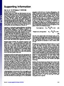
A
1. Burn (1.2cm)
2. Wound Excision
Dressing
B
4. Inoue T, Meyer T (2008) Synthetic activation of endogenous PI3K and Rac identifies an AND-gate switch for cell polarization and migration. PLoS One 3:e3068. 5. Ismail JA, et al. (2003) Immunohistologic labeling of murine endothelium. Cardiovasc Pathol 12:82–90. 6. Hanjaya-Putra D, et al. (2011) Controlled activation of morphogenesis to generate a functional human microvasculature in a synthetic matrix. Blood 118:804–815.
3. Apply hydrogel
Control
4. Cover to protect
Hydrogel
Fig. S1. Dextran hydrogel for burn wound healing. (A) Surgery procedure: We placed wounds on the posterior-dorsum of each mouse and performed burn wound excisions after 48 h. We covered wounds with either dextran hydrogels or control scaffold, followed by their coverage with dressing. We covered the control wounds only with dressing. (B) Photo image of wound healing within 21 d demonstrate a more complete wound healing in burn wounds treated with dextran hydrogel than in wounds treated with control scaffolds or dressing alone.
Control
60/40
100µm
Fig. S2.
80/20
20µm
20µm
Scaffold porosity. Representative SEM images demonstrate scaffold porosity.
F4/80
CD3
Day 7
Day 5
MPO
Fig. S3. Inflammatory cell infiltration in control wounds covered with dressing. Neutrophils (MPO) and macrophages (F4/80) were clearly observed on day 5, and T cells (CD3) were observed on day 7. Scale bars, 100 μm.
Sun et al. www.pnas.org/cgi/doi/10.1073/pnas.1115973108
2 of 4
A Scaffold hydrogel
α-SMA
H&E
B W
H
W H
Fig. S4. Illustration of angiogenic response at the interface between treatment and wounded skin. (A) Schematic illustrating the location of the interface between the wound and hydrogel and (B) H&E-stained histologic sections and α-SMA staining showing the interface between the wound and hydrogel. The dotted line indicates the interfaces. W, wound area; H, hydrogel scaffold. Scale bars, 100 μm.
vWF
α -SMA
Masson’s trichrome
Hydrogel
Control
Dressing
H&E
Fig. S5. Skin regeneration within 21 d. Representative images of control dressing, control scaffold and hydrogel stained with H&E, vWF, α-SMA, and Masson’s trichrome. Scale bars, 100 μm.
Sun et al. www.pnas.org/cgi/doi/10.1073/pnas.1115973108
3 of 4
Fig. S6.
Normal mouse skin. H&E-stained histologic section of a 129S1/SvImJ mouse skin. Scale bar, 100 μm.
Sun et al. www.pnas.org/cgi/doi/10.1073/pnas.1115973108
4 of 4