Invest Clin 52(1): 23 - 34, 2011
Diagnostic accuracy of immunological methods in patients with tuberculous pleural effusion from Venezuela. Carlos Fernández de Larrea1, Aglae Duplat1, Francesca Giampietro2, Jacobus H. de Waard3, Julieta Luna4, Mahavir Singh5 y Zaida Araujo2. Servicio de Medicina Interna, Hospital Vargas de Caracas. Caracas, Venezuela, Laboratorio de Inmunología de Enfermedades Infecciosas, 3Laboratorio de Tuberculosis, Instituto de Biomedicina, Universidad Central de Venezuela. Caracas, Venezuela, 4Escuela Nacional de Ciencias Biológicas, Instituto Politécnico Nacional, México y 5LIONEX Diagnostics and Therapeutics GMBH, Braunschweig, Germany. 1 2
Key words: Tuberculous pleural effusion (TPE), IFN-g, IL-12p40, sensitivity, specificity. Abstract. In recent years, better diagnostics for tuberculosis (TB) has received increasing attention, especially the diagnosis of tuberculous pleural effusion, which is difficult and at present the main tool in TPE diagnostic is pleural effusion smear and culture, but unfortunately, sensitivities are low, therefore better TPE diagnostic tools are needed. The aim of this study was to find a diagnostic algorithm to assess the progress in TPE diagnostic at the Hospital Vargas de Caracas, that permits identification of the majority of patients, at a satisfactory cost-benefit ratio, evaluating the levels of IFN-g and IL-12p40 in pleural effusion and serum, as well as the antibody reactivity in order to compare it with microbiological tests. A total of 60 individuals with pleural effusion were studied; 20 patients with tuberculous pleural effusion (TPE) formed the patient group and 40 patients with non-tuberculous pleural effusion (NTPE) formed the control group. The levels of IFN-g and IL-12p40 in effusion and serum and class and subclasses of IgG reactivity to Mycobacterium tuberculosis antigens were measured by ELISA. The utility of these methods for diagnosis of TPE was evaluated using receiver operating characteristic (ROC) curve analysis. The results of the 11 immunological methods evaluated showed that the anti-PPD IgG2 method was able to reach the highest specificity of 95% (CI: 88.3-101.8), positive predictive value (PPV)=75 (at 30% sensitivity); while that the overall sensitivity of methods was between 95% and 30%, of these, two methods reached higher sensitivities; increased levels of pleural IFN-g, with a sensitivity of 95% (CI: 85.5-104.5) with the highest negative pre-
Corresponding author: Zaida Araujo. Laboratorio de Inmunología de Enfermedades Infecciosas, Instituto de Biomedicina, Universidad Central de Venezuela. Apartado 4043. Caracas 1010A, Venezuela. Teléf. +58-212860-4636/414-248-0215; fax: +58-212-861-1258. E-mail:
[email protected]
24
Fernández y col. dictive value (NPV)=97, (at 82.5% specificity), followed by decreased levels of serum IL-12p40 with a sensitivity of 95% (CI: 85.5-104.5), NPV=95.2 (at 50% specificity). In contrast, microbiological methods showed that smear had a sensitivity of only 20%, while smear plus culture had, a sensitivity of 70%. Considering that TPE represents approximately 15 percent of all the TB clinically diagnosed at the Hospital Vargas de Caracas, in those patients with preliminary microbiology negativity in the effusion, the combined analysis of pleural IFN-g and anti-PPD IgG2 could represent a fast and effective diagnostic algorithm for improving the diagnosis previous to obtain culture results. In this way treatment against TB could be initiated or the need to cytological and pleural biopsy could be considered.
Precisión diagnóstica de métodos inmunológicos en pacientes con tuberculosis pleural de Venezuela.
Palabras clave: líquido pleural tuberculoso, IFN-g, IL-12p40, sensibilidad, especificidad. Resumen. Recientemente existe un gran interés hacia un mejor y más rápido diagnóstico de tuberculosis (TB), especialmente de tuberculosis pleural, el cual es difícil. Al presente las principales herramientas diagnósticas son la baciloscopia y el cultivo de líquido pleural; desafortunadamente, las sensibilidades de estos métodos son bajas, por lo que el desarrollo de nuevas herramientas diagnósticas es necesario. El objetivo del presente estudio consistió en encontrar un algoritmo que permita la rápida identificación de la mayoría de los pacientes con TB pleural que ingresan en el Hospital Vargas de Caracas a un buen costo-beneficio. Para esto se evaluaron los niveles de las citocinas Interferón-gamma (IFN-g) y la Interleucina 12p40 (IL-12p40) en líquido pleural y suero, así como la reactividad de anticuerpos contra antígenos de Mycobacterium tuberculosis. Se estudiaron 60 individuos con derrame pleural; 20 individuos con líquido pleural tuberculoso (LPT) conformaron el grupo de pacientes y 40 individuos con líquido pleural no tuberculoso (LPNT) el grupo de controles. La técnica de inmunoensayo de ELISA fue utilizada para medir los niveles de IFN-g y IL-12p40; así como las reactividades de los diversos isotipos y subclases de inmunoglobulina G (IgG) frente a antígenos del bacilo. La utilidad de los métodos fue evaluada utilizando el análisis de las curvas ROC (receiver operating characteristic). Los resultados de los 11 métodos inmunológicos evaluados mostraron que el método IgG2 anti-PPD alcanzó la mayor especificidad de 95%, (CI: 88,3-101,8) con un valor predictivo positivo (VPP) de 75. La sensibilidad de los métodos estuvo entre 30% y 95%; dos métodos alcanzaron altas sensibilidades: los altos niveles de IFN-g en líquido pleural, con sensibilidad de 95% (CI: 85,5-104,5), con un valor predictivo negativo (VPN) de 97, seguido de los bajos niveles de IL-12p40 en suero, con una sensibilidad de 95% (CI: 85,5-104,5) con un VPN de 95,2. En contraste, los métodos mi-
Investigación Clínica 52(1): 2011
Immunological methods and tuberculous pleural effusion
25
crobiológicos mostraron una baja sensibilidad, en el caso de la baciloscopia sólo del 20%; mientras que la combinación baciloscopia más el cultivo alcanzó 70%. Considerando que las LPT representan el 15% de todos los casos de TB diagnosticados en el Hospital Vargas de Caracas; aquellos pacientes con una microbiología preliminar negativa en líquido pleural, la realización del análisis de IFN-g en líquido y el método serológico IgG2 anti-PPD podrían constituir un algoritmo diagnóstico rápido y efectivo, previo al resultado del cultivo para micobacteria, lo cual permitiría iniciar el tratamiento antituberculoso de forma más precoz o considerar la realización de la biopsia. Received: 27-06-2010. Accepted: 28-10-2010.
INTRODUCTION Pleural tuberculosis represents approximately 5 percent of all the associated diseases caused through infection by Mycobacterium tuberculosis (1). The gold standard for tuberculosis (TB) diagnosis is based on a combination of clinical and radiological examination, epidemiological investigation, microbiological testing such as culture and smear and appropriate response to anti-tuberculosis therapy. Diagnostic difficulties in pleural tuberculosis exist; as the results of the pleural fluid culture take several weeks, in many cases diagnosis takes a long time (2). This delay in diagnosis leads to patients being lost in follow up. In addition, conventional tests are not always helpful in making a differential diagnosis including infectious, inflammatory and neoplastic etiologies. The latter are especially important in the context of lymphocyte pleural effusion, with a totally different prognosis and treatment. Other tests like pleural biopsy are painful or, as in the case of the polymerase chain reaction (PCR)-based method, which in previous studies, have not shown a consistent advantage (3). It has been reported that fluid PCR alone should not be relied on as a single test; rather, combined analysis with other methods could be more useful in the diagnosis of tuberculous effusions (3). Various strategies such as the determinaVol. 52(1): 23 - 34, 2011
tion of enzymes, like adenosine deaminase (ADA), lysozyme and cytokines like interferon gamma (IFN-g) and tumor necrosis factor-alpha (TNF-a), in pleural effusion and serum, have been designed in order to improve the cost-benefit performance of the diagnosis. Nevertheless, these are not yet of routine use in many developing countries (4-6). Extensive immunological studies have been carried out in order to explain the various aspects of cytokine dynamics in patients with active pulmonary and extrapulmonary tuberculosis (7-9). However, the measurement of the cytokine level was found to be useful in the diagnosis of the tuberculous pleural effusion (10). These studies are mainly focused on the cellular response type Th1 that produces interleukin 2 (IL-2), IFN-g and interleukin 12 (IL-12). In this context, it has been investigated the diagnostic value of type Th1 cytokines, mainly IFN-g, TNF-a and IL-12 in TPE (11). With regard to estimate the humoral immune response in tuberculosis, it has been reported that serum IgG antibodies are produced against a variety of M. tuberculosis antigens and that, the vast majority of sera from pulmonary tuberculosis patients contained antibodies against one or more M. tuberculosis antigens (12, 13). Few studies have addressed the importance of antibody response in the diagnosis of ex-
26
Fernández y col.
tra-pulmonary tuberculosis (12). A recent study about the diagnosis of pleural tuberculosis reported that the specific IgA-ELISA method using MPT-64 and MT-10.3 antigens showed a sensitivity of 78% as compared to the PCR (sensitivity of 82%) and ADA (sensitivity of 91%) methods (14). Around 7,000 new cases of TB are identified each year in Venezuela, of which 15% are associated with TPE (15). The aim of this study was to assess the progress in diagnosis of pleural tuberculosis evaluating the levels of IFN-g and IL-12p40 in pleural effusion and serum, as well as the antibody reactivity in order to compare them with the gold standard for TB diagnosis based on criteria such as clinical and radiological examinations, microbiological testing and appropriate response to anti-tuberculosis treatment and to find a diagnostic algorithm in the Hospital Vargas de Caracas for TPE, that permits identification of the majority of patients at a satisfactory cost-benefit ratio. MATERIALS AND METHODS Study groups A descriptive transversal study was conducted in the Department of Internal Medicine of the Hospital Vargas de Caracas. A total population of 60 individuals for management of pleural effusion were enrolled into the study and classified as individuals with TPE and with NTPE. The average age was 34.7±16.9 and 51.0±19.4 for TPE and NTPE, respectively. The sex ratio (male/female) was 15/5 in TPE and 25/15 in NTPE. The study group consisted of diagnosed cases of TB whereas the non-TB effusion formed the control group. Sixty sera and pleural effusions from patients with pleural discharge were tested. They included 20 TPE patients and 40 NTPE controls; the latter group included: malignant pleural effusions (n=14), parapneumonic exudates (n=11), transudates (n=10) and
miscellaneous exudates (n=5). All the patients were HIV negative. An informed consent form was voluntarily signed by all patients and individual controls before blood and pleural effusion samples were selected. Individuals with the presence or absence of BCG scars were included. The tuberculin skin test (TST) was performed according to existing norms in the Venezuelan Program of Tuberculosis Control, on all the individuals of this study using two tuberculin units of purified protein derivative (PPD) of M. tuberculosis RT-23, from the Statens Serum Institute in Copenhagen, Denmark. A positive TST was defined for an induration with a diameter equal or superior to 10 mm after 48 hours of the test. This study was approved by the Ethical Committee of the Hospital Vargas de Caracas. Diagnosis of tuberculous pleural effusions and therapeutic conduct All patients included in the study underwent thoracentesis for diagnostic purpose by the Department of Pneumonology. The pleural fluid aspirate was subjected to cytological, microbiological (including mycobacteriological) culture and cytokine assays. Pleural biopsy was performed only when results of staining for acid-fast bacillus (smear) or culture of pleural fluid for M. tuberculosis were negative. The TPE was diagnosed using one of the following criteria as suggested in the national program of TB (15). 1) M. tuberculosis isolated from pleural effusion by culture. 2) Pleural biopsy showing granulomatous inflammation together with stainable acid-fast bacilli. 3) Pleural biopsy showing granulomatous inflammation, but no stainable acid-fast bacilli, together with a sputum culture positive for M. tuberculosis or a good radiographic response to anti-tuberculous treatment. 4) No histological or bacteriological confirmation, but with other likely alternative diagnoses excluded, together with a
Investigación Clínica 52(1): 2011
Immunological methods and tuberculous pleural effusion
27
Biologic parameters White blood cell (leucocytes) and hemoglobin (Hb, g dL–1) were studied according to routine use in the central laboratory of the Hospital Vargas de Caracas. In sera from all individuals, hepatic proteins were tested by different commercial kits: Total Proteins (TP), Lactate Dehydrogenase (LDH), Albumin (Alb) and Reactive C Protein (RCP) by Teco Diagnostic Kit (Anaheim, CA, US).
of the standard curve (pg mL–1)]. Microtiter plates (Immunolon, Birmingham, UK) with 96 wells were coated with anti-human IL-12p40 overnight at 4°C. Excess protein binding sites were blocked by incubation with 1% BSA in PBS at 37°C for 1 h. Then the plates were washed four times with PBS containing 0.1% Tween 20. Samples diluted 1:50 in PBS containing 0.5% BSA were added and plates were incubated for 1 h at 37°C. The plates were washed four times, then incubated with the second antibody diluted 1:1000 in blocking solution for 1 h at 37°C and washed four times. After incubating with streptavidin-HRP and washing, substrate solution (30 µL of 30% H2O2 and 10 mg o-phenylenediamine (OPD) dihydrochloride, Sigma-Aldrich, US) in 25 mL citrate buffer, pH 5 was added and incubated at room temperature. Colour development was measured in an ELISA reader at 492 nm. This assay was sensitive to 7.8 pg of IL-12p40 activity per mL. The IFN-g levels were also determined following the instructions of the human IFN-g kit (DuoSet, DY285 R&D Systems, Minneapolis, MN, USA). Mouse anti-human IFN-g was used as capture antibody and biotinylated goat anti-human IFN-g as the second antibody. This assay was sensitive to 15.6 pg of IFN-g activity per mL.
Determination of cytokines Cytokine assays were carried out according to the manufacturer’s protocol. For human IL-12 test, the IL-12p40 (DuoSet, DY1240 R&D System Minneapolis, MN, USA) kit was used. ELISA contained: mouse anti-human IL-12p40 (capture antibody), and IL-12p40 detection antibody (biotinylated goat anti-human IL-12p40). Sera and pleural effusions isolated from TPE patients and NTPE controls were always tested in duplicate [a quality control of duplicate variation was performed for looking standard deviations of the each different points
Antibody assays The antibody assays were performed with antigens and isotypes previously tested (16). Enzyme-linked immunosorbent assays (ELISA) were performed blindly by a lab technician, and developed and standardized in our laboratory for the measurement of IgG, IgG1, IgG2 and IgG3 against PPD (Statens Serum Institute, Copenhagen, Denmark) and IgG4 against H37Rv (Instituto Politécnico Nacional de México). Each assay included positive and negative sera and blanks to control non-specific binding. Briefly, sera were isolated from ve-
good clinical and radiographic response to anti-tuberculous treatment. The latter was initiated in all identified cases of TB, where microbiological evidence suggestive of TB and bacteriological confirmation by bacilloscopy or culture were found. Clinical and nutritional follow-up occurred for six months after anti-TB drugs treatment in order to evaluate the improvement of these aspects as therapeutic confirmation, permitting corroboration of the diagnosis. All TB patients were assessed for residual pleural scarring in thorax X-rays after completing anti-TB treatment. Malignant pleural effusions were diagnosed either by pleural effusion cytology or biopsy specimen histology. Miscellaneous pleural effusions were those not linked to either infection or malignant disease.
Vol. 52(1): 23 - 34, 2011
28 nous blood obtained from controls and TB patients. Microtiter plates (Immunolon, Birmingham, UK) with 96 wells were coated with 1µg/well diluted antigen PPD or H37Rv in 100 µL/well of carbonate-bicarbonate buffer pH 9.6 solution, overnight at 4°C. Excess protein binding sites were blocked by incubation with PBS-BSA 1% at 37°C for 1 h. Then plates were washed four times with PBS-Tween 20 0.1%. Sera were added at 1:400 (IgG) and 1:50 (IgG subclasses) dilutions, respectively, in PBS-BSA 0.5% and plates were incubated for 1 hr at 37°C. Plates were washed four times and then incubated for 1 hr at 37°C with 1:10.000 and 1:1000 diluted peroxidaseconjugated monoclonal antibody anti-IgGgamma chain (Promega Corp., USA) anti-IgG1, IgG2, IgG3 and IgG4, respectively (AP006, AP007, AP008 and AP009, The Binding Site, UK). After this period, plates were washed four times and the substrate solution composed by citrate buffer pH 5.0, H2O2 30% and 10 mg orthophenylenediamine dihydrochloride (OPD, Sigma-Aldrich) was added and plates incubated for 6 min at room temperature. Color development was measured in an ELISA microplate reader at 492 nm. The levels of serum IgA against PPD antigen (anti-PPD IgA) and saliva IgA (sIgA) against 38 kDa (GBF and LIONEX GmbH, Braunschweig, Germany), (anti-38 kDa sIgA) were determined by an ELISA similar to that described for anti-PPD IgG. Sera and saliva were diluted at 1:100 and 1:10 respectively, and detection was made with 1:2500 diluted peroxidase-conjugated anti-IgA monoclonal antibody (Pierce Biotechnology, USA) and 1:2000 peroxidase-conjugated anti-IgA-alpha chain monoclonal antibody (Sigma-Aldrich, USA), respectively. Statistical analysis Receiver operating characteristics (ROC) curves were constructed to compare
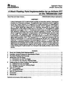
Fernández y col. the overall diagnostic information of the methods by comparing the areas under the curves (AUCs), obtaining the cut-off, sensitivity, specificity and predictive values. The t-Student test was used to compare differences cytokine concentrations between groups and for pairwise comparison of ROC curves. RESULTS Bacteriologic and biologic parameters Table I shows bacteriologic and biologic parameters. The TPE was confirmed by smear in 4/20 patients and bacterial culture in 14/20 patients. The parameters that reached statistical significance were: the median of hemoglobin (Hb) was significantly decreased in the NTPE group (11.4±2.4), as compared to the TPE group (12.8±1.9), p<0.002 (Table I). The median of total protein (TP) was significantly increased in the TPE group (7.2±1.1), as compared to the NTPE group (6.4±1.0), p<0.006 (Table I), the median of Albumin (ALB) was also significantly increased in TPE group (3.54±0.8), as compared to NTPE group (3.11±0.8), p<0.05 (Table I). Of the 60 individuals, 55 presented BCG scars (data not shown). Of the 20 TPE patients, 13 individuals were positive for the TST; while of the 40 NTPE controls, 7 individuals were positive for the TST (data not shown). Cytokine concentrations Fig. 1 shows the medians of the concentration of pleural IL-12p40. Similar levels of pleural IL-12p40 were found in both groups, TPE patients (988.8±686.4) and NTPE controls (1030.9±1294.5) (Fig. 1A). Concerning serum IL-12p40 levels, the TPE patients presented significantly low concentrations of serum IL-12p40 (294.5±127.5) as compared to NTPE controls (705.5± 768.07), p<0.02 (Fig. 1B). Regarding the
Investigación Clínica 52(1): 2011
Immunological methods and tuberculous pleural effusion
29
TABLE I BACTERIOLOGIC AND BIOLOGIC PARAMETERS BETWEEN PATIENTS AND CONTROLS Parameter
TPE
NTPE
Smear + (%)
20.0
0.0
Smear + and culture (%)
70.0
0.0
12.8 ± 1.9
11.4 ± 2.4
7.2 ± 1.1
6.4 ± 1.0
3.5 ± 0.8
3.1 ± 0.8
10032 ± 6788.2
10989.8 ± 5562.4
Hb (g/dL–1) (a) TP (6.2-8.5 g dL–1) (b) ALB (3.5-5.3 g
dL–1) (c)
Leucocytes (cells
mm3–1)
LDH (UI L–1)
197.3 ± 59.5
295.9 ± 233.5
90.0
87.5
RCP (%)
A 3500
*
3000
TPE NTPE
2500 2000 1500 1000 500 0 Pleural Effusion IL-12
Pleural Effusion IFN-gamma
Cytokine Concentration (pg/mL)
Cytokine Concentration (pg/mL)
Patients with tuberculous pleural effusion (TPE). Patients with non-tuberculous pleural effusion (NTPE). Median values of the hemoglobin (Hb), total protein (TP), albumin (ALB), the absolute levels of leucocytes, lactate dehydrogenase (LDH), and reactive C protein (RCP), (Pos>0.8 mg/dL). Analysis of the t Student test: (a) represents the significance of Hb increase between TPE and NTPE groups (p<0.002), (b) represents the significance of TP increase between TPE and NTPE groups (p<0.006), (c) represents the significance of ALB increase between TPE and NTPE groups (p<0.05).
1800 1600 1400
B *
TPE NTPE
1200 1000 800 600 400 200 0 Serum IL-12
Serum IFN-gamma
Fig. 1. Pleural effusion and serum IL-12p40 and IFN-g in patient and control groups. TPE: Patients with tuberculous pleural effusion (n), NTPE: Controls with non-tuberculous pleural effusion (o). (A) Data representing the concentration medians of pleural IL-12p40 as compared to pleural IFN-g, (*) represents the significance between the pleural IFN-g in TPE and NTPE groups. (B) Data representing the concentration medians of serum IL-12p40 as compared to serum IFN-g, (*) represents the significance between the serum IL-12p40 in TPE and NTPE groups.
concentration medians of pleural IFN-g showed that there was a significant difference between levels of IFN-g produced in pleural effusion by the TPE patients (1872.8±1219.4) as compared to NTPE controls (498.3±983.2), p<0.0001 (Fig. 1A). Similar concentration medians of serum IFN-g were observed in the TPE paVol. 52(1): 23 - 34, 2011
tients (225.7±233.3) and the NTPE controls (296.7±404.9) (Figure 1B). Comparison of accuracy according to ROC analysis In order to show the diagnostic accuracy of immunological methods, comparison of sensitivity and specificity of different
30
Fernández y col.
methods are showed in Table II. The cut-off value according to analysis ROC curves for pleural IFN-g was 0.612 pg mL–1. The pleural IFN-g showed higher sensitivity of 95% (Negative Predictive Value, NPV= 97) and specificity of 82.5% (Positive Predictive Value, PPV= 73) (Table II). The specificity of this test within NTPE group was of 92.9% in malignant pleural effusions, 72.7% in parapneumonic exudates, 80% in trasudates and 100% in miscellaneous effusions (Data not shown). However, the serum IFN-g method (cut-off 0.162 pg mL–1) was less sensitive and specific, [65% (NPV= 63.6) and 55% (PPV= 41.9) respectively] (Table II).
The cut-off value according to analysis ROC curves for pleural IL-12p40 was 0.890 pg mL–1. The pleural IL-12p40 method showed sensitivity of 55% (NPV = 74.3) and specificity of 65% (PPV= 44) (Table II). The cut-off value for serum IL-12p40 method was 0.497 pg mL–1. Low levels of serum IL-12p40 in TPE group correlated with a method highly sensitive (95%, NPV= 95.2) and less specific (50%, PPV= 48.7) (Table II). Concerning sensitivity and specificity of the antibody methods, it was found that the anti-H37Rv IgG4 (cut-off 0.122) method, which correlated with no reactivity of IgG4 against H37Rv in the TPE group
TABLE II SENSITIVITY AND SPECIFICITY OF THE DIFFERENT METHODS Methods
Cut Off
Sensitivity (%)
CI (95%)
Specificity (%)
CI (95%)
NPV/PPV
Pleural IFN- g (pg mL–1)
0.612
95
85.45/104.6
82.5
70.7/94.3
97.0/73.0
Serum IFN- g (pg mL–1)
0.162
65
44.1/85.90
55
39.6/70.4
63.6/41.9
Pleural IL-12p40 (pg mL–1)
0.890
55
33.2/76.8
65
50.2/79.8
74.3/44.0
Serum IL-12p40 (pg/mL)
0.497
95
85.5/104.6
50
34.5/65.5
95.2/48.7
Anti-PPD IgG (OD)
0.738
50
28.1/71.91
72.5
58.7/86.3
74.4/47.6
Anti-PPD IgG1 (OD)
0.127
65
44.1/85.9
62.5
47.5/77.5
78.1/46.4
Anti-PPD IgG2 (OD)
0.037
30
9.9/50.1
95
88.3/101.8
73.0/75.0
Anti-PPD IgG3 (OD)
0.054
80
62.5/97.5
42.5
27.2/57.8
80.9/41.0
Anti-H37Rv IgG4 (OD
0.122
95
75.01/99.2
42.5
27.1/59.1
94.4/45.2
Anti-PPD IgA (OD)
0.114
90.0
68.3-98.5
30.0
16.6-46.5
85.7/39.1
Anti-38 kDa sIgA (OD)
0.126
84.2
60.4/96.4
42.5
27.1/59.1
85.0/41.0
Antigens: PPD, 38 kDa and H37Rv. Confidence Interval (CI), Negative Predictive Value (NPV), Positive Predictive Value (PPV).
Investigación Clínica 52(1): 2011
Immunological methods and tuberculous pleural effusion reached the highest sensitivity (95%, NPV=94.4) but the lowest specificity (42.5%, PPV=45.2), followed by the antiPPD IgA method (90%, NPV= 85.7) at 30% specificity (Table II); while the anti-PPD IgG2 was found to be the most specific (95%, PPV=75) at 30% sensitivity, followed by pleural IFN- g (82.5%, PPV= 73) at 95% sensitivity (Table II). Analysis of the ROC curves showed significances between pleural IFN-g and serum IFN-g (p<0.0001), pleural IL-12p40 (p<0.0001), serum IL-12p40 (p<0.004), and anti-38kDasIgA (p< 0.0001). DISCUSSION In this study, the overall sensitivities of the 11 immunological methods in TPE group were between 30% and 95%, three methods showed higher sensitivities; increased levels of pleural IFN-g (sensitivity of 95%) followed by decreased levels of serum IL-12p40 (sensitivity of 95%), and decreased production of anti-H37Rv IgG4 (sensitivity of 95%), while two methods were able to show higher specificities, anti-PPD IgG2 (specificity of 95%) and pleural IFN-g (specificity of 82.5%). The pleural IFN-g was shown to be of greater diagnostic utility, which is concordant with previous studies where findings showed that the determination of the total levels of IFN-g in TPE was of greater yield and diagnostic utility (8). In this context, studies about different methods, such as the pleural levels of adenosine deaminase (ADA), IFN-g, IL12p40, IL-18 and sIL-2R have been reported as a tool in the diagnosis of tuberculous pleuritis due to the fact that these levels are all significantly higher in TPE than in NTPE cases (5, 6, 13). It has been demonstrated that other conditions can elevate the levels of adenosine deaminase in pleural effusions (for e.g. empyema, rheumatoid arthritis
Vol. 52(1): 23 - 34, 2011
31 and some malignant conditions). In order to improve the cost/benefit performance of the diagnosis of patients with TPE, the determination of both ADA and IFN-g have been suggested; the latter could be recommended in those cases where the levels of ADA are not high (17). In particular, IFN-g has been shown to be a diagnostic tool, it has been reported that levels greater than 140 pg mL–1 have a sensitivity of 94% and specificity of 92% to detect TPE similar to the levels of ADA over 40 UI L–1. In addition, it has been found that the change in the levels of TNF-a and IFN-g after two weeks of treatment can help to predict the resolution of the pleural tuberculosis (8, 11, 18). Other studies have confirmed that the levels of TNF-a and IFN-g are greater in patients with tuberculous effusions that those with another etiology (19-22). It has also been postulated that IFN-g and TNF-a retain predictive ability when the malignant effusions are the only conditions to consider. The authors reported that when IFN-g and IL-12p40 concentrations in all the specimens were determined the results showed that, both cytokines could be used as valuable parameters for the differentiation of tuberculous from malignant effusion, the sensitivity and specificity of TPE IFN-g were 84.4% and 95.9%, respectively; while that for TPE IL-12p40 were 85.1% and 65.3%, respectively, the latter correlated with the specificity of 65% found in the present study for TPE IL-12p40, even though the sensitivity was lower (55%). In addition, comparison of the pleural IFN-g levels between TPE and the malignant pleural group included in NTPE, showed that TPE IFN-g was significantly higher than in malignant diseases, confirming that pleural IFN-g could be used as a valuable parameter for the differentiation of tuberculous from malignant effusion. In contrast, pleural IFN-g was less specific, probably due to the different types of patients NTPE
32 included in the control group, which contained malignant effusions and parapneumonic exudates, transudates and miscellaneous exudates. Many studies have investigated the usefulness of IFN-g measurements in pleural fluid for the early diagnosis of tuberculous pleurisy. It has been reported a metanalysis to determine the accuracy of IFN-g measurements in the diagnosis of tuberculous pleurisy (23). The results confirmed that the measurement of IFN-g levels in pleural effusions is thus likely to be a useful tool for diagnosis of tuberculous pleurisy. The results of IFN-g assays should be interpreted in parallel with clinical findings and the results of conventional tests (23). It has been accepted that, in patients with TPE, a systemic diminution of the Th1 response exists, favoring the same one at local level in pleura (11, 18). These reports correlate with our findings in relation to the pleural IFN-g, which showed a better correlation than serum IFN-g. On the other hand, in our study, pleural IL-12p40 was decreased, showing a significant low sensitivity and specificity. The latter probably due to the most potent enhancer of IFN-g release of pre-activated T-cells is interleukin IL-12; continuous IL-12 production is required for maintenance of the IFN-g production (24). As regards to serum cytokines utility, it has been reported that neither serum IL-12 nor serum IFN-g in patients with pulmonary and extra-pulmonary TB correspond to disease activity (24). These findings correlate with our results where similar serum IFN-g concentrations were found in both the TPE and NTPE groups. However, low serum IL-12p40 levels were found in TPE; while those serum IL-12p40 levels were significantly higher in NTPE group. These findings suggest that probably the serum IL-12p40 levels could be of diagnostic utility in those cases of NTPE such as malignant effusions. Since several determinations of biologic parameters such as ALB, RCP and
Fernández y col. LDH have been claimed to assist in the decision to study the biochemical characteristics of the pleura fluid (25), we wanted to determine whether some biologic parameters such as TP, ALB, RCP and LDH could contribute as additional accurate parameters to identify TPE. The findings showed that these biologic parameters lacked sensitivity. On the other hand, the use of PCR in the detection of M. tuberculosis has been compared to others tests, it is a controversial but potentially useful diagnostic method (14). In the present study it was not performed since the test was not available in our laboratory; further studies using PCR are necessaries to perform in the future. On the other hand, several studies have shown that patients with active pulmonary tuberculosis clearly had higher levels of IgG antibody to PPD antigen than healthy controls (26-28). Furthermore, it has been reported that the specific IgG subclasses are elevated with increased bacillary load and also in relapse and chronic TB cases (29, 30). However, there are few studies on the serologic diagnosis of TPE. It has been reported that the estimation of TB-associated glycolipid antigen (TBGL) in TPE had a definitive role in establishing tuberculous etiology in those patients in whom bacteriologic methods do not demonstrate M. tuberculosis and also, in those in whom the etiology is not possible based on the clinical and radiologic features of the thorax. The method showed a sensitivity of 87% and specificity of 100% (31). Focussing on the utility of the antibody response in tuberculous pleuritis according to specific reactivity, there is a recent study, reporting that a specific IgA-ELISA method using MPT-64 and MT-10.3 antigens showed a sensitivity of 78% (14). In the present work, the anti-PPD IgA method yielded 90% sensitivity. This higher sensitivity could allow us to eliminate the risk of false-negative results; at the same time, false-positive results could be safely
Investigación Clínica 52(1): 2011
Immunological methods and tuberculous pleural effusion eliminated by using the anti-PPD IgG2 method, which achieved a maximum specificity of 95%. In addition, even though that anti-H37Rv IgG4 showed sensitivity of 95%, it was correlated with no reactivity of IgG4 against H37Rv in the TPE group. Using the point estimates for triple performance; microbiological methods, such as smear and culture and pleural biopsy as the actual diagnosis of active infection versus the maximum false-negative and false-positive cases of double performance; pleural IFN-g and anti-PPD IgG2, there is an estimated cost of US$ 5/test if double, instead of triple performance, is done; so double performance is better cost/beneficial than triple performance. The results suggest that at the Hospital Vargas de Caracas, the patients with negative preliminary microbiology in the effusion, a panel of immunological markers such as pleural IFN-g and anti-PPD IgG2 methods are useful in guiding the selection of those patients in the initial diagnostic approach to TPE that might benefit from further invasive procedures. Thus these methods could be a diagnostic algorithm to develop and validate for improving the diagnosis previous to culture and collaborating in the rapid differential diagnosis, particularly with neoplastic disease, so treatment against TB could be initiated or the need to cytological and pleural biopsy considered. Finally, this algorithm may be useful to tuberculosis regional programs by providing a starting point for development of their own context specific diagnostic guidelines. ACKNOWLEDGEMENTS This work has been financed by the FONACIT and the Central University of Venezuela (Projects No. S1-2000000667 and CDCH/UCV No. 09-6256-2006/6645-2008, respectively).
Vol. 52(1): 23 - 34, 2011
33 REFERENCES 1.
Dye C. Global epidemiology of tuberculosis. Lancet. 2006; 367:938-994. 2. Honore S, Vincensini JP, Hocqueloux L, Noguera ME, Farge D, Lagrange P, Herrmann, JL. Diagnostic value of a nested polymerase chain reaction assay on peripheral blood mononuclear cells from patients with pulmonary and extra-pulmonary tuberculosis. Int J Tuberc Lung Dis 2001; 5:754-762. 3. Pai M, Flores LL, Hubbard A, Riley LW, Colford Jr. JM. Nucleic acid amplification tests in the diagnosis of tuberculous pleuritis: a systematic review and metaanalysis. BMC Infect Dis. 2004; 4:6-19. 4. Mishra OP, Kumar R, Ali Z, Prasad R, Nath G. Evaluation of polymerase chain reaction and adenosine deaminase assay for the diagnosis of tuberculous effusions in children. Arch Dis Chile 2006; 91:985-989. 5. Valdés L, San José E, Álvarez D, Sarandeses A, Pose A, Chomón B, AlvarezDobaño JM, Salgueiro M, RodríguezSuárez JR. Diagnosis of tuberculous pleurisy using the biologic parameters adenosine deaminase, lysozyme and interferon gamma. Chest 1993; 103:458-465. 6. Giusti E. Adenosine Deaminase. In Methods of enzymatic analysis, 2nd edn. Edited by H.U. Bergmayer. 1974. New York: Academic Press Inc, p. 1092. 7. Rivas-Santiago B, Vieyra-Reyes P, Araujo Z. Cell immunity response in human pulmonary tuberculosis. Invest Clin 2005; 46: 391-412. 8. Aoe K, Hiraki A, Murakami T, Eda R, Maeda T, Seguí K. Diagnostic significance of interferon-gamma in tuberculous pleural effusions. Chest 2003; 123:740-744. 9. Sharma SK, Mitra DK, Balamurugan A, Pandey RM, Mehra NK. Cytokine polarization in miliary and pleural tuberculosis. J Clin Immunol 2002; 22:345-352. 10. Strieter RM, Belperio JA, Keane MP. Cytokines in innate host defense in the lung. J Clin Invest 2002; 109:699-705. 11. Wong CF, Yew WW, Leung S, Chan CY, Hui M, Au-Yeang C, Cheng AF. Assay of pleural fluid interleukin-6, tumour necro-
34
12.
13.
14.
15.
16.
17.
18.
19.
20.
21.
22.
Fernández y col. sis factor-alpha and interferon-gamma in the diagnosis and outcome correlation of tuberculous effusion. Respir Med 2003; 97:1289-1295. Bothamley GH. Serological diagnosis of tuberculosis. Eur Respir J 1995; 20:676s688s. Pottumarthy S, Wells VC, Morris AJ. A comparison of seven tests for serological diagnosis of tuberculosis. J Clin Microbiol 2000; 38:2227-2231. Trajman A, Kaisermann C, Luiz RR, Sperhacke RD, Rossetti ML, Féres-Saad MH, Sardella IG, Spector N, Kritski AL. Pleural fluid ADA, IgA-ELISA and PCR sensitivities for diagnosis of pleural tuberculosis. Scand J Clin Lab Invest 2007; 67:877-884. MPPS (Ministerio del Poder Popular para la Salud de Venezuela), 2006. website: http:// www.minsalud.com. Araujo Z, Giampietro F, CanVado L, Singh M, Wide A. Comparison of serological responses in two different populations with pulmonary tuberculosis. Mem Inst Oswaldo Cruz 2008; 99:517-524. Greco S, Girardi E, Masciangelo R, Capoccetta GB, Saltini C. Adenosine deaminase and interferon gamma measurements for the diagnosis of tuberculous pleurisy: a meta-analysis. Int J Tuberc Lung Dis 2003; 7:777-786. Kim YK, Lee SY, Kwon SS, Kim KH, Moon HS, Song JS, Park SH. Gamma-interferon and soluble interleukin 2 receptor in tuberculous pleural effusion. Lung 2001; 179:175-184. Hiraki A, Keisuke A, Ryosuke E, Tadashi M, Tomoyiki M, Kasuro S, Hiroyasu T. Comparison of six biological markers for the diagnosis of tuberculous pleuritis. Chest 2004; 125:987-989. Light RW. Tumor markers in undiagnosed pleural effusions. Chest. 2004; 126:17211725. Porcel JM, Vives M, Esquerda A. Tumor Necrosis Factor-a in Pleural Fluid. Chest 2004; 125:160-164. Porcel JM, Vives M, Cao G, Bielsa S, Ruiz-González A., Martínez-Iribarren A. Esquerda A. Biomarkers of infection for
23.
24.
25.
26.
27.
28.
29.
30.
31.
the differential diagnosis of pleural effusions Eur Respir 2009; 34:1383-1389. Jiang J, Shi HZ, Liang QL, Qin SM, Qin XJ. Diagnostic value of interferon- g in tuberculous pleurisy. Chest 2007; 131:11331141. Feng CG, Jankovic D, Kullberg M, Cheever A, Scanga CA, Hieny S, Caspar P, Yap GS, Sher A. Maintenance of pulmonary Th1 effector function in chronic tuberculosis requires persistent IL-12 production. J Immunol 2005; 174:4185-4192. Chierakul N, Kanitsap A, Chaiprasert A, Viriyataveekul R. A simple C-reactive protein measurement for the differentiation between tuberculous and malignant pleural effusion. Respirology 2004; 9:66-69. Conde MB, Suffys P, Lapa e Silva, JR, Kritski AL, Dorman SE. Immunoglobulin A (IgA) and IgG immune responses against p-90 antigen for diagnosis of pulmonary tuberculosis and screening for Mycobacterium tuberculosis infection. Clin Diagn Lab Immunol 2004; 11:94-97. Raja A, Uma-Devi KR, Ramalingam B, Brennan PJ. Immunoglobulin G, A and M responses in serum and circulating immune complex elicited by the 16kilodalton antigen of tuberculosis. Clin Diagn Lab Immunol 2002; 9:308-312. Radin RC, Zeiss CR, Phair JP. Antibodies to purified protein derivative in different immunoglobulin classes in the diagnosis of tuberculosis in man. Int Arch Allergy Immunol 1983; 70:25-29. Gupta S, Shende N, Bhatia AS, Kumar S, Harinath BC. IgG subclass antibody response to mycobacterial serine protease at different stages of pulmonary tuberculosis. Med Sci Monit 2005; 11:585-588. Anie Y, Sumi S, Varghese P, Madhavi LGK, Sathish M, Radhakrishnan VV. Diagnostic approaches in patients with tuberculous pleural effusion. Diagn Microbiol Infect Dis 2007; 59:389-394. Silva VMC, Kanaujia G, Gennaro ML, Menzies D. Factors associated with humoral response to ESAT-6, 38 kDa and 14 kDa in patients with a spectrum of tuberculosis. Int J Tuberc Lung Dis 2003; 7: 478-484.
Investigación Clínica 52(1): 2011