~
Journal of Orthopaedic Research 19 (2001) 6&69
Journal of Orthopaedic Research www.elsevier nl/locate/orthres
Development of tensile strength during distraction osteogenesis in a rat model J. Aronson *, W.R. Hogue, C.M. Flahiff, G.G. Gao, X.C. Shen, R.A. Skinner, T.M. Badger, C.K. Lumpkin Jr. Depurtrntvt of 0rthopuedir.r und Pediatrics, Unioersiiy of Arkan.ws fbr hlrdical Sciences and Luhorutory f o r Limh, Regenrrution Research, Arkansas Children's Hospikd Research Institute. Slot 539, 800 Marshal Street. Little Rock, A R 72205, USA
Received 22 March 1999; accepted 16 February 2000
Abstract These studies were designed to determine the reliability of in vitro tensile testing to measure the temporal development of regenerate bone strength in rats during limb lengthening (distraction osteogenesis, DO). External fixators were placed on the right tibiae of 36 virus-free, 4 0 0 4 5 0 g male Sprague Dawley rats, and osteotomies (n = 33) were performed. Distraction was initiated the following morning (0 day latency) at 0.4 mmlday and continued to day 20. The 8 mm gap was allowed to consolidate for up to 50 days (day 70 postop). Contralateral unoperated and operated (fixator only) controls were included. On days 20, 30, 50 and 70 postop, the rats were anesthetized, and their tibiae were radiographed prior to undergoing sacrifice for histological or tensile analysis. On day 70, an additional group was tested by three-point bending. Radiodensity measurements demonstrated progressive mineralization of the DO gap, and histology confirmed typical intramembranous ossification of collagen bundles oriented parallel to the distraction force. Tensile stiffness increased significantly between days 20 and 30 postop, this increase correlated with initial radiographic and histologic bridging of the D O gap. Energy to failure and ultimate tensile strength increased progressively to day 70. At day 70, the force to failure for three-point bending was 65% of control tibiae. In conclusion, in vitro tensile testing provides a reliable method to test the development of structural integrity during the early stages of DO. Therefore, the biomechanical effects of postulated modulators of bone repair can be measured during early stages (bone formation, bridging, early consolidation) of DO in a rat model. 0 2001 Orthopaedic Research Society. Published by Elsevier Science Ltd. All rights reserved.
Introduction Distraction osteogenesis (DO) is a unique clinical method of bone formation. Ilizarov [6] first introduced this method both experimentally and clinically over his 40 year career in Siberia. DO results from slowly pulling apart the edges of a low-energy osteotomy with an external fixator; this permits rapid formation of new bone in the expanding gap. This procedure has been used successfully to regenerate bones in patients with: (1) congenital conditions such as hemimelias and other failures of formation; (2) acquired shortening secondary to fractures or infections during childhood that prevented normal bone growth; and ( 3 ) intercalary bone defects where large segments of a bone have been lost due to open fracture, .-
..
*Corresponding author. Tel.: + 1-501-320-1467; fax: +I-501-3201522. E-muil utldrcss: aronsonjames@)exchange.uams.edu (J. Aronson).
osteomyelitis, or a variety of local tumors or dysplasias of bone. Bone deformities and discontinuities (e.g., nonunions) have also been corrected with this technique. The procedure has been successful in patients from early childhood to middle-age. The actual length of new bone produced from a single procedure can be as much as 20 cm per limb segment, sometimes extending an individual bone by over loo%, of its initial baseline length. The new bone lengths are normally of equivalent crosssection and quality to the local site in the host bone. In a clinical review, structural failure of the new bone segment by compression fracture, bending, or shortening has been cited as a major complication occurring in about 10Y0of patients [2]. The standard methods to test the mechanical integrity of a healing bone fracture, torsion and four point bending, may not be appropriate for testing the earliest stages of DO, when the distraction gap is bridged by parallel collagen bundles, resembling tendon or ligament more than bone itself. Since tensile testing has been used to test healing of
0736-0266/01/$ - see front matter 0 2001 Orthopaedic Research Society. Published by Elsevier Science Ltd. All rights reserved. PII: SO 7 3 6 - 0 2 6 6 ( 0 0 ) O 0 0 0 2 - 4
J. Aronson et
a/.
I Journal o j Orthopardic ~ Reseurch 19 (2001I 64 69
tendons and ligaments, we decided to evaluate the early healing of the distraction gap tissues using tensile testing. Our goal was to measure the transition from unmineralized collagen to actual bone. In vivo mechanical load measurements in dogs and humans, which have been used to monitor the development of new bone within the distraction gap, have demonstrated increasing resistance to mechanical distraction over time [3]. These resulting loads have been correlated to the cross-sectional area of the collagen bridge and the degree of mineralization of the distraction gap [l]. Limb lengthening by DO results in progressive mineralization of the parallel bundles of collagen bridging the distraction gap [ 121; however, during the early stages of formation the new bone may be too soft to test by torsion, bending or compression. An alternative in vitro method, tensile testing, may represent a viable means for mechanical monitoring of the progression from soft to hard callus. Tensile testing has been recently employed to measure physical properties of lengthened bone in a canine model and fractured bone in a rat model [8,9]. We are not aware of tensile analysis of the biomechanical properties of the developing regenerate bone in a rat DO model [4,5,7]. We hypothesized that (1) tensile testing could be reproducibly performed in vitro in a rat model with the same external fixators used for distraction and (2) that the changes in tensile strength would parallel the previously published increasing mechanical loads measured during DO in vivo [3]. Therefore, we initiated this study using tensile tests to measure the temporal development of the new bone strength and to correlate the mechanical data to radiodensity and histology measures. At day 70 of consolidation, the tensile test data were correlated to three point bending strength, a more standardized method to specifically focus the test force at the central distraction site.
Methods Study design External fixators were placed on the right tibiae of 36 virus-free, 4month old (400450 g) male Harlan Sprague Dawley rats and osteotomies were performed on 33 as previously reported [3]. At this age, the rats have achieved approximately 75% of their body weight at the average lifespan of 24 months. The remaining three rats had external fixation without osteotomy. To promote the predominance of intramembranous bone formation, distraction was initiated ( n = 33) the following morning (0 day latency) at 0.4 mmlday (0.2 mm bid-I0 AM and 4 PM) and continued to day 20 [4]. The 8 mm gap was allowed to consolidate for up to 50 days (day 70 postop). On days 20 ( n = 8), 30 ( n = S ) , 50 ( n = 8). and 70 (n = 9) post-osteotomy, the rats were anesthetized with Nembutal, sacrificed and their tibiae were radiographed. The specimens from days 20, 30 and 50 were randomly designated for either histology ( n = 3 ) or tensile tests ( n = 5 ) . On day 70 the specimens were designated for tensile tests ( n = 5). three-point bending ( n = 3) or histology ( n = 1). In addition on day 70 the operated (fixator only) tibia1 controls ( n = 3) underwent three-point
65
bending. Contralateral unoperated tibiae ( n = 6) were also tested in three-point bending.
Rudiography and histology After gross examination, the specimens underwent standardized high-resolution radiography with Kodak X-OMAT film in a Xerox Micro 50 radiography unit as previously described [3,4]. Tibiae, for histological analysis, were fixed in 10% neutral buffered formalin, decalcified in 5% formic acid, embedded in paraffin, cut, and stained with hematoxylin and eosin. These procedures were performed as previously published [3,4]. By using NIH Image analysis, both the radiographs and histology slides were videocaptured from a light microscope and analyzed. The relative areas of bone (radiodense columns or osteoid matrix), fibrous tissue (radiolucent space or collagen matrix), and gap (bounded by the four cortices) were quantified as described previously [4]. The percent of new bone within the gaps was also calculated [4]. Strength testing
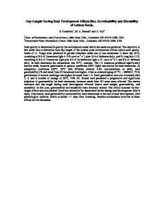
After sacrifice, the legs were disarticulated at the knee and fresh frozen with the soft tissue and fixators attached in order to maintain pre-existing tensile forces in the tissue bridging the distraction gap. This test configuration was used to avoid introduction of new bending forces. Since the rat tibia is naturally curved, tensile testing of the individual bone would add a bending moment. During in vivo distraction of the rat tibia, these bending moments are neutralized by the external fixator-wire -bone construct as evidenced by histological studies demonstrating collagen and bone formation parallel to the fixator distraction rods and by in vivo strain gage testing [l-31. By testing the distraction gap tissues within the original external fixation device, we approach pure axial tension, since any bending moments have already been dissipated. Prior to testing, the tibiae, with fixators attached, were thawed and macerated under a dissecting microscope. Methyl methacrylate (MMA) was allowed to polymerize to a doughy consistency. Two portions of MMA were rolled and wrapped into a donut shape around each end of the tibia proximally and distally (exactly 20 mm apart and within the fixation wires) to secure tubing for the differential variable reluctance transducer (DVRT). A custom-made tool was used to insert two pieces of plastic tubing (2 mm diameter, 5 mm length) into each MMA donut. The MMA was allowed to harden for 15 min at which time the tool was removed leaving the tubing. Tensile testing was carried out using the upper and lower fixator rings attached to a custom-made materials testing machine by four one-inch hex standol? nuts (Fig. I ) . Before the fixated tibia was mounted, the testing machine load cell was set to zero. After securing the fixator rings, the distraction rods connecting the rings were removed to isolate the new bone segment for tensile testing. The prongs of the DVRT, used to measure displacement, were inserted into the MMA-fixed tubing. The tensile load was readjusted to zero and the specimen position recorded. A LabVIEW program (National Instruments, Austin, TX) and computer were used to control the axial distraction (0.1 mm increments) of the ringsltibia and to record the load-deformation curves. Stiffness (slope), ultimate tensile strength (UTS, load at failure), and energy to failure (area under the curve) were calculated by computer. For three-point bending analyses, selected tibiae were tested to failure in the mid-sagittal plane with an MTS machine (BionixTM). operated at a constant rate of 0.25 m d s and load to failure was recorded and bending moment calculated (Fig. 2). We have considered the use of four-point bending for rat DO studies, however, we have chosen three-point bending previously and in this study for the following reasons. First, as illustrated in Fig. 2, we use roller points which contact the bone over a space of 2 mm in the MTS apparatus which distributes the stress over the consolidation gap of the distracted tibiae. Second, this setup was designed to match the size requirements for distracted or control tibiae, with the fulcrum localized to the distraction gap and the two outer rollers equidistant at a standardized moment arm of 18 mm. Third, the mid-sagittal plane can be tested in standardized fashion, since the tibia fits into the rollers in a reproducible alignment (Fig. 2). Fourth, the results we have obtained using this method demonstrate small variances associated with this
J. Aronson et ul. I Journal of Orthopuedic Reseurch 19 (2001 i 6 4 4 9
66
!
Distraction Motor
I
technique, especially after 70 days, and in fact the variances are small enough to allow the use of n = 3 per time point with appropriate statistical power. Stutistic,s Although the sample size for the three-point bending comparisons appears small ( n = 3), a retrospective power analysis demonstrates that the p values obtained are valid with a power of greater than 80%. Statistical significance was determined by the unpaired Student’s t test for the data presented in Figs. 3(a), 4 and 7, by linear regression analysis for Fig. 5 , and by ANOVA for Figs. 6(a) and (b). All values are given and illustrated as the mean fthe standard error of the mean.
Results
Fig. 1. Schematic of the custom-made tensile testing apparatus (dark gray) designed to use the original lengthening fixator (light gray) to test the tibiae. Note attachments are to the lengthening rings with only the DVRT bridging the distraction gap, after the two distraction hexagonal supports are detached. Scale: each hexagonal support is exactly 18 mm long and the tibia is approximately 40 mm long.
Radiodensitometric and histologic evidence demonstrated progressive bridging (between days 20 and 30) and remodeling (days 30-70) of the bone in the distraction gap (Figs. 3(a) and (b)).At day 20, histological analysis re-confirmed primarily intramembranous ossification bridging the distraction gap with a central fibrous interzone of collagen bundles that progressively coalesce into osteoid columns parallel to the distraction force [4]. The microscopic structures paralleled the radiographic findings, such that the central fibrous interzone was radiolucent and the microscopic osteoid columns appeared as hazy, radiodense longitudinal trabeculae of bone. At day 30, both the radiographic and histologic microcolumns coalesced centrally, to bridge the previous fibrous interzone [4]. Between days 30 and 70 the microcolumns of new bone remodeled into cortical bone and cancellous trabeculae. Tensile testing demonstrated increasing ultimate tensile strength (UTS) from day 20 to 70 (9.9 f 10.39 4 . 2 f 18.8 N, P < 0.02) (Fig. 4). UTS was positively correlated (9= 0.82) with radiographic gap densities (Fig. 5). Both UTS and energy to failure (0.17f0.064 . 7 f 2 . 0 N mm) increased from day 20 to 70 (Figs. 4 and 6(b)). A significant increase in stiffness was demonstrated (Fig. 6(a)) during the period of bone bridging from day 20 to 30 (8.0f3.9-27.9f5.0 N/mm, P < 0.05). Tensile tests could not be properly analyzed for the control tibiae or day 70 distracted tibiae since these tibiae all failed at the proximal pin sites at -1300 N mm rather than in the healing gap. Three-point bending tests at day 70 demonstrated that the distracted tibiae had regained 65% of both contralateral and operated (fixator only) tibia1 strength (Fig. 7). Discussion
Fig. 2. Standardized alignment for three-point bending with roller balls on the MTS machine for day 70 experimental and control tibiae. The central roller is exactly 18 mm from each end roller.
These studies suggest that tensile test data correlates well to histological and radiographic data during distraction osteogenesis. Both UTS and energy to failure increase steadily from days 20 to 70 which reflects
67
J. Aronson et ul. / Journal of Orthopuedic Research 19 (2001 i 6 4 4 9
Ultimate Tensile Strength
X-ray Density I2O 100
200.
g
11
T I
150.
Day20
(a)
Day30
Day50
Day70
Control
Day 20
P
Day 30
Day 50
Day 70
P < 0.02 Day 20 vs. 70
Fig. 4. Ultimate tensile strength (average in N ) of distracted tibiae, n = 51time interval, measured on days 20 ( 9 . 9 f 10.3), 30 (42.1 f 10.6), 50 (62.5&8.5) and 70 (94.2f 18.8). Values shown are the m e a n i
standard error of the mean.
Concurrent Increase in X-ray Density and Ultimate Tensile Strength
2oo 150
1
100
50
0 Day20
[-
Day30
Day50
- X-ray Density
Day70
-+ UTS
I
Fig. 5. Graphic correlation of radiographic density (gray scale) to ultimate tensile strength (N).Values shown are the mean &standard error of the mean. Ultimate tensile strength and radiodensity were correlated at r' = 0.818 by linear regression (not shown).
(b)
Fig. 3. (a) Radiographic video analysis, using a gray scale from 0 (black) to 255 (white), of n = 8-9 tibiae harvested per time interval after osteotomy; day 20= 87.5 and day 70= 170, P < 0.01. Values shown are the mean fstandard error of the mean. Control = contralateral tibiae. (b) Radiographs from day 20 (upper left), day 30 (upper right), day 50 (lower left) and day 70 (lower right).
similar increases in radiodensity and histological evidence of remodeling to normal structure. The results of tensile testing demonstrate that radiographic and histologic evidence for bridging of the distraction gap is accompanied by a significant increase in tensile stiffness. In this 25% tibia1 lengthening model, the lengthened tibia reaches 65Y0 of normal three-point bending bone strength by day 70. These data demonstrate that a combination of tensile and three-point bending measures can, in a rat model, characterize the gain of bone strength during the distraction and consolidation phases of DO. Both tensile
68
J . Avonson et al. I Journal of Orthopaedic Research 19 (20011 6 4 4 9
Tensile Stiffness 40
-
35
.
30
-
E 25
-
E
Three Point Bending to Failure
700
T
600
T
-
E
2 20. 2
1
s 400.
H
15.
300.
Zoo. loo.
5. 0,
0,
Day 20
(a)
Day 30
Day 50
Day 70
P < 0.05 Day 20 VS. 30
7. 6.
f
5,
E.
T Day 20
(b)
Control Day 70 (IefI)
Normal (Right)
Normal
(Len)
P < 0.05 Day 70 Dlsfraction vs. Control
Energy to Failure
E E
Distraction Day 70 (right)
Day 30
Day 50
Day 70
P < 0.05 Day 20 vs. 70 and Day 30 vs. 70
Fig. 6. (a) Development of tensile stiffness (average in Nimm) on day 7 0 ( 8 . 0 ? ~ 3 . 9 3) ,0 ( 2 7 . 9 f 5 . 0 ) ,50(32.2-+3.l)and70(26.6&1.7),with a significant increase between day 20 and 30. (b) Energy to failure (average in N mm) on day 20 (0.17-+0.06), 30 (0.9 +0.29), 50 ( I .6 f 0.49) & 70 (4.7 4~2.0), with significant differences noted. Values shown are the mean fstandard error of the mean. Note that despite an apparent decrease in tensile stiffness at day 70, the energy to failure increases significantly up to day 70.
and bending tests are recommended because neither one can be used at all time points. Three-point bending is not applicable at the early time points (up to day 20 and perhaps 30) while tensile testing is not possible with this protocol when the new bone strength exceeds 1300 N mm (attained after bony bridging by day 70). It is possible that both tensile and three-point bending could be performed at day 50, although we did not compare specimens at this time point. Tensile testing during distraction and through the early consolidation phase appears to provide a reliable means of evaluating significant changes in stiffness and UTS and these changes correlate to radiodensity and histology. The reliability of the tensile tests are demon-
Fig. 7. Results of three-point bending tests ( n = 3/group) at day 70 on distracted right tibiae, contralateral left tibiae (control), externally fixed-only right tibiae (normal), and unoperated left tibiae (normal) presented as bending moments, N mm. No significant differences were found between the controls. Although the distracted tibiae at day 70 were 65% of normal, they were still significantly lower than controls. Values shown are the mean fstandard error of the mean.
strated by the small standard errors of the relatively small samples ( n = 5) and by comparison to the standard errors associated with the bending tests ( n = 3). The significant increase in stiffness coinciding with bone bridging is an expected finding since native collagen and bone exhibit major differences in their modulus of elasticity. The results of in vitro tensile testing mimic the data from in vivo strain gauges used to measure distraction forces in line with external fixators [3]. Therefore, it appears that both in vivo and in vitro tensile forces rise in proportion to mineralization of the gap collagen [3]. In addition, a decrease in both X-ray density and tensile stiffness from day 50 to 70 was observed and, though not significant statistically, we believe these values may reflect either stress shielded osteoporosis or increasing osteoclastic activity which will result in the eventual reformation/remodeling of the marrow cavity. In conclusion, in vitro tensile testing employing external fixators provides a useful method to test the development of structural integrity during D O in a rat model. The tensile test results parallel the radiodensity and histological findings. Therefore, the effects of inhibitors or stimulators of bone repair on strength development can be reliably measured in a rat model during the early stages (bone formation, bridging, early consolidation) of DO.
Acknowledgements
NIH grant #AR44987, USDA grant #9402890, Brooks Medical Research Fund, Laboratory for Limb
J. Aronson et al. I Journal of' Orthopavdir Reseurch 19 (2001i 64-69
Regeneration Research - Arkansas Children's Hospital Research Institute, and the Departments of Orthopaedics and Pediatrics, University of Arkansas for Medical Sciences. References [l] Aronson J, Good B, Stewart C, Harrison B, Harp J. Preliminary studies of mineralization during distraction osteogenesis. Clin Orthop 1990;250:43-9. [2] Aronson J. Experimental and clinical experience with distraction osteogenesis. Cleft Palate-Craniofacial J 1994;31:473-81, [3] Aronson J, Harp J. Mechanical forces during tibia1 lengthening. Clin Orthop 1994;301:73-9. [4] Aronson J, Shen XC, Skinner RA, Hogue WR. Badger TM, Lumpkin Jr CK. Rat model of distraction osteogenesis. J Orthop Res 1997;15(2):221-6.
69
[5] Aronson J, Shen XC. Gao GG, Miller F, Quattlebaum T, Skinner
RA, Badger TM, Lumpkin Jr CK. Sustained proliferation accompanies distraction osteogenesis in the rat. J Orthop Res 1997;15(4):563-9. [6] Ilizarov GA. Part I. The influence of stability of fixation and softtissue preservation: the tension-stress effect on the genesis and growth of tissues. Clin Orthop 1989;?38:249-81. [7] Lumpkin Jr CK, Aronson J , Shen XC. Gao GG, Skinner RA, Badger TM. The impact of total enteral nutrition on distraction osteogenesis in a rat model. J Bone Miner Res 1996;11:962-9. [8] Walsh WR, Hamdy RC, Ehrlich MG. Biomechanical and physical properties of lengthened bone in a canine model. Clin Orthop 1994;306:230-8. [9] Walsh WR, Sherman P, Howlett CR, Sonnabend DH. Erlich MG. Fracture healing in a rat osteopenia model. Clin Orthop 1997;342:218--27.