Concise Definitive Review
Critical care of the burn patient: The first 48 hours Barbara A. Latenser, MD, FACS Objective: The goal of this concise review is to provide an overview of some of the most important resuscitation and monitoring issues and approaches that are unique to burn patients compared with the general intensive care unit population. Study Selection: Consensus conference findings, clinical trials, and expert medical opinion regarding care of the critically burned patient were gathered and reviewed. Studies focusing on burn shock, resuscitation goals, monitoring tools, and current recommendations for initial burn care were examined. Conclusions: The critically burned patient differs from other critically ill patients in many ways, the most important being the necessity of a team approach to patient care. The burn patient is best cared for in a dedicated burn center where resuscitation and
M
ajor strides in understanding the principles of burn care over the last half century have resulted in improved survival rates, shorter hospital stays, and decreases in morbidity and mortality rates due to the development of resuscitation protocols, improved respiratory support, support of the hypermetabolic response, infection control, early burn wound closure, and early enteral nutrition (1). Critical care of the burn patient requires the participation of every discipline in the hospital.
Resuscitation Goals Effective fluid resuscitation is one of the cornerstones of modern burn care and perhaps the advance that has most directly improved patient survival. Proper fluid resuscitation aims to anticipate and prevent rather than to treat burn shock (2– 4). Resuscitation of burn shock cannot hope to achieve complete normalization of physiologic variables because the
From the Carver College of Medicine, University of Iowa Hospitals & Clinics, Department of Surgery, Iowa City, Iowa. The author has not disclosed any potential conflicts of interest. For information regarding this article, E-mail:
[email protected] Copyright © 2009 by the Society of Critical Care Medicine and Lippincott Williams & Wilkins DOI: 10.1097/CCM.0b013e3181b3a08f
Crit Care Med 2009 Vol. 37, No. 10
monitoring concentrate on the pathophysiology of burns, inhalation injury, and edema formation. Early operative intervention and wound closure, metabolic interventions, early enteral nutrition, and intensive glucose control have led to continued improvements in outcome. Prevention of complications such as hypothermia and compartment syndromes is part of burn critical care. The myriad areas where standards and guidelines are currently determined only by expert opinion will become driven by level 1 data only by continued research into the critical care of the burn patient. (Crit Care Med 2009; 37:2819 –2826) KEY WORDS: burn resuscitation; burn monitoring; critical care; burn edema; burn complications; burn shock
burn injury leads to ongoing cellular and hormonal responses. The obvious challenge is to provide enough fluid replacement to maintain perfusion without causing fluid overload (3, 5–17). Without effective and rapid intervention, hypovolemia/shock will develop if the burns involve ⬎15% to 20% total body surface area (TBSA) (18). Delay in fluid resuscitation beyond 2 hrs of the burn injury complicates resuscitation and increases mortality (7, 16). The consequences of excessive resuscitation and fluid overload are as deleterious as those of under-resuscitation: pulmonary edema, myocardial edema, conversion of superficial into deep burns, the need for fasciotomies in unburned limbs, and abdominal compartment syndrome (5, 19 –22). A Lund-Browder chart should be completed at the time of admission to calculate the TBSA burn (1).
Burn Shock Pathophysiology Burn shock is a unique combination of distributive and hypovolemic shock (5, 22–26) manifested by intravascular volume depletion, low pulmonary artery occlusion pressures, elevated systemic vascular resistance, and depressed cardiac output (23, 27). Reduced cardiac output is a combined result of decreased plasma volume, increased afterload, and decreased contractility (4). Studies suggest that impaired myocardial contractility is likely caused by circulating mediators
such as tumor necrosis factor-␣ (28, 29), however impaired Ca⫹2 at the cellular level is most likely involved as well (30). The exact mechanisms of altered cardiac mechanical function remain unclear and are most likely multifactorial (5, 30, 31). Virtually all components that control fluid and protein loss from the vascular space are altered after a burn (25). Immediately after burn injury, the systemic microcirculation loses its vessel wall integrity and proteins are lost into the interstitium (5, 17, 32). This protein loss causes the intravascular colloid osmotic pressure to drop precipitously and allows fluid to escape from the circulatory system (5, 32). There is a marked transient decrease in interstitial pressure caused by the release of osmotically active particles, causing a vacuum effect that sucks in fluid from the plasma space. There is a marked increase in fluid flux into the interstitium caused by a combination of the sudden decrease in interstitial pressure, an increase in capillary permeability to protein, and a further imbalance in hydrostatic and oncotic forces favoring the fluid movement into the interstitium (25). The outcome is a dramatic outpouring of fluids, electrolytes, and proteins into the interstitium with rapid equilibrium of intravascular and interstitial compartments (17). These changes are reflected in loss of circulating plasma volume, hemoconcentration, massive edema formation, decreased urine output, and 2819
depressed cardiovascular function (23). What actually changes is the volume of each fluid compartment, with intracellular and interstitial volumes increasing at the expense of plasma and blood volume (17). Functional plasma volume in burn tissue can be restored only with expansion of the extracellular space (33). Most edema occurs locally at the burn site and is maximal at 24 hrs postinjury (5, 14, 17, 18, 25, 33, 34). The rate and extent of edema formation in major burn injury far exceed the intended beneficial effect of inflammatory system activation (21, 25). The edema itself results in tissue hypoxia and increased tissue pressure with circumferential injuries. Aggressive fluid therapy can correct the hypovolemia but will accentuate the edema process (21, 25, 35, 36).
Resuscitation Formulas Adequate resuscitation from burn shock is the single most important therapeutic intervention in burn treatment. Due to a paucity of evidence-based literature, burn resuscitation remains an area of clinical practice driven primarily by local custom of treating burn units (20). The only issue exempt from debate is that fluid administration is universally advocated (22, 37). Each patient will react uniquely to burn injury depending on age, depth of burn, concurrent inhalation injury, preexisting comorbidities, and associated injuries. Formulas should be regarded as a resuscitation guideline; fluid administration has to be adjusted to individual patient needs. Of the numerous formulas for fluid resuscitation, none is optimal regarding volume, composition, or infusion rate (2, 4 – 6, 12, 15, 17, 32). Lactated Ringer’s solution most closely resembles normal body fluids. Factors that influence fluid requirements during resuscitation besides TBSA burn include burn depth, inhalation injury, associated injuries, age, delay in resuscitation, need for escharotomies/fasciotomies, and use of alcohol or drugs (34). The Parkland formula has been renamed the Consensus formula because it is the most widely used resuscitation guideline. The Advanced Burn Life Support curriculum supports the use of the Consensus formula for resuscitation in burn injury (32). Simply put, it is 4 mL/kg per percentage TBSA, describing the amount of lactated Ringer’s solution required in the first 24 hrs after burn injury, where kg represents patient 2820
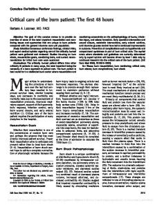
weight, and percentage TBSA is the size of the burn injury. Starting from the time of burn injury, half of the fluid is given in the first 8 hrs and the remaining half is given over the next 16 hrs. The rapid determination of percentage TBSA burn and calculation of the fluid requirements can be difficult and often incorrect when the person treating these burns is an inexperienced clinician. The substantial errors in estimating burn extent and depth result in significant under- or overcalculation of fluid requirements (17, 18, 38, 39). Most doctors outside burn centers have infrequent experience with major burn management and a relative lack of sufficient knowledge regarding such management (3, 17, 36, 38). Even among burn center physicians, there is considerable variability in determining the amount of fluids to be administered during the resuscitation period. There has not been a clinical advantage with colloids (5, 12, 40). One study showed a decreased risk of death when albumin was used during resuscitation (20), but the difference did not achieve statistical significance. A meta-analysis comparing albumin to crystalloid showed a 2.4-fold increased risk of death with albumin (24). Hypertonic saline has also had disappointing results, with a fourfold increase in renal failure and twice the mortality of patients given lactated Ringer’s solution(41). Hypertonic saline does not routinely have a place in burn resuscitation (22). Fresh frozen plasma should not be used as a volume expander, according to new policies on blood product delivery (24). Due to the risk of bloodborne infectious transmission (5), the American Burn Association Practice Guidelines for Burn Shock Resuscitation do not recommend the use of fresh frozen plasma without active bleeding or coagulopathy outside of a clinical trial, when other choices are available (4). Depletion of limited blood bank reserves is another deterrent to using fresh frozen plasma in burn resuscitation (5). Although there are many resuscitation protocols, the University of Utah has a simple, easy-to-follow protocol for adult burn patients (39) that is based on the Consensus formula (Fig. 1). During resuscitation, development of unstable vital signs, inadequate response to fluids, or persistently high fluid requirements should prompt a call to an experienced burn care physician. Resuscitation Nonresponders. It is not possible to accurately predict who will fail
resuscitation, but patients who routinely require additional fluid include those with inhalation injury, electrical burns, those in whom resuscitation is delayed, and those using alcohol or illicit drugs (42). Patients making methamphetamine have larger, deeper burns (43) and often require two to three times the standard Consensus formula resuscitation (43, 44). There is significantly increased inhalation injury, nosocomial pneumonia, respiratory failure, and sepsis (43, 44). Vascular Access/Other Tubes and Catheters. No factor other than airway protection is as critical in the early postburn period as vascular access. Ideally, obtain peripheral intravenous access away from burned tissues (36). Most patients with small to medium-sized burns do not require central catheters. If no intravenous access is available, intraosseous catheters may safely be placed in patients of any age. These tools obviate the need for cutdowns in burn patients. A patient undergoing resuscitation should have a Foley catheter placed. Nasogastric tubes should be considered in patients with ⬎20% TBSA burns, as they will experience gastroparesis and probable emesis (1).
First-Line Monitoring Although urine output and heart rate are the primary modalities for monitoring, the current standards for monitoring fluid therapy in patients with large burns are not supported by data (12, 15, 37, 45). Reliance on hourly urine output as the sole index of optimum resuscitation sharply contrasts with the lack of clinical studies demonstrating the ideal hourly urine output during resuscitation (4). The American Burn Association Practice Guidelines for Burn Shock Resuscitation recommend 0.5 mL/kg/hr urine output in adults and 0.5–1.0 mL/kg/hr in children weighing ⬍30 kg (5, 26, 32, 34). Lesser hourly urinary outputs in the first 48 hrs post burn almost always represent inadequate resuscitation (35). Hemodynamic monitoring and treatment of deviation from normovolemia are the fundamental tasks in intensive care (46). A pulse rate ⬍110 beats/min in adults usually indicates adequate volume, with rates ⬎120 beats/min usually indicative of hypovolemia. Narrowed pulse pressure provides an earlier indication of shock than systolic blood pressure alone (5). Arterial Catheter Versus Blood Pressure Cuff. Noninvasive blood pressure measurements by cuff are rendered inacCrit Care Med 2009 Vol. 37, No. 10
Figure 1. Fluid resuscitation algorithm for the adult burn patient, reprinted with permission by Jeffrey R. Saffle, MD (39). HR, heart rate; BP, blood pressure; MD, physician; IV, intravenous; LR, lactated Ringer’s solution.
curate because of the interference of tissue edema and read lower than the actual blood pressure (32). An arterial catheter placed in the radial artery is the first choice, followed by the femoral artery. Pulmonary Artery/Central Venous Catheters. The decision to perform invasive hemodynamic monitoring requires careful consideration (45). The lack of benefit associated with goal-directed supranormal therapy has resulted in waning enthusiasm for the use of pulmonary artery catheters (47, 48). The most applicable cardiac output-related variable to manipulate in burn patients is preload. Crit Care Med 2009 Vol. 37, No. 10
Pulmonary artery occlusion pressure and central venous pressure are not good indicators of preload (5). As long as other signs of adequate tissue perfusion are normal, the temptation to normalize filling pressures should be avoided (32). The use of end points demonstrating the adequacy of oxygen delivery has not yet found a place in the management of burn shock (11, 23, 49). Laboratory Studies. Although the initial lactate is a strong predictor of mortality (5, 20, 50), it is not clear how serum lactate can be used as a resuscitation end point (32, 50, 51). Although lactate and
base deficit (BD) are resuscitation markers that act as independent variables (50 – 52), there is a low correlation between urinary output, mean arterial pressure, serum lactate, and base deficit (51). Serum lactate trends provide greater information regarding the homeostatic status (53, 54). Determinations of BD do not demonstrate the same predictive power; the effect of specific correction of the BD during fluid resuscitation is unknown (13, 50, 52). There are insufficient data to make recommendations on the use of BD or lactate as resuscitation guidelines during burn resuscitation or as independent predictors of outcome in patients with large burns (5, 32, 51, 55). Hematocrits of 55% to 60% are not uncommon in the early postburn period and cannot be used to monitor fluid resuscitation. Resuscitation End Points. End points of resuscitation have been the subject of numerous strategies with conflicting results (5, 13, 15, 16, 19, 22–24). Many authors feel that urine output (34) and traditional vital signs (heart rate and mean arterial pressure) are too insensitive to ensure appropriate fluid replacement in burn injuries (11, 32, 49, 51). In children, trends in heart rate, blood pressure, and capillary refill toward normal are more reasonable therapeutic end points (19). In adults, arterial blood pressure is relatively insensitive to the adequacy of fluid replacement; pulse rate is more helpful. In older patients, pulse rate becomes less reliable. Urine output can be taken to reflect organ perfusion; however, urine must be nonglycosuric to be accurate (36). Hypertonic saline can increase urine output due to an osmotic diuresis that does not accurately reflect volume status (33). Although urine output does not precisely mirror renal blood flow, it remains the most readily accessible and easily monitored index of resuscitation (35, 56). Fluid Creep. The use of excessive volumes for resuscitation is being documented with increasing frequency in many burn centers (39, 57). Burn care providers have become more aggressive with the administration of benzodiazepines and narcotics, which may result in additional fluid demands (18, 20, 56, 58 – 60). Outreach education in burn care has contributed to a now-common problem of excessive resuscitation given by first responders and non-burn physicians. Thus, many patients arrive at a burn center having received most of their first 8-hr Consensus formula requirements in just an hour or two (39). 2821
Vitamin C Resuscitation. The landmark study by Tanaka et al showed that high dose ascorbic acid during the initial 24 hrs post burn reduced fluid requirements by 40%, reduced burn tissue water content 50%, and reduced ventilator days (61, 62). The clinical benefits led to a clear reduction in edema and body weight gain and were associated with reduced respiratory impairment and reduced requirement for mechanical ventilation (36, 61, 62). Although not in mainstream use, the findings are meaningful to experienced burn care practitioners. Inhalation Injury. The combination of a body burn and smoke inhalation produces a marked increase in mortality and morbidity (63, 64). Burn patients with inhalation injury have been shown to require increased fluids during resuscitation (1, 8, 15, 37, 65). Navar et al (66) found that the presence of inhalation injury was associated with a 44% increase in fluid requirements, which was remarkably uniform across all age groups and burn sizes. The degree of lung dysfunction caused by a smoke inhalation injury is accentuated by the presence of even a small body burn (25, 36, 64, 65). Acute upper airway obstruction occurs in 20% to 33% of hospitalized burn patients with inhalation injury and is a major hazard because of the possibility of rapid progression from mild pharyngeal edema to complete upper airway obstruction (67). Patients presenting with stridor should be intubated on presentation. Patients at risk of requiring early intubation include those with a history of being in an enclosed space with or without facial burns, history of unconsciousness, carbonaceous sputum, voice change, or complaints of a “lump in the throat.” In isolation, these factors do not predict the need for intubation, but the more signs present, the more elevated the risk. A carboxyhemoglobin level taken within 1 hr after injury is strongly indicative of smoke inhalation if ⬎10% (3). If there is a significant cutaneous burn requiring resuscitation, the need for intubation will be greater. The small cross-sectional diameter of the pediatric airway places children at higher risk of requiring emergent intubation. If intubation is needed, the most experienced clinician in airway management should perform endotracheal intubation (67). Intubation itself is not without risk so should not be undertaken routinely simply because there are facial burns. The care of inhalation injury remains supportive. Even the gold standard of 2822
bronchoscopy within the first 24 hrs of admission cannot accurately predict the severity of inhalation injury. For patients with inhalation injury, no ideal ventilator strategy has emerged (67). According to the American College of Chest Physicians, recommendations for mechanical ventilation serve as general guidelines: Use a ventilator mode that is capable of supporting oxygenation and ventilation that the clinician has experience using, limit plateau pressures to ⬍35 cm H2O, allow PCO2 to increase if needed to minimize plateau pressures, and use the appropriate level of positive end-expiratory pressure (68). Roughly 70% of patients with inhalation injury will develop ventilator-associated pneumonia. Routine pneumonia prevention strategies should include elevating the head of the bed 30°, turning the patient side to side every 2 hrs, oral care every 6 hrs, and gastrointestinal prophylaxis. Prophylactic antibiotics have no role and actually increase infection rates. For patients who fail to respond to maximal conventional therapy, consider extracorporeal membrane oxygenation as a rescue therapy for patients with acute respiratory failure who are expected to die otherwise (69).
Preventable Complications Hypothermia. The profoundly adverse effects of hypothermia cannot be overstated. Strategies to vigorously prevent hypothermia include a warmed room, warmed inspired air, warming blankets, and countercurrent heat exchangers for infused fluids. Metabolic responses can be minimized by treating the patient in a thermoneutral environment (32°C) (3). During hydrotherapy, in the operating room, and in the burn unit, keep the room temperature at ⱖ85°F to minimize heat loss and decrease metabolic rate. Compartment Syndromes. A lifethreatening complication caused by highvolume resuscitation is abdominal compartment syndrome (ACS) (24), defined as intra-abdominal pressure ⱖ20 mm Hg plus at least one new organ dysfunction (70). ACS has been associated with renal impairment, gut ischemia, and cardiac and pulmonary malperfusion. Clinical manifestations include tense abdomen, decreased pulmonary compliance, hypercapnia, and oliguria. Simply monitoring urine output is insufficiently sensitive or specific to diagnose ACS (36, 71, 72). Vigilant monitoring and aggressive treat-
ment should be instituted to avoid this deadly complication (71, 72). Appropriate intravascular volume, appropriate body positioning, pain management, sedation, nasogastric decompression if appropriate, chemical paralysis if required, and torso escharotomy are all interventions to increase abdominal wall compliance and decrease intra-abdominal pressures (72, 73). Bladder pressure monitoring should be initiated as part of the burn fluid resuscitation protocol in every patient with ⬎30% TBSA burn (5, 24, 73). Patients who receive ⬎250 mL/kg of crystalloid in the first 24 hrs will likely require abdominal decompression (15). Percutaneous abdominal decompression is a minimally invasive procedure that should be performed before resorting to laparotomy (71, 74). The International Conference of Experts on Intra-abdominal Hypertension and Abdominal Compartment Syndrome recommends that if less invasive maneuvers fail, decompressive laparotomy should be performed in patients with ACS that is refractory to other treatment options (72). The reported mortality rates for decompressive laparotomy for ACS can be as high as 88% (71) to 100% (74). Extremity compartment syndromes can also result from extensive edema formation. Patients may require escharotomies, fasciotomies, or both for the release of extremity compartment syndrome (36, 75). Patients with circumferential fullthickness burns are also at risk of requiring escharotomies (35). Impaired capillary refill, paresthesia in the involved extremity, and increased pain develop earlier than decreased pulses. The orbit is a compartment limited to expansion and may require lateral canthotomy to successfully reduce intraocular pressure to normal (76). Deep Venous Thrombosis. The incidence of deep venous thrombosis in burn patients is estimated to be 1% to 23% (77). In the absence of level 1 evidence, deep venous thrombosis chemoprophylaxis is routinely practiced in many burn centers. Heparin-Induced Thrombocytopenia. Early thrombocytopenia occurs in the postburn course in patients with extensive injury. Problems after burn injury such as pulmonary infections, multiorgan failure, sepsis, and bleeding disorders accentuate this trend. As in nonburn patients, careful observance for thrombocytopenia after the first week of hospitalization will alert the practitioner to make Crit Care Med 2009 Vol. 37, No. 10
the diagnosis in burn patients (78, 79). Although the incidence of heparininduced thrombocytopenia was relatively low (1.6%) in one study (79), the complications in those patients were profound, including arterial and deep venous thromboses and increased number of surgical procedures (79). Neutropenia. Transient leukopenia is common, primarily due to a decreased neutrophil count. Maximal white blood cell depression occurs several days after admission with rebound to normal a few days later. Use of silver sulfadiazine has been associated with this transient leukopenia; resolution is independent of continued silver sulfadiazine (1). Stress Ulcers. Level 1 data exists that patients with major burn injuries are at risk for stress ulcers and should receive routine prophylaxis beginning at admission (80). Adrenal Insufficiency. Although absolute adrenal insufficiency occurs in up to 36% of patients with major burns, there is no correlation between response to corticotropin stimulation and survival. Those with massive burns have higher cortisol levels but may be resistant to serum cortisol increases in response to stimulation. The clinical relevance of this finding has not been established (81, 82).
Infection/Inflammation/Sepsis Consensus Paper on Sepsis and Infection-Related Diagnoses. Current definitions for sepsis and infection have many criteria routinely found in patients with extensive burns without infection/sepsis (e.g., fever, tachycardia, tachypnea, leukocytosis). Burn experts recently developed standardized definitions for sepsis and infection-related diagnoses in burn patients from which I will summarize key discussion points and recommendations (78). Patients with large burns have a baseline temperature reset to 38.5°C, and tachycardia and tachypnea may persist for months. Continuous exposure to inflammatory mediators leads to significant changes in the white blood cell count, making leukocytosis a poor indicator of sepsis. Use other clues as signs of infection or sepsis such as increased fluid requirements, decreasing platelet counts ⬎3 days after burn injury, altered mental status, worsening pulmonary status, and impaired renal function. The term systemic inflammatory response syndrome should not be applied to burn patients Crit Care Med 2009 Vol. 37, No. 10
because patients with large burns are in a state of chronic systemic inflammatory stimulation (78). Any infection in a burn patient should be considered to be from the central venous catheter until proven otherwise (78). Central catheters should be changed to a new site every 3 days to minimize bloodstream infections (83). Although prophylactic systemic antibiotics have no role in thermal injury, topical antimicrobial therapy is efficacious (1). Systemic antibiotic therapy should be culture directed and administered for the shortest time possible.
Metabolism/Nutrition Enteral Nutrition. As hypermetabolism can lead to doubling of the normal resting energy expenditure, enteral nutrition should be started as soon as resuscitation is underway with a transpyloric feeding tube. Patients with burns ⬎20% TBSA will be unable to meet their nutritional needs with oral intake alone. Patients fed early have significantly enhanced wound healing and shorter hospital stays (84). In the rare case that precludes use of the gastrointestinal tract, parenteral nutrition should be used only until the gastrointestinal tract is functioning. Endocrine and Glucose Monitoring. Strict glucose control of 80 –110 mg/dL can be achieved using an intensive insulin therapy protocol, leading to decreased infectious complications and mortality rates (85, 86). Anabolic Steroids. Severe burn injuries induce a hypermetabolic response, which leads to catabolism. Anabolic androgenic steroids such as oxandrolone promote protein synthesis, nitrogen retention, skeletal muscle growth, and decreased wound healing time. Burn patients receiving oxandrolone regain weight and lean mass two to three times faster than with nutrition alone (87). -Blockade. -blockers after severe burns decrease heart rate, resulting in reduced cardiac index and decreased supraphysiologic thermogenesis (3, 88). In children with burns, treatment with propranolol during hospitalization attenuates hypermetabolism and reverses muscle-protein catabolism. Propranolol is given to achieve a 20% decrease in heart rate of each patient compared with the 24-hr average heart rate immediately before administration (88).
Additional Therapies Wound Management. The primary goal for burn wound management is to close the wound as soon as possible, beginning at the time of injury. Burn centers are uniquely set up to provide optimal wound care. Beginning on admission and then daily, hydrotherapy is routine, involving washing the entire patient with chlorhexidine and warm tap water. The goal is to gently debride the nonviable tissue while leaving any newly formed dermis/epidermis. The practice of immersion in large tanks or other standing bodies of water has fallen out of favor, as bacteria from the fecal fallout zone quickly colonize the entire burn wound. Once the wound is clean, topical antimicrobial agents limit bacterial proliferation and fungal colonization in the burn wound (26). Silver sulfadiazine is the most commonly used topical antimicrobial, being readily available, affordable, and well tolerated by the patient. There are also silver-containing sheets and compounds that may be placed on partial thickness burns and remain in place for up to 7 days. For patients with fullthickness burns, prompt surgical excision of the eschar and allografting in patients with large burns, or autografting in patients with smaller burns, contributes to reduced morbidity and mortality (26). A host of temporary wound coverage products are available. Pain Management. Burn patients may experience pain that is multifaceted and constantly changing as the individual undergoes repeated procedures and wound manipulation. Inconsistent and inadequate pain management has been well documented. Although there is no universal treatment standard for pain management, opioid doses often significantly exceed recommended standard dosing guidelines (60, 89). Practice Management Guidelines for the Management of Pain by the Committee on the Organization and Delivery of Burn Care of the American Burn Association recommends that once intravenous access is obtained and resuscitation started, intravenous opioids should be administered. Background pain is best managed through the use of longacting analgesic agents. Breakthrough pain is addressed with short-acting agents via an appropriate route (89). Ketamine can be used for extensive burn dressing changes and procedures such as escharotomies. Anxiolytics such as benzodiazepines decrease background and 2823
procedural pain (89). For patients requiring mechanical ventilation, a propofol infusion will provide sedation but not analgesia. All medications should be given intravenously, orally, or rectally due to erratic absorption with intramuscular/ subcutaneous administration. Physiotherapy. Rehabilitation therapy begins at admission to maximize functional recovery. Burn patients require special positioning and splinting, early mobilization, strengthening and endurance exercises to promote healing (1). Transfer Criteria. The American Burn Association has established criteria for burn patients who should be acutely transferred to a burn center: ⬎10% TBSA partial thickness burns, any size fullthickness burn, burns to special areas of function or cosmesis, inhalation injury, serious chemical injury, electrical injury including lightning, burns with trauma where burns are the major problem, pediatric burns if the referring hospital has no special pediatric capabilities, and smaller burns in patients with multiple comorbidities (90).
CONCLUSIONS Not many topics in acute burn care are more hotly debated than fluid resuscitation and monitoring. Burn management is still not evidence based as in many areas of acute medicine (24). However, there does seem to be agreement among burns surgeons that: 1) the Consensus formula provides for a hypovolemic resuscitation; 2) patients with inhalation injury will require more fluid than that prescribed by the Consensus formula; and 3) over-resuscitation leads to excessive burn edema, abdominal compartment syndrome, need for fasciotomies on unburned limbs, pulmonary edema, and prolongation of mechanical ventilation. Type of monitoring to use during the early resuscitation period remains controversial in part because current end points have not yet been demonstrated to reflect tissue perfusion status independently and accurately (5, 91). Vital signs and urine output in burn patients do not fulfill these criteria (14). Defining better end points of resuscitation to avoid excessive volume administration is a high priority for future investigations (4). Future improvements in managing burn shock will include a complex ballet that includes pharmacologic interventions, rapid surgical removal of necrotic tissue, and a dynamic range of 2824
fluid types and rates of delivery. The continuing challenge for burn clinicians and researchers is to collaborate in large multicenter studies to critically evaluate and establish resuscitation end points and therapies (5, 36).
17. 18.
19.
REFERENCES 20. 1. Herndon DN (Ed): Total Burn Care. Philadelphia, Elsevier Saunders, 2007 2. Evans EI, Purnell OJ, Robinett PW, et al: Fluid and electrolyte requirements in severe burns. Ann Surg 1952; 135:804 – 815 3. Judkins K: Current consensus and controversies in major burns management. Trauma 2000; 2:239 –251 4. Pham TN, Cancio LC, Gibran NS: American Burn Association Practice Guidelines Burn Shock Resuscitation. J Burn Care Res 2008; 29:257–266 5. Ahrns KS: Trends in burn resuscitation: Shifting the focus from fluids to adequate endpoint monitoring, edema control, and adjuvant therapies. Crit Care Nurs Clin N Am 2004; 16:75–98 6. Bang RL, Ghoneim IE: The constant factor for fluid resuscitation in major burns. Ann Medit Burns Club 1994; VII:1– 8 7. Barrow RE, Jeschke MG, Herndon DN: Early fluid resuscitation improves outcomes in severely burned children. Resuscitation 2000; 45:91–96 8. Cancio LC, Cha´vez S, Alvarado-Ortega M, et al: Predicting increased fluid requirements during the resuscitation of thermally injured patients. J Trauma 2004; 56:404 – 414 9. Choi J, Cooper A, Gomez M, et al: The relevance of base deficits after burn injuries. J Burn Care Rehabil 2000; 21:499 –505 10. Cocks AJ, O’Connell A, Martin H: Crystalloids, colloids and kids: A review of paediatric burns in intensive care. Burns 1998; 24: 717–724 11. Elliott DC: An evaluation of the end points of resuscitation. J Am Coll Surg 1998; 187: 536 –547 12. Fodor L, Fodor A, Ramon Y, et al: Controversies in fluid resuscitation for burn management: Literature review and our experience. Injury, Int J Care Injured 2006; 37: 374 –379 13. Holm C, Melcer B, Ho¨rbrand F, et al: Intrathoracic blood volume as an end point in resuscitation of the severely burned: An observational study of 24 patients. J Trauma 2000; 48:728 –734 14. Holm C, Tegeler J, Mayr M, et al: Effect of crystalloid resuscitation and inhalation injury on extravascular lung water. Chest 2002; 121:1956 –1962 15. Klein MB, Hayden D, Elson C, et al: The association between fluid administration and outcome following major burn: A multicenter study. Ann Surg 2007; 245:622– 628 16. Schiller WR, Bay RC: Hemodynamic and ox-
21.
22. 23.
24.
25.
26.
27.
28.
29.
30.
31.
32.
33. 34. 35.
36.
ygen transport monitoring in management of burns. New Horiz 1996; 4:475– 481 Warden GD: Burn shock resuscitation. World J Surg 1992; 16:16 –23 Mitra B, Fitzgerald M, Cameron P, et al: Fluid resuscitation in major burns. ANZ J Surg 2006; 76:35–38 Carvajal HF: Fluid resuscitation of pediatric burn victims: a critical appraisal. Pediatr Nephrol 1994; 8:357–366 Cochran A, Morris SE, Edelman LS, et al: Burn patient characteristics and outcomes following resuscitation with albumin. Burns 2007; 33:25–30 Holm C, Mayr M, Tegeler J, et al: A clinical randomized study on the effects of invasive monitoring on burn shock resuscitation. Burns 2004; 30:798 – 807 Ipaktchi K, Arbabi S: Advances in burn critical care. Crit Care Med 2006; 34:S239 –S244 Barton RG, Saffle JR, Morris SE, et al: Resuscitation of thermally injured patients with oxygen transport criteria as goals of therapy. J Burn Care Rehabil 1997; 18:1–9 Berger MM, Bernath M-A, Chiole´ro RL: Resuscitation, anaesthesia and analgesia of the burned patient. Curr Opin Anaesthesiol 2001; 14:431– 435 Demling RH: The burn edema process: Current concepts. J Burn Care Rehabil 2005; 26:207–227 Rose JK, Herndon DN: Advances in the treatment of burn patients. Burns 1997; 23(Suppl 1):S19 –S26 Csontos C, Foldi V, Fischer T, et al: Factors affecting fluid requirement on the first day after severe burn trauma. ANZ J Surg 2007; 77:745–748 Willis MS, Carlson DL, DiMaio JM. et al: Macrophage migration inhibitory factor mediates late cardiac dysfunction after burn injury. Am J Physiol Heart Circ Physiol 2005; 288:H795–H804 Maass DL, White J, Horton J: IL-1 beta and IL-6 act synergistically with TNF-alpha to alter cardiac contractile function after burn trauma. Shock 2002; 18:360 –366 Lee MA, Yatani A, Sambol JT, et al: Role of gut-lymph factors in the induction of burninduced and trauma-shock-induced acute heart failure. Int J Clin Exp Med 2008; 1:171–180 Batchinsky AI, Wolf SE, Molter N, et al: Assessment of cardiovascular regulation after burns by nonlinear analysis of the electrocardiogram. J Burn Care Res 2008; 29:56 – 63 Ahrns KS, Harkins DR: Initial resuscitation after burn injury: Therapies, strategies, and controversies. AACN Clin Issue 1999; 10: 46 – 60 Demling RH: Fluid replacement in burned patients. Surg Clin North Am 1987; 67:15–30 Greenhalgh DG. Burn resuscitation. J Burn Care Res 2007; 28:555–565 Pruitt BA: Advances in fluid therapy and the early care of the burn patient. World J Surg 1978; 2:139 –150 Oliver RI, Spain D, Stadelmann W: Burns,
Crit Care Med 2009 Vol. 37, No. 10
37.
38.
39.
40.
41.
42.
43.
44.
45.
46.
47.
48.
49.
50.
51.
resuscitation and early management. Available at: http://www.emedicine.com/plastic/ topic159.htm. Accessed November 2004 Holm C: Resuscitation in shock associated with burns: Tradition or evidence-based medicine? Resuscitation 2000; 44:157–164 Bhat S, Humphries YM, Gulati S, et al: The problems of burn resuscitation formulas: A need for a simplified guideline. Available at: http://www.journalofburnsandwounds.com. Accessed February 8, 2009 Saffle JR: The phenomenon of “fluid creep” in acute burn resuscitation. J Burn Care Res 2007; 28:382–392 Gore DC, Dalton JM, Gehr TW: Colloid infusions reduce glomerular filtration in resuscitated burn patients. J Trauma 1996; 40: 356 –360 Huang PP, Stucky FS, Dimick AR, et al: Hypertonic sodium resuscitation is associated with renal failure and death. Ann Surg 1995; 221:543–557 Cancio LC, Reifenberg L, Barillo DJ, et al: Standard variables fail to identify patients who will not respond to fluid resuscitation following thermal injury: Brief report. Burns 2005; 31:358 –365 Burke BA, Lewis RW, Latenser BA, et al: Methamphetamine-related burns in the cornbelt. J Burn Care Res 2008; 29:574 –579 Santos AP, Wilson AK, Hornung CA, et al: Methamphetamine laboratory explosions: A new and emerging burn injury. J Burn Care Rehabil 2005; 26:228 –232 Ku¨ntscher MV, Czermak C, Blome-Eberwein S, et al: Transcardiopulmonary thermal dye versus single thermodilution methods for assessment of intrathoracic blood volume and extravascular lung water in major burn resuscitation. J Burn Care Rehabil 2003; 24: 142–147 Holm C, Melcer B, Ho¨rbrand F, et al: Arterial thermodilution: an alternative to pulmonary artery catheter for cardiac output assessment in burn patients. Burns 2001; 27:161–166 Venkatesh B, Meacher R, Muller MJ, et al: Monitoring tissue oxygenation during resuscitation of major burns. J Trauma 2001; 50: 485– 494 Mansfield MD, Kinsella J: Use of invasive cardiovascular monitoring in patients with burns greater than 30 per cent body surface area: A survey of 251 centers. Burns 1996; 22:549 –551 Dries DJ, Waxman K: Adequate resuscitation of burn patients may not be measured by urine output and vital signs. Crit Care Med 1991; 19:327–329 Jeng JC, Jablonski K, Bridgeman A, et al: Serum lactate, not base deficit, rapidly predicts survival after major burns. Burns 2002; 28:161–166 Jeng JC, Lee K, Jablonski K, et al: Serum lactate and base deficit suggest inadequate resuscitation of patients with burn injuries: Application of a point-of-care laboratory instrument. J Burn Care Rehabil 1997; 18: 402– 405
Crit Care Med 2009 Vol. 37, No. 10
52. Kamolz L-P, Andel H, Schramm W, et al: Lactate: Early predictor of morbidity and mortality in patients with severe burns. Burns 2005; 31:986 –990 53. Pal JD, Victorino GP, Twomey P, et al: Admission serum lactate levels do not predict mortality in the acutely injured patient. J Trauma 2006; 60:583–589 54. Vincent J-L: End-points of resuscitation: arterial blood pressure, oxygen delivery, blood lactate, or . . .? Intensive Care Med 1996; 22:3–5 55. Cartotto R, Choi J, Gomez M, et al: A prospective study on the implications of a base deficit during fluid resuscitation. J Burn Care Rehabil 2003; 24:75– 84 56. Blumetti J, Hunt JL, Arnoldo BD, et al: The Parkland formula under fire: Is the criticism justified? J Burn Care Res 2008; 29:180 –186 57. O’Mara MS, Slater H, Goldfarb IW, et al: A prospective, randomized evaluation of intraabdominal pressures with crystalloid and colloid resuscitation in burn patients. J Trauma 2005; 58:1011–1018 58. Friedrich JB, Sullivan SR, Engrav LH, et al: Is supra-Baxter resuscitation in burn patients a new phenomenon? Burns 2004; 30: 464 – 466 59. Pruitt BA: Protection from excessive resuscitation: “Pushing the pendulum back.” J Trauma 2000; 49:567–568 60. Sullivan SR, Friedrich JB, Engrav LH, et al: “Opioid creep” is real and may be the cause of “fluid creep.” Burns 2004; 30:583–590 61. Dubick MA, Williams C, Elgjo GI, et al: Highdose vitamin c infusion reduces fluid requirements in the resuscitation of burninjured sheep. Shock 2005; 24:139 –144 62. Tanaka H, Matsuda T, Miyagantani Y, et al: Reduction of resuscitation fluid volumes in severely burned patients using ascorbic acid administration. Arch Surg 2000; 135: 326 –331 63. Holt J, Saffle JR, Morris SE, et al: Use of inhaled heparin/N-acetylcystine in inhalation injury: Does it help? J Burn Care Res 2008; 29:192–195 64. Darling GE, Keresteci MA, Iban˜ez D, et al: Pulmonary complications in inhalation injuries with associated cutaneous burn. J Trauma 1996; 40:83– 89 65. Demling RH, Knox J, Youn Y-K, et al: Oxygen consumption early postburn becomes oxygen delivery dependent with the addition of smoke inhalation injury. J Trauma 1992; 32: 593–599 66. Navar PD, Saffle JR, Warden GD. Effect of inhalation injury on fluid resuscitation requirements after thermal injury. Am J Surg 1985; 150:716 –720 67. Mlcak RP, Suman OE, Herndon DN: Respiratory management of inhalation injury. Burns 2007; 33:2–13 68. Slutsky AS: Mechanical ventilation. American College of Chest Physicians’ Consensus Conference. Chest 1993; 104:1833–1859 69. Pierre EJ, Zwischenberger JB, Angel C, et al: Extracorporeal membrane oxygenation in
70.
71.
72.
73.
74.
75.
76.
77.
78.
79.
80.
81.
82.
83.
the treatment of respiratory failure in pediatric patients with burns. J Burn Care Rehabil 1998; 19:131–134 Malbrain MD, Cheatham MD, Kirkpatrick S, et al: Results from the International Conference of Experts on Intra-abdominal Hypertension and Abdominal Compartment Syndrome, I: Definitions. Intensive Care Med 2006; 32:1722–1732 Hershberger RC, Hunt JL, Arnoldo BD, et al: Abdominal compartment syndrome in the severely burned patient. J Burn Care Res 2007; 28:708 –714 Cheatam ML, Malbrain ML, Kirkpatrick A, et al: Results from the International Conference of Experts on Intra-abdominal Hypertension and Abdominal Compartment Syndrome, II: Recommendations. Intensive Care Med 2007; 33:951–962 Ivy ME, Atweh NA, Palmer J, et al: Intraabdominal hypertension and abdominal compartment syndrome in burn patients. J Trauma 2000; 49:387–391 Latenser BA, Kowal-Vern A, Kimball D, et al: A pilot study comparing percutaneous decompression with decompressive laparotomy for acute abdominal compartment syndrome in thermal injury. J Burn Care Rehabil 2002; 23:190 –195 Greenhalgh DG, Warden GD: The importance of intra-abdominal pressure measurements in burned children. J Trauma 1994; 36:685– 690 Sullivan SR, Ahmadi AJ, Singh CN, et al: Elevated orbital pressures: Another untoward effect of massive resuscitation after burn injury. J Trauma 2006; 60:72–76 Faucher LD, Conlon KM: Practice guidelines for deep venous thrombosis prophylaxis in burns. J Burn Care Rehabil 2007; 28: 661– 663 Greenhalgh DG, Saffle JR, Holmes JH, et al: American Burn Association consensus conference to define sepsis and infection in burns. J Burn Care Res 2007; 28:776 –790 Scott JR, Klein MB, Gernsheimer T, et al: Arterial and venous complications of heparin-induced thrombocytopenia in burn patients. J Burn Care Res 2007; 28:71–75 Guillamondegui OD, Gunter OL Jr, Bonadies JA, et al: Practice management guidelines for stress ulcer prophylaxis. Chicago, Eastern Association for the Surgery of Trauma (EAST), 2008. Available at: http://www. guideline.gov/summary/. Accessed February 8, 2009 Fuchs PC, Groger A, Bozkurt A, et al: Cortisol in severely burned patients: Investigations on disturbance of the hypothalamicpituitary-adrenal axis. Shock 2007; 28: 662– 667 Reiff DA, Harkins CL, McGwin G Jr, et al: Risk factors associated with adrenal insufficiency in severely injured burn patients. J Burn Care Res 2007; 28:854 – 858 King B, Schulman CI, Pepe A, et al: Timing of central venous catheter exchange and fre-
2825
quency of bacteremia in burn patients. J Burn Care Res 2007; 28:859 – 860 84. Venter M, Rode H, Sive A, et al: Enteral resuscitation and early enteral feeding in children with major burns: Effect on McFarlane response to stress. Burns 2007; 33:464 – 471 85. Hemmila MR, Taddonio MA, Arbabi S, et al: Intensive insulin therapy is associated with reduced infectious complications in burn patients. Surgery 2008; 144:629 – 637 86. Pidcoke HF, Wolf SE, Loo F, et al: Decreased
2826
mortality in burns with improved glucose control. Burns 2007; 33(1 Suppl):S33 87. Demling RH, Desanti L: Oxandrolone induced lean mass gain during recovery from severe burns is maintained after discontinuation of the anabolic steroid. Burns 2003; 29:793–797 88. Herndon DN, Hart DW, Wolf SE, et al: Reversal of catabolism by beta-blockade after severe burns. N Engl J Med 25 2001; 345: 1223–1229 89. Faucher LD, Furukawa K: Practice guide-
lines for the management of pain. J Burn Care Rehabil 2006; 27:659 – 668 90. American Burn Association. Available at: http://www.ameriburn.org. Accessed February 8, 2009 91. Samuelsson A, Steinvall I, Sjo¨berg F: Microdialysis shows metabolic effects in skin during fluid resuscitation in burn-injured patients. Crit Care 2006. Available at: http:// ccforum.com/content/10/6/R172. Accessed February 8, 2009
Crit Care Med 2009 Vol. 37, No. 10