THE ANATOMICAL RECORD 267:177–189 (2002)
Comparative Study of the Innervation Patterns of the Hyobranchial Musculature in Three Iguanian Lizards: Sceloporus undulatus, Pseudotrapelus sinaitus, and Chamaeleo jacksonii JAY J. MEYERS,1* ANTHONY HERREL,2 AND KIISA C. NISHIKAWA1 Physiology and Functional Morphology Group, Department of Biological Sciences, Northern Arizona University, Flagstaff, Arizona 2 Laboratory of Functional Morphology, Biology Department, University of Antwerp, Antwerp, Belgium 1
ABSTRACT The neuroanatomy and musculature of the hyobranchial system was studied in three species of iguanian lizards: Sceloporus undulatus, Pseudotrapelus sinaitus, and Chamaeleo jacksonii. The goal of this study was to describe and compare the innervation and arrangement of the hyobranchial musculature in the context of its function during tongue protrusion. A comparison of the hyobranchial innervation patterns revealed a relatively conserved innervation pattern in S. undulatus and P. sinaitus, and a modified version of this basic layout in C. jacksonii. All three species show anastomoses between sensory neurons of the trigeminal nerve and motor neurons of the hypoglossal nerve, suggesting that feedback may be important in coordinating tongue, jaw, and hyoid movements. The hyobranchial musculature of S. undulatus is very similar to that of P. sinaitus; however, there are minor differences, including the presence of an M. genioglossus internus (GGI) muscle in S. undulatus. Further differences are found mainly in functional aspects of the hyobranchial musculature, such as changes in the muscle lengths and the origins and insertions of the muscles. In C. jacksonii the hyobranchial system is comprised of largely the same components, but it has become highly modified compared to the other two species. Based on the innervation and morphological data gathered here, we propose a revision of the terminology for the hyobranchial musculature in iguanian lizards. Anat Rec 267:177–189, 2002. ©
2002 Wiley-Liss, Inc.
Key words: lizard; Sceloporus; Pseudotrapelus; Chamaeleo; nerve innervation; hyobranchium
Because of the systematic value of the hyobranchial apparatus in lepidosaurs (Camp, 1923; McDowell, 1972; Presch, 1971; Schwenk, 1988) and its importance in behaviors such as chemoreception (Gove, 1979; Cooper, 1995; Herrel et al., 1998; Toubeau et al., 1994; Schwenk, 2000a), feeding (Schwenk and Throckmorton, 1989; Delheusy and Bels, 1992; Herrel et al., 1995, 1996, 1997, 2000), and drinking (Bels et al., 1992; Bels et al., 1994; Delheusy, 1996), numerous authors have investigated its morphology over the past century (Mivart, 1867, 1870; Sanders, 1870, 1872, 1874; Minot, 1880; De Vis, 1883; Gnanamuthu, 1930, 1937; Zoond, 1933; George, 1948; Oelrich, 1956; Sondhi, 1958; De la Serna de Esteban, 1959, 1965; Jenkins and Tanner, 1968; Tanner and Avery, 1982; ©
2002 WILEY-LISS, INC.
Grant sponsor: NSF; Grant number: 9809942; Grant sponsor: NIH; Grant number: R25GM56931; Grant sponsor: Fund for Scientific Research (Flanders, Belgium). *Correspondence to: Jay Meyers, Physiology and Functional Morphology Group, Department of Biological Sciences, Northern Arizona University, Flagstaff, AZ 86011-5640. Fax (928) 5237500. E-mail:
[email protected] Received 25 July 2001; Accepted 1 March 2002 DOI 10.1002/ar.10096 Published online 00 Month 2002 in Wiley InterScience (www.interscience.wiley.com).
178
MEYERS ET AL.
Smith, 1984, 1988; Schwenk, 1986, 1988; Bell, 1989; Delheusy et al., 1994; Herrel et al., 1997; see Schwenk, 2000a, for an overview). Although in lizards the hyobranchial apparatus is often referred to as the hyoid or hyoid apparatus, a more appropriate term is the “hyobranchial” apparatus, since in lizards a larger portion of the apparatus (compared to that in mammals) is comprised of the branchial elements (visceral arches 3–5) (Schwenk, 2000a). Although the hyobranchial apparatus has been fairly well studied, remarkably little consensus regarding the homology of the different skeletal and muscular elements of the system has been reached. Consequently, there is no uniform terminology, which makes comparative morphological and functional studies of the hyobranchial apparatus extremely difficult. Even within closely related groups (e.g., within the iguania) there is no consensus regarding the homology of the different hyobranchial muscles, thus complicating evolutionary studies. A component of the hyobranchial system that has received disproportionately little attention up to now is the innervation pattern of the hyobranchial musculature. In contrast to the large body of literature cited above, only a handful of studies have looked at the innervation of the tongue and hyobranchial muscles in lizards in any detail (Bendz, 1840; Willard, 1915; Poglayen-Neuwall, 1954; Oelrich, 1956; Sondhi, 1958; Barbas-Henry, 1982; BarbasHenry and Lohman, 1984, 1986, 1988). Yet this information can provide crucial insights into the homology of the hyobranchial musculature among different groups (Kardong, 1998) and might be of great systematic value as well (Julien, 1967; Le´ curu, 1968; Julien and Renous-Le´ curu, 1972; Arnold, 1989). Moreover, with so few descriptions of the peripheral innervation, it is extremely hard to create valid biomechanical models of tongue function, or to predict how motor and sensory components of the system integrate during complex behaviors, such as tongue protrusion during prey capture (Chiel et al., 1992; Van Leeuwen, 1997; Mu and Sanders, 1999; McClung and Goldberg, 2000). Because of its importance during prey capture, the iguanian hyobranchial system has received far more attention than that in any other group of lizards (see Schwenk, 2000a, for an overview). As chameleons possess the unique ability to ballistically project the tongue out of the mouth, they have been the subject of numerous morphological studies over the past two centuries (Houston, 1828; Rice, 1973; Bell, 1989; see Herrel et al., 2001, for an overview). Although it is generally assumed that chameleons are most closely related to agamids (Moody, 1980; Estes et al., 1988; Schwenk, 1988; Macey et al., 2000a, b), until relatively recently the morphological changes that accompany the evolution of this unique behavior had drawn relatively little attention (Smith, 1988; Schwenk and Bell, 1988; Herrel et al., 1995; Meyers and Nishikawa, 2000). Despite these numerous studies devoted to the hyobranchial system in iguanians, no consensus has been reached regarding the homology and, hence, the terminology of the elements of the hyobranchial apparatus or the associated hyobranchial musculature. Although the study of innervation patterns of the hyobranchial musculature holds great potential in elucidating functional questions regarding the evolution from protrusible to projectile tongues, to date no comparative studies have been performed.
The goal of this study was to provide a detailed description of the innervation and arrangement of the hyobranchial musculature in an iguanid (Sceloporus undulatus), an agamid (Pseudotrapelus sinaitus), and a chamaeleonid (Chamaeleo jacksonii) lizard. Based on the innervation patterns we discuss the homology of the different hyobranchial muscles, and propose a uniform nomenclature for the hyobranchial musculature in iguanians. Moreover, we use the data gathered in this study to discuss functional hypotheses regarding the evolution and function of the hyobranchial system in iguanians.
MATERIALS AND METHODS The S. undulatus used in this study were collected in Coconino County, Arizona, between 1996 and 1998 (Arizona Game and Fish permit #SP839083). P. sinaitus (Moody, 1980; Macey et al., 2000a, b) and C. jacksonii were purchased from commercial animal dealers. All animals were of similar size, with the S. undulatus ranging from 5.3– 6.3 cm, the P. sinaitus 6.7– 8.6 cm, and the C. jacksonii 6.9 – 8.0 cm in snout–vent length (SVL). At least two preserved individuals each of S. undulatus, P. siniatus, and C. jacksonii were dissected to examine the arrangement of tongue and hyobranchial muscles. In addition to gross dissection, histological sections of the lower jaw and tongue were made of each species in the transverse and sagittal planes. Specimens were decalcified, embedded in paraffin, and serially sectioned at 10 m. Sections were stained using Milligan’s Trichrome stain (Humason, 1972). Two more individuals of each species were cleared and their nerves were stained with Sudan black B (Filipski and Wilson, 1985; Nishikawa, 1987) to visualize innervation of the tongue and hyobranchial muscles. To determine innervation points for each muscle, the preserved, cleared, and stained specimens were dissected and the nerves were traced to each muscle. One drawback to this method is that it is difficult to trace the branches back into the brainstem. This makes it difficult to discern motor and sensory fibers that may run together in a nerve bundle, as well as to determine which cranial or spinal nerve is innervating a given muscle. The descriptions of Willard (1915) and Oelrich (1956) were used to determine the cranial and spinal nerves innervating specific muscle groups. In addition, we cleared and stained the bones and cartilage of several specimens (Taylor and Van Dyke, 1978) to look at the ossification of the hyobranchial elements. Drawings of hyobranchial muscles and innervation were made via camera lucida on a Zeiss dissecting microscope.
RESULTS Hyobranchial Anatomy In all three species, the tongue is supported by a large, robust hyobranchial apparatus. The hyobranchial apparatus is made up of several components, including the entoglossal process (EP) (⫽ lingual process), paired anterior processes, the ceratohyals (CHs) and two pairs of posterior processes, and the ceratobranchials I (CBI) and II (CBII). In both the agamid and phrynosomatid lizards, the EP is directed anteriorly and is surrounded by the body of the tongue. In both S. undulatus and P. sinaitus, it extends approximately two-thirds of the resting length of the tongue. The paired anterior processes run anterolaterally
HYOBRANCHIAL MUSCULATURE IN IGUANIAN LIZARDS
from the basihyoid (base of the EP) before connecting to the CHs that run postero-laterally. Both CBI and CBII run posteriorly from the basihyoid. Whereas the second, more medially situated pair of CBs is continuous with the basihyoid, the first pair (situated lateral to the second pair) is connected to the basihyoid by a well developed synovial joint. The highly modified bauplan of chameleons is also reflected in the structure of the hyobranchial apparatus. As the homology of the elements remains a subject of debate, different names have been used by different authors (Gnanamuthu, 1937). Here we use the terminology suggested by Fu¨ rbringer (1922). In C. jacksonii, the EP is longer and more robust, extending nearly to the tip of the tongue. In contrast to the other two species, the hyobranchial apparatus of chameleons is characterized by the reduction of the CHs and the loss of the second pair of CBs (see also Schwenk, 2000a; Herrel et al., 2001). The basihyal is largely reduced, and gives rise to the small anterior processes at its anterolateral aspect. These anterior processes are usually well developed and articulate, with largely reduced cartilaginous CHs. More posteriorly, a saddleshaped articulation between the basihyoid and the first pair of CBs is present. The first CBs are ossified and directed dorso-ventrally, nearly perpendicular to the EP (at rest). The muscles of the tongue and hyobranchial apparatus of lizards are innervated by four cranial nerves: the facial (VII), trigeminal (V), glossopharyngeal (IX), hypoglossal (XII), and first spinal nerve (Bendz, 1840; Oelrich, 1956; Sondhi, 1958; Willard, 1915). The pattern of innervation in S. undulatus and P. sinaitus is relatively conserved between the two taxa. A description of the number and position of innervations points for each of the hyobranchial muscles is given. For each muscle, we first give a description of the arrangement of S. undulatus, then of P. sinaitus as it differs from S. undulatus, and finally that of C. jacksonii. To facilitate the descriptions of the innervation patterns of the hyobranchial muscles, we first provide a general description of the position, orientation, and pathway of the major cranial nerves in S. undulatus. The hypoglossal nerve (XII) provides most of the motor input to the tongue and hyobranchial apparatus muscles. This nerve emerges from the brainstem as several separate branches that quickly converge with branches of the glossopharyngeal and first spinal nerves. The main stem of the hypoglossal is the largest nerve running into the floor of the mouth. It runs ventrally, deep to the neck musculature crossing the dorsal surface of the M. sternohyoideus profundus (SHP), and runs ventral to the glossopharyngeal nerve near the insertion of the SHP onto the first CB. The hypoglossal nerve continues anteriorly on the ventral side of the M. hyoglossus (HG) and begins to put off small branches into the surrounding musculature. As it proceeds anteriorly, small branches diverge into the HG and M. mandibulohyoideus (MH) complex. Midway up the length of the HG, near the insertion of the M. genioglossus medialis (GGM) onto the HG, the hypoglossal nerve trifurcates into three main branches. The medial branch (ramus lingualis medialis) innervates musculature surrounding the EP, the middle branch (ramus lingualis intermedius) runs deep into the HG, and the lateral branch (ramus lingualis lateralis) runs forward to innervate the GGI, GGL, and GGM. The mandibular branch of the trigeminal nerve provides motor input to the superficial throat musculature and
179
receives sensory information from the mandible, buccal epithelium, and tongue pad (Oelrich, 1956; Sondhi, 1958). Usually, two to three branches of the trigeminal nerve emerge midway along the medioventral side of the dentary. In all three species, at least one branch forms an anastomosis, with a branch of the hypoglossal terminating in the tongue pad. The glossopharyngeal nerve (IX) has both motor and sensory components that enter the buccal floor (Oelrich, 1956; Willard, 1915). Two small nerves emerge from the brainstem anterior to the hypoglossal nerve and run postero-ventrally before merging and then joining the hypoglossal nerve. As the hypoglossal complex reaches the M. omohyoideus (OH), the glossopharyngeal nerve diverges dorsally and runs across the ventral side of the M. branchiohyoideus (BH). The glossopharyngeal provides motor input to the BH and passes forward to receive sensory information from the posterior third of the tongue. The first spinal nerve makes up another portion of the descending hypoglossal complex. Two branches emerge posterior to the ramus hypoglossus and quickly merge with the hypoglossal nerve. This first spinal nerve splits from the ramus hypoglossus dorsal to the branching of the ramus glossopharyngeus. It proceeds ventrally before sending off anterior and posterior branches innervating the hyobranchial retractors. The ventral ramus of the first spinal nerve innervates the OH, M. sternohyoideus superficialis (SHS), and SHP (Willard, 1915). The ramus hyoideus of the facial nerve innervates the M. depressor mandibulae, the M. constrictor colli, and the M. intermandibularis posterior in chameleons (Gnanamuthu, 1937) and Anolis (Willard, 1915). The musculature and innervation patterns of S. undulatus are presented in Figure 1, of P. sinaitus in Figure 2, and of C. jacksonii in Figure 3. Since both S. undulatus and P. sinaitus have a more generalized iguanid morphology, the initial description refers to this “generalized morphology,” after which distinct differences among the species are mentioned. Table 1 summarizes the origins, insertions, and innervations of these muscles.
Superficial Musculature M. intermandibularis anterior (IMA). This muscle originates medially on the middle third of the mandible between the origins of the M. genioglossus lateralis (GGL) and the M. mandibulohyoideus I (MHI). It runs toward the midline ventral to the GG and MHII to insert onto the midventral fascia. In S. undulatus, these fibers run perpendicular to the midline. However, in P. siniatus the anterior-most fibers run anteriad. In C. jacksonii, this muscle originates from a similar position and its fibers insert at the midventral fascia. The posterior fibers of the IMA in C. jacksonii run obliquely posteriad to the midline and insert dorsal to the IMP at the midventral fascia. Innervation (V). Two large branches of the ramus mandibularis of the trigeminal nerve emerge ventro-medially at the level of the middle third of the mandible. The posterior of these two branches runs into the body of the IMA and gives off anterior and posterior branches. In P. sinaitus, there are three main branches of the trigeminal nerve with the most posterior one running into the IMA. In C. jacksonii, there are also three branches of trigeminal nerve emerging medially on the mandible, with the anterior and posterior branches innervating the IMA.
180
MEYERS ET AL.
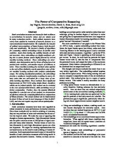
Fig. 1. Ventral view of the hyobranchial muscle innervation in Sceloporus undulatus. As the muscle layers were removed, so were the nerve branches innervating each muscle. Not all nerve branches are displayed in A and B. A: The left side is the superficial layer; the right side is the first layer. B: The left side is the third layer; the right side shows deep musculature. C: Camera lucida drawing of the nerves innervating the hyobranchial musculature of a cleared and stained S. undulatus. The mandible (black) and the hyobranchial apparatus (stippled) are shown as reference points. IMA, M. intermandibularis anterior; IMP, M intermandibularis posterior; CC, M. constrictor colli; OH, M. omohyoideus; MHI, M. mandibulohyoideus I; MHII, M. mandibulohyoideus II; SHS, M. sternohyoideus superficialis; SHP, M. sternohyoideus profundus; GGM, M. genioglossus medialis; GGL, M. genioglossus lateralis; HG, M. hyoglossus; V, M. verticalis; EP, entoglossal process; CH, ceratohyal; CBI, ceratobranchial I; CBII, ceratobranchial II; hypoglossal nerve (brown), trigeminal nerve (red), glossopharyngeal nerve (green), first spinal nerve (blue). 1) rami of the omohyoideus; 2) rami of the sternohyoideus profundus; 3) rami of the sternohyoideus superficialis; 4) rami of the branchiohyoideus; 5) rami of the mandibulohyoideus I; 6) ramus mandibulohyoideus II; 7) ramus mandibulohyoideus III; 8) rami of the hyoglossus; 9) ramus verticalis; 10) ramus genioglossus medialis; 11) rami of the genioglossus lateralis; 12) rami of the genioglossus internus; 13) ramus intermandibularis anterior; 14) ramus intermandibularis posterior.
M. intermandibularis posterior (IMP). Anteriorly, this muscle originates on the medial side of the mandible and is dissected by four slips of the MH as it proceeds toward the midline. Posteriorly, this muscle originates at a connective tissue sheath covering the M. ptery-
goideus. Throughout the length of the muscle, it runs medially to insert at the midventral fascia. In Pseudotrapelus siniatus, all the fibers insert superficially to the MHI. In C. jacksonii, this muscle originates at the connective tissue covering the M. pterygoideus. From its origin, it
HYOBRANCHIAL MUSCULATURE IN IGUANIAN LIZARDS
181
Fig. 2. Ventral view of the hyobranchial muscle innervation in Pseudotrapelus sinaitus. As the muscle layers were removed, so were the nerve branches innervating that muscle. Not all nerve branches are displayed in A and B. A: The left side is the superficial layer; the right side is the first layer. B: Left side is the third layer; the right shows deep musculature. C: Camera lucida drawing of the nerves innervating the hyobranchial musculature in P. sinaitus. The mandible (black) and the hyobranchial apparatus (stippled) are shown as reference points. IMA, M. intermandibularis anterior; IMP, M intermandibularis posterior; CC, M. constrictor colli; OH, M. omohyoideus; MHI, M. mandibulohyoideus I; MHII, M. mandibulohyoideus II; SHS, M. sternohyoideus superficialis; SHP, M. sternohyoideus profundus; GGM, M. genioglossus medialis; GGL, M. genioglossus lateralis; HG, M. hyoglossus; V, M. verticalis; EP, entoglossal process; CH, ceratohyal; CBI, ceratobranchial I; CBII, ceratobranchial II; hypoglossal nerve (brown), trigeminal nerve (red), glossopharyngeal nerve (green), first spinal nerve (blue). 1) rami of the omohyoideus; 2) rami of the sternohyoideus profundus; 3) rami of the sternohyoideus superficialis; 4) rami of the branchiohyoideus; 5) rami of the mandibulohyoideus I; 6) ramus mandibulohyoideus II; 7) ramus mandibulohyoideus III; 8) rami of the hyoglossus; 9) ramus verticalis; 10) ramus genioglossus medialis; 11) rami of the genioglossus lateralis; 12) rami of the genioglossus internus; 13) ramus intermandibularis anterior; 14) ramus intermandibularis posterior.
runs superficially over the MH complex. As it passes around the mandible, its anterior fibers run antero-medially and insert at the midline deep to the IMA. Its posterior fibers run perpendicular to the midline and insert at the midventral fascia deep to the CC. Innervation (V). In S. undulatus this muscle is innervated by a small, posterior branch of the ramus mandibularis of the trigeminal nerve. This branch emerges ventrally at the level of the posterior fourth of the mandible
and gives off one main branch that runs to the middle of the muscle. In P. sinaitus this branch is also present, but it emerges much closer to the trigeminal complex described above. In neither of these species was the presence of rami from the first spinal nerve observed. In C. jacksonii innervation of the IMP is also provided by the trigeminal. However, a branch splits from the ramus mandibularis of the trigeminal before this nerve enters the mandible, and runs medioventrally to the lower jaw to innervate the IMP.
182
MEYERS ET AL.
Fig. 3. Ventral and lateral views of the hyobranchial muscle innervation in Chamaeleo jacksonii. As the muscle layers were removed, so were the nerve branches innervating the muscle. A: Lateral view of the first layer. B: Top half shows the superficial musculature, and the lower half the first layer. C: Top half shows the first layer, while the bottom half shows the deep musculature. Note that there are no lines showing fiber orientation in the M. hyoglossus. This is because the muscle is coiled and runs in all three dimensions within its surrounding connective tissue. D: Camera lucida drawing of the nerves innervating the hyobranchial musculature in C. jacksonii. The mandible (black) and the hyobranchial apparatus (stippled) are shown as reference points. Note that the hyobranchial apparatus has been pulled to the right side to allow a better view of the deeper nerve branches. The ramus hypoglossal is not shown in its entirety in part C, and the point where it runs through the anterior M. hyoglossus and into the M. verticalis and intrinsic tongue muscles is not shown. IMA, M. intermandibularis anterior; IMP, M intermandibularis posterior; CC, M. constrictor colli; OH, M. omohyoideus; MHI, M. mandibulohyoideus I; MHII, M. mandibulohyoideus II; SHS, M. sternohyoideus superficialis; SHP, M. sternohyoideus profundus; GG, M. genioglossus; HG, M. hyoglossus; V, M. verticalis; EP, entoglossal process; CH, ceratohyal; CBI, ceratobranchial I; hypoglossal nerve (brown), trigeminal nerve (red), first spinal nerve (blue). 1) ramus omohyoideus; 2) ramus sternohyoideus profundus; 3) rami of the sternohyoideus superficialis; 4) ramus mandibulohyoideus I; 5) ramus mandibulohyoideus II; 6) ramus mandibulohyoideus III; 7) ramus lingualis of the hypoglossal; 8) ramus genioglossus; 9) rami of the intermandibularis anterior; 10) ramus intermandibularis posterior; 11 ramus lingualis of the trigeminal.
M. constrictor colli (CC). This consists of a superficial band of muscle that originates at the dorsal cervical fascia. It runs ventrally and then medially to insert at the midventral fascia just posterior to the CBIII. In S. undulatus, it is difficult to differentiate between the CC and the IMP near their insertion on the midventral fascia. In P.
sinaitus, this muscle runs postero-ventrally before turning toward the midline, where it is clearly distinguishable from the IMP. In C. jacksonii, this muscle is no longer strap-like, forming instead a thick, broad sheet extending across much of the throat musculature. It originates at the dorsal cervical fascia. As it runs ventrally, it spreads into
183
HYOBRANCHIAL MUSCULATURE IN IGUANIAN LIZARDS
TABLE 1. Origin, insertion and innervation of hyobranchial musculature in Sceloporus undulatus, Pseudotrapelus sinaitus and Chamaeleo jacksonii Muscle M. intermandibularis anterior M. intermandibularis posterior M. constrictor colli M. mandibulohyoideus I
Origin/insertion Middle third of the mandible/midventral fascia Posterior third of the mandible/midventral fascia
M. genioglossus internus
Dorsal cervical fascia on either side Posterior half of the mandible/posterior portion of the ceratobranchial I Near the mandibular symphysis/anterior half of the ceratobranchial I Posterior half of the mandible ventral to MH I/anteriorly on the ceratohyal Connected to the sternum by a tendinous band/dorsolateral edge of the ceratobranchial II Same origin as M. sternohyoideus superficialis/dorsal surface of the ceratobranchial I Scapula and or clavicle/ceratobranchial II and basihyoid Posterior third of the ceratobranchial I/ceratohyal Near the mandibular symphysis/M. hyoglossus by a band of fascia Near the mandibular symphysis/lateral edge of the M. hyoglossus Near the mandibular symphysis/tongue tip
M. hyoglossus
Posterior half of the ceratobranchial I/body of the tongue
M. verticalis
Borders or surrounds the entoglossal process of the hyobranchium Intrinisic, lies ventral to the lingual epithelium and dorsal to the M. hyoglossus
M. mandibulohyoideus II M. mandibulohyoideus III M. sternohyoideus superficialis M. sternohyoideus profundus M. omohyoideus M. branchiohyoideus M. genioglossus medialis M. genioglossus lateralis
M. transversalis
a sheet that connects anteriorly to the midventral fascia (over the IMP) and posteriorly to the connective tissue covering the SHS and SHP. Innervation (VII). The facial nerve is thought to provide motor input to this muscle (Willard, 1915; Oelrich, 1956). However, we were not able to trace the innervation of this muscle in any of these species.
Hyobranchial Musculature The hyobranchial muscles lie dorsal to the M. intermandibularis and can be divided into two groups: the hyobranchial apparatus protractors and retractors. The protractors originate on the mandible and insert at the basihyoid, CHs, or CBs, whereas the retractors originate on the sternum, clavicle, scapula, and suprascapula to insert at the posterior aspect of the elements of the hyobranchial apparatus. M. mandibulohyoideus I (MHI). In S. undulatus and P. sinaitus, the MHI lies deep to the IMA and IMP. The muscle originates on the posterior half of the mandible and inserts ventrally on the posterior half of the first CB (CBI). In P. sinaitus, this muscle is more strap-like and proceeds almost parallel to the midline, whereas in S. undulatus this is a larger sheet of muscle running medially and inserting on a greater portion of the CBI. In C. jacksonii, this muscle is typically referred to as the MH lateralis (Gnanamuthu, 1930). This muscle originates at the ventral aspect of the mandible, runs posteriad to the anterior process, and then dorsally to insert onto the dis-
Innervation Ramus mandibularis of the trigeminal nerve (V) Ramus mandibularis of the trigeminal nerve (V) Facial nerve (VII) (Willard, 1915) Hypoglossal nerve (XII) Hypoglossal nerve (XII) Hypoglossal nerve (XII) First spinal nerve First spinal nerve First spinal nerve Glossopharyngeal nerve (IX) Ramus lingualis lateralis of the hypoglossal nerve (XII) Ramus lingualis lateralis of the hypoglossal nerve (XII) Ramus lingualis lateralis of the hypoglossal nerve (XII) Hypoglossal nerve (XII), including ramus lingualis intermedius Ramus lingualis medialis of the hypoglossal nerve (XII) Ramus lingualis lateralis of the hypoglossal nerve (XII), also possibly ramus lingualis medialis (XII)
tal third of the CBI. Besides attachment of the muscle to the CBI, it is also connected proximally to the anterior process by a short aponeurosis. Innervation (XII). This muscle is innervated dorsally by two to three lateral branches off of the ramus hypoglossus. Each of these branches innervates the muscle farther along its length. In P. sinaitus there is only one branch that innervates the muscle near its insertion on the CBI. In C. jacksonii, the MHI and MHII are innervated by the same branch of the hypoglossal nerve. The hypoglossal nerve in chameleons runs deep to the superficial neck musculature and splits into two branches as it passes the tip of the CBI. The smaller of the two branches continues anteriad and soon diverges into three branches, the most posterior of which runs towards the EP. It puts off small branches into the MHII as it passes this muscle. Before this branch turns anteriorly to run between the MHI and MHII, it puts off another small branch into the latter muscle. It continues to run forward between the two muscles before bifurcating and giving off a single branch toward the origin of each slip of the MHI and MHII. M. mandibulohyoideus II (MHII). This muscle originates near the mandibular symphysis by a narrow aponeurosis, and runs posteriorly superficial to the GGM and the M. verticalis to insert onto the lateral edge of the basihyoid and the anterior third of the CBI. In C. jacksonii, this muscle is referred to as the MH medialis (MHM) (Gnanamuthu, 1930), but it has a largely similar position and course. Here too, the muscle originates tendinously at
184
MEYERS ET AL.
the symphysis and runs posteriorly to insert onto the basihyoid by means of a short aponeurosis. Innervation (XII). The branch innervating this muscle slip is anterior to the other two branches of the MH. This muscle is innervated near its insertion by a single medial branch. The nerve runs anteriorly into the posterior third of the muscle before branching into the muscle itself. It is also innervated by a single medial branch in P. sinaitus; however, it branches off the ramus hypoglossal more posteriorly, near the branches supplying the MHI. As noted above, the MHI and MHII are innervated by the same branch of the hypoglossal nerve in chameleons. The hypoglossal nerve splits into two branches as it passes the tip of the CBI. The smaller of the two branches continues anteriad and soon diverges into three branches, the most posterior of which runs towards the EP. It puts off small sprigs into the MHII as it passes this muscle. This branch then turns anteriorly to run between the MHI and MHII, where it continues to run forward between the two muscles before bifurcating and giving off a single branch toward the origin of each slip of the MHI and MHII.
M. mandibulohyoideus III (MHIII). In all of the species examined in this study, there is a third slip of the MH. The MHIII is a small, thin band of muscle situated slightly lateral and deep to the HG. It originates on the mandible deep to the origin of the MHI, just anterior to the insertion of the M. pterygoideus, and runs posteriad to insert anteriorly on the CH in S. undulatus and more posteriorly in P. sinaitus. In C. jacksonii, there is a thin band of muscle that originates on the posterior third of the mandible just anterior to the M. pterygoideus, and runs posteriorly to insert medially on the dorsal tip of the anterior process. Innervation (XII). This muscle is innervated by the third or fourth lateral branch of the hypoglossal nerve. This is a thin muscle that has a correspondingly small nerve branch. It runs dorso-laterally into the middle of the muscle before sending a large branch to the origin and one toward the insertion of the muscle. In P. sinaitus, this slip is innervated by the second lateral branch of the hypoglossal nerve. In C. jacksonii the MHIII is innervated by an anteriorly running branch that separates from the branch running into the M. genioglossus. M. branchiohyoideus (BH). In S. undulatus, this muscle runs from the posterior three-fourths of the CBI to insert distally on the anterior process and anteriorly on the CH. In P. sinaitus, this muscle runs from the posterior third of the CBI to insert at most of the posterior aspect of the CH. This muscle was not noted in C. jacksonii. Innervation (IX). The BH is innervated by the pharyngeal branch of the ramus glossopharyngeus. This nerve branches from the bundle of nerves including the M. hypoglossus just before its contact with the SHP. It branches dorsally and runs along the ventral side of the BH. At least one small branch runs laterally to innervate the BH while the rest of the nerve runs forward to innervate the posterior portion of the tongue. In P. sinaitus, the ramus glossopharyngeus runs deep to the CBI and sends off a small branch into the BH near the muscle’s origin on the posterior third of the CBI. M. sternohyoideus superficialis (SHS). At their origin the SHS and SHP merge and it is difficult to see a
discrete separation; however, the difference between both muscles becomes more apparent as they diverge toward their insertions. Along with the SHP, this muscle originates on the sternum via a tendinous band. The SHS runs in a band of fibers that parallel the trachea and inserts anteriorly on the dorso-lateral edge of the CBIII and the posterior most lateral aspect of the basihyoid. In C. jacksonii, the SHS also originates at the sternum by a sheet of connective tissue and inserts on the posterior aspect of the basihyoid by a short aponeurosis. Innervation (first spinal). The most medial branch of the first spinal nerve innervates the SHP, continues to run toward the midline, and innervates the SHS in the middle portion of the muscle. The SHS of P. sinaitus is also innervated by the terminal branches of the nerves. In C. jacksonii a similar arrangement is observed (see below).
M. sternohyoideus profundus (SHP). This is the deepest layer ventral to the OH and runs along the buccal floor. It arises from a tendinous band connected to the sternum and fans out to insert on the posterior dorsal surface of the CBI in both S. undulatus and P. sinaitus. In C. jacksonii, the SHP originates on the sternum anterior to the origin of the SHS and runs as a thin band anteriad and dorsad to insert distally onto the posterior cornua. Innervation (first spinal). In both S. undulatus and P. sinaitus, the first spinal nerve runs between the OH and the SH. Midway through the length of these muscles, it splits into at least seven branches. Several of the anteriorly and posteriorly running branches innervate the SHP. C. jacksonii shows a similar arrangement, with the SHP being innervated first, after which the branches continue to terminate in the SHS. M. omohyoideus (OH). This muscle is the largest of the hyobranchial retractors and runs deep to the M. episternocleidomastoideus and the CC and superficial to the SHP. In S. undulatus it originates on the scapula and distal portion of the clavicle and runs forward as a broad sheet to insert the entire length of the CBI as well as the basihyoid. P. sinaitus shows the same orientation except that it does not share a clavicular origin and more of its fibers insert on the basihyoid than on the CBII. In C. jacksonii, this muscle originates on the suprascapula. Initially it runs antero-ventrally to pass beneath the M. episternocleidomastoideus, but then turns postero-ventrally to insert on the ventral aspect of the basihyoid near its articulation with the CBI. Innervation (first spinal). In all three species, the OH is innervated by several branches of the first spinal nerve. In each species, the branches innervating this muscle diverge from the point where the first spinal nerve splits into numerous branches. Again, the innervation of C. jacksonii is similar to that in the other two species. Here too, the OH is innervated by two small branches just before the first spinal nerve diverges into numerous branches innervating the sternohyoideus superficialis and profundus muscles. “Extrinsic” Tongue Musculature Although the extrinsic and intrinsic musculatures of the tongue do not represent a true dichotomy (Schwenk, 2000b), it is an easy way to grossly differentiate most of the muscles originating on the hyobranchial apparatus or mandible from those with no external connection. Lizards
HYOBRANCHIAL MUSCULATURE IN IGUANIAN LIZARDS
usually have at least three extrinsic muscles associated with the tongue. The M. genioglossus is usually comprised of two slips: the M. genioglossus medialis (GGM) and lateralis (GGL). In addition, there is sometimes a small band of fibers reaching into the anterior tip of the tongue (the GGI) in agamids and some iguanids. The HG is a large muscle that makes up most of the body of the tongue.
M. genioglossus medialis (GGM). The GGM is well developed in S. undulatus and P. sinaitus. The GGM is a thick but somewhat narrow band of muscle originating at the mandibular symphysis deep to the MHII. As it runs posteriorly across the HG, the muscle spreads to insert across the entire width of the ventral surface of the HG. It is connected to the latter by a thin band of connective tissue. The GGM is much reduced, and has lost its insertion on the HG in chameleons. The GG of C. jacksonii is a small, thin muscle originating deep to the MHII at the mandibular symphysis. Its fibers run postero-dorsally around the tongue sheath surrounding the accelerator and meet with the other side of the GGM at the dorsal surface of the sheath. Innervation (ramus lateralis of XII). In both S. undulatus and P. sinaitus, this muscle is innervated by a small branch of the ramus lingualis. This branch is the first medial branch that splits off the main branch, just before the latter passes the ascending branch of the trigeminal nerve. It runs antero-medially and dives into the dorsal surface of the GGM. In C. jacksonii, this muscle is innervated by the most medial of the three branches diverging from the main hypoglossal nerve. It is the largest branch of the three and runs towards the M. accelerator before turning anteriorly and merging with a descending branch of the trigeminal nerve. It runs with (against) the trigeminal before giving off three separate branches along the length of the GGM. M. genioglossus lateralis (GGL). The GGL originates on the mandible lateral to the GGM. As it runs posteriorly its fibers turn sideways so that the muscle runs along the lateral surface, rather than on the dorsal surface of the HG. It inserts onto the lateral side of the HG, with some of the fibers running around to the dorsal side of the HG. In C. jacksonii, a slip of muscle originating lateral to the GGM runs posteriad to insert at the dorsolateral aspect of the tongue sheath. Innervation (ramus lateralis of XII). As the ramus lingualis runs forward, it splits into three branches. The lateral branch (the smaller of the two), runs forward to pass ventral to the ascending ramus mandibularis of the trigeminal nerve. Once it passes the ramus mandibularis of the trigeminal nerve, it innervates the posterior end of the GGL. The intermediate branch runs forward past the trigeminal nerve and gives off numerous small branches along the length of the GGL. At its anterior end, this branch forms an anastomosis with the ramus medialis in P. sinaitus. In C. jacksonii, the muscle is innervated by the most medial of the three branches diverging from the main hypoglossal nerve. As mentioned above, it runs along the trigeminal nerve before giving off a number of branches along the length of the GG. M. genioglossus internus (GGI). The GGI is present in both S. undulatus and P. sinaitus. Due to its size and position, the muscle is hard to see in gross dis-
185
section but is obvious in serial sagittal sections through the tongue. It originates at the mandibular symphysis, runs ventrad, but quickly recurves dorsad and runs into the body of the tongue towards the tongue tip. The GGI is absent in C. jacksonii. Innervation (ramus lateralis of XII). In both S. undulatus and P. sinaitus, this muscle is innervated by the intermediate branch of the ramus lingualis which also innervates the GGL. At its distal portion, it turns anterodorsally to terminate in the GGI in S. undulatus. In P. sinaitus, it sends a small branch anteriorly into the GGI before turning posteriorly to combine with the ramus lingualis of the trigeminal nerve.
M. hyoglossus (HG). The HG is the largest muscle of the tongue in S. undulatus and P. sinaitus. Its fibers originate on the posterior half of the first CBI and run anteriorly, ventral to the CH, and then turn dorsally into the body of the tongue. The hyoglossus tapers as it enters the tongue, forming a bundle that does not completely extend to the tongue tip. The HG of C. jacksonii retains some of these general characteristics. This muscle originates medially at the tip of the posterior cornua and runs along its length to the basihyoid. At the basihyoid, it turns anteriorly and runs forward to insert, via connective tissue, postero-laterally on the M. accelerator. Throughout most of its length, the HG forms a densely coiled structure that is surrounded by a sheath of epimysium. Innervation (ramus intermedius of XII). Posteriorly, near the innervation of the MH slips, this muscle is innervated by several small branches running dorsally into the muscle. However, the ramus lingualis intermedius of the hypoglossal is by far the largest nerve branch innervating the HG. After the branching of the lingual rami, it runs dorsally into the body of the muscle and puts off many smaller branches into the HG. At its distal end, the ramus lingualis intermedius forms an anastomosis with the ramus lingualis lateralis/trigeminal (ramus lingualis) complex. In C. jacksonii, the hypoglossal nerve runs through the body of the muscle throughout almost its entire length. As it winds through the coiled muscle, it puts off numerous small branches before continuing to run along the lateral edges of the M. accelerator. “Intrinsic” Tongue Musculature The intrinsic tongue musculature consists of several muscles that have their origin and insertion on the muscles or connective tissue in the tongue. In iguanians, the largest of these is the muscle surrounding the EP of the hyoid. The other intrinsic tongue muscles are not described in detail in the present study, as the homology of the different muscles in the different groups remains unclear. For detailed descriptions, see Delheusy et al. (1994) for the intrinsic muscle arrangement in iguanids, Smith (1988) for agamids, and Bell (1989) and Herrel et al. (2001) for chameleons.
M. verticalis (V). In S. undulatus, this muscle is called the M. verticalis. It has dorso-ventrally arranged fibers that run parallel to the EP. In Pseudotrapelus siniatus, this muscle is generally referred to as the ring muscle. The muscle fibers are also dorso-ventrally arranged, and in addition there are fibers that curl dorsally and ventrally to form a ring around the EP. This arrangement is most pronounced in the hind tongue of P. sinaitus. In C.
186
MEYERS ET AL.
jacksonii, this ring-like muscle is typically referred to as the accelerator muscle. It consists of helically wound fibers surrounding the posterior part of the EP (Van Leeuwen, 1997). Innervation (ramus medialis of XII). The ramus lingualis medialis of the hypoglossal nerve innervates this muscle along its entire length. It runs ventrally between the M. verticalis and the HG, and gives off numerous small branches into the M. verticalis. As it travels forward, it puts off a branch that heads laterally to form an anastomosis with the ramus lingualis lateralis/trigeminal complex. In chameleons the M. verticalis is also innervated by the hypoglossal nerve. As the hypoglossal nerve passes through the insertion of the HG onto the M. accelerator, the nerve begins to send branches medially, deep into the M. accelerator.
M. transversalis (T). The dorsal transverse fibers are most easily seen in histological section. The fibers lie ventral to the lingual epithelium and dorsal to the HG and M. verticalis, and run parallel to the long axis of the tongue. In both S. undulatus and P. sinaitus this muscle occurs throughout most the length of the tongue. Innervation (ramus lingualis lateralis of XII). In S. undulatus and P. sinaitus, the M. transversalis is innervated by the ramus lingualis lateralis of the hypoglossal nerve, which sends out branches that run around the dorsal surface of the HG. Some fibers of the M. transversalis are also innervated by the ramus lingualis medialis of the hypoglossal nerve and the ramus mandibularis of the trigeminal nerve. DISCUSSION Muscle Anatomy and Homology The general arrangement of the hyobranchial musculature of these three lizards showed no major departures from previous descriptions of other phrynosomatids (Sanders, 1874; Jenkins and Tanner, 1968; Secoy, 1971; Tanner and Avery, 1982; Meyers and Nishikawa, 2000), agamids (Tanner and Avery, 1982; Smith, 1988; Herrel et al., 1995), or chamaeleonids (Houston, 1828; Gnanamuthu, 1930; Lubosch, 1932, 1933; Zoond, 1933; Bell, 1989; Herrel et al., 2001). However, we did note the presence of the GGI in S. undulatus, which has not been previously noted in other iguanid (sensu lato) or phrynosomatid lizards (Oelrich, 1956; Secoy, 1971; Tanner and Avery, 1982; Schwenk, 1988; Delheusy et al., 1994; see Schwenk, 2000a, for an overview). We suggest that, based on its arrangement and similar innervation pattern to that of the agamid P. sinaitus, this muscle is in fact homologous to that seen in agamids. One problem in comparing the hyobranchial musculature and function between chameleons and other lizards is the confusion regarding the terminology of the sternohyoid complex (sensu Zavattari, 1910). Originally, Sanders (1870, 1872, 1874), Zavattari (1909, 1910), and Richter (1933) described an omohyoideus muscle and a superficial and a deep (epi)sternohyoideus as the three components of the sternohyoid complex. Avery and Tanner (1971) later used a different terminology, referring to Camp (1923), who considered M. sternothyroideus to be the deepest layer of the sternohyoid complex. However, Avery and Tanner (1971) reversed the names used by Camp (1923) and referred to the medial-most (superficial) muscle as the M. sternothyroideus. Lubosch (1933) and subsequent au-
thors (Gnanamuthu, 1937; Schwenk, 2000a; Herrel et al., 2001) typically referred to the medial-most muscle in chameleons as the SH. As most authors agree that there are two well separated muscles in most lizards (Zavattari, 1909; Richter, 1933; Avery and Tanner, 1971) different names for both slips are indeed appropriate. Given the position of the muscles, and their innervation pattern (the medial-most muscle being innervated by the terminal branches of the spinal nerve in all species examined), we homologize the muscle typically referred to as the sternohyoideus in chameleons (Lubosch, 1933; Wainwright and Bennett, 1992; Herrel et al., 2001) with the sternothyroideus of agamids and iguanids (sensu lato), and the chameleon sternothyroideus with the agamid and iguanid sternohyoid muscle (Table 1). The name M. sternothyroideus, however, is derived from mammalian studies wherein a medially situated muscle was observed running from the sternum to the thyroid cartilage, and from there to the basihyoid (Zavattari, 1910). Since in mammals this muscle is innervated by the first spinal nerve (Zavattari, 1910), we consider it homologous to the medial-most muscle of the sternohyoid complex. However, as this nomenclature was originally conceived for mammals, and as no thyroid cartilage is present in lizards, this name cannot be used here. In accordance with the first descriptions of this muscle complex (Zavattari, 1909, 1910; Richter, 1933), we thus refer to the medial-most muscle of the sternohyoid complex, innervated by the terminal branches of the first spinal nerve, and attaching at the posterior aspect of the basihyoid, as the SHS, and to the deeper, lateral part as the SHP. One other source of potential confusion in comparative studies is the homology of the verticalis muscle in iguanids with the muscles described as the ring muscle (Gandolfi, 1908) and the accelerator muscle (Lubosch, 1933) in agamids and chameleons, respectively. However, based both on position and innervation, these structures must be regarded as homologous in the three species of lizards examined in this study. Previous descriptions of this muscle and its innervation in other species of lizards indicate that its presence must be considered a plesiomorphic character for lizards (Zavattari, 1909, 1910; Richter, 1933; Gnanamuthu, 1937). Similarly, the muscle slips originating on the lower jaw and inserting onto the lingual sheet in chameleons are most likely homologous to the M. genioglossi in other lizards. A similar arrangement of the genioglossus muscle attaching to the mouth floor is observed in Sphenodon (Osawa, 1898; Lubosch, 1933; Rieppel, 1978; Schwenk, 1986). This muscle thus seems to have reverted to a more primitive state in chameleons.
Functional Implications In S. undulatus and P. sinaitus, the hyobranchial apparatus and associated musculature are very similar. However, two important differences between these species can be noted. The first, which was documented previously (Gandolfi, 1908; Smith, 1988), is the development of the M. verticalis from dorsoventral fibers bordering the EP to fibers that form a ring around the EP. In S. undulatus, the vertical fibers surrounding the EP work to compress the tongue. This may translate into some forward movement of the tongue during protrusion. In P. sinaitus, the formation of a complete ring around the EP allows this muscle to produce a centrally directed force that would allow the tongue to slide forward on the process, resulting in greater
HYOBRANCHIAL MUSCULATURE IN IGUANIAN LIZARDS
lingual protrusion (Smith, 1988). Meyers and Nishikawa (2000) found that after denervation of this muscle, tongue protrusion distance was not affected in S. undulatus but was reduced for P. sinaitus. Although the arrangement of the M. verticalis is superficially similar to what is observed in chameleons, the fibers of the chameleon M. verticalis form a complex, radially arranged matrix surrounding the EP. Unlike in the agamid lizard P. sinaitus, this muscle is almost exclusively responsible for tongue projection (Houston, 1828; Gnanamuthu, 1930; Zoond, 1933; Wainwright and Bennett, 1992b; Meyers and Nishikawa, 2000). The second most notable difference is a change in the relative length of the muscles, and their origins and insertions. These muscles include the MHI, II, and III; the GGM and GGL; and the HG. In P. sinaitus, the muscles are relatively longer and narrower, and are attached across smaller portions of the elements upon which they originate and insert. The same muscles in S. undulatus form broader sheets that attach across larger areas on the mandible and hyobranchial apparatus. In addition, muscles such as the MH lie more in line with the midline of the body than do those of S. undulatus. Physiologically, if these muscles shorten to the same extent as in S. undulatus, then the greater lingual protrusion seen in some agamids may be due solely to the longer muscles of P. sinaitus. These observations may explain some of the results obtained by nerve transections experiments in S. undulatus and P. sinaitus (Meyers and Nishikawa, 2000). In these species, transection of the nerve innervating the MH muscles reduced tongue protrusion distance considerably in P. sinaitus, but not in S. undulatus (Meyers and Nishikawa, 2000). Similarly, in some myrmecophagous phrynosomatids (horned lizards of the genus Phrynosoma), the hyobranchial muscles are more elongated (compared to more generalized species), which seems to be related to an increase in tongue protrusion distance (about 50% of mandible length (Schwenk, 2000a)).
Neuroanatomy The relative scarcity of neuroanatomical studies makes comparisons of the different species difficult, although the general findings seem to be consistent with those of previous workers (Bendz, 1840; Willard, 1915; Oelrich, 1956). One major problem in describing innervation patterns is the numerous plexi, and anastomoses between the various cranial and spinal nerves. Ideally, histochemical tracers could be used to map the cranial nerves (Anderson and Nishikawa, 1997). However, as this is a time-consuming and extremely invasive procedure requiring numerous animals, it has been performed infrequently in lizards (but see Barbas-Henry, 1982; Barbas-Henry and Lohman, 1984, 1986, 1988). As such an approach was beyond the scope of this study, the innervation patterns described here are based on the assumption that the nerves (hypoglossal, glossopharyngeal, trigeminal, facial, and first spinal) do indeed connect to the brainstem as described in previous studies (Bendz, 1840; Watkinson, 1906; Willard, 1915; Gnanamuthu, 1937; Oelrich, 1956; Islam and Ashiq, 1972; Barbas-Henry and Lohman, 1984, 1986, 1988). Like the hyobranchial apparatus and muscle anatomy, the gross innervation patterns of S. undulatus and P. sinaitus are largely conserved. The hypoglossal nerve provides most of the motor input to the tongue and hyobranchial muscles. In both species, it emerges in the
187
throat region near the CBI and runs along the superficial side of the HG. It first innervates the MH muscles before splitting into three main branches. The innervation of the MH muscles occurred relatively farther posterior in P. sinaitus, probably to accommodate for the proportionally more posterior insertions of the muscles. In both species, the hypoglossal nerve trifurcates into a ramus lingualis medialis, ramus lingualis intermedius, and ramus lingualis lateralis. For Anolis, Willard (1915) stated that the lateral branch was the largest of the three branches; in S. undulatus and P. sinaitus, on the other hand, the medial branch is the largest of the three. It is important to note that in both species there are numerous anastomoses between the three large rami of the hypoglossal and the lingual ramus of the trigeminal. Many of the trigeminal nerve fibers terminate in or near the tongue pad, most likely providing sensory input from the taste buds. These anastomoses are likely important in feeding sensory information about the prey item (e.g., position, mass, and state of reduction) back to the brain stem, and may thus be important in coordinating tongue and jaw movements during prey capture and transport (Herrel et al., 2000). Like the musculature, the innervation patterns of C. jacksonii are modified from the general iguanid pattern. The hypoglossal nerve still runs along the HG, but it runs through the body of the muscle instead of its ventral surface. During its course through the HG it forms a highly coiled structure, which allows it to stretch during tongue projection without being damaged. It runs forward into the tongue pad to innervate the M. accelerator and the “intrinsic” lingual muscles. Before the hypoglossal nerve runs into the HG it gives off a medium-sized branch that innervates the MH and GG muscles. As observed in the other two species, several anastomoses between the lingual ramus of the trigeminal nerve and rami of the hypoglossal nerve are present and likely provide sensory information from the tongue pad. Moreover, it may be involved in coordinating tongue, hyoid, and jaw movements during tongue projection. In fact, Meyers and Nishikawa (2000) suggested that reduction in tongue projection distance after MH denervation may be due to loss of sensory feedback between the hyoid and the tongue muscles. It is clear that anatomical studies are crucial in providing the foundation necessary for making sound functional predictions and interpretations of previous hypotheses regarding control of the hyobranchial apparatus. Moreover, innervation patterns can be extremely helpful in elucidating problems regarding homologies of muscles. Given that the innervation patterns of the hyobranchial musculature show variability among higher-level taxa yet show a large within-group similarity, we suggest that they would be useful as systematic characters in lizards.
ACKNOWLEDGMENTS We thank Dr. Ted Papenfuss for identification of the Pseudotrapelus sinaitus used in the study. We also thank S. Hoyer, K. Lappin, J. Monroy, D. Pierotti, J.T. Laitman, and an anonymous reviewer for comments on earlier drafts of this manuscript. This research was supported by NSF grant 9809942 and NIH grant R25GM56931 to K.C.N., and by the Fund for Scientific Research, Flanders, Belgium to A.H.
188
MEYERS ET AL.
LITERATURE CITED Anderson CW, Nishikawa KC. 1997. The functional anatomy and evolution of hypoglossal afferents in the leopard frog, Rana pipiens. Brain Res 771:285–291. Arnold EN. 1989. Towards a phylogeny and biogeography of the Lacertidae: relationships within an Old-World family of lizards derived from morphology. Bull Br Mus Nat Hist 55:209 –257. Avery DF, Tanner WW. 1971. Evolution of the iguanine lizards (Sauria, Iguanidae) as determined by osteological and myological characters. Brigham Young Univ Sci Bull Biol Ser 12:1–79. Barbas-Henry HA. 1982. The motor nuclei and primary projections of the facial nervein the monitor lizard Varanus exanthematicus. J Comp Neurol 207:105–113. Barbas-Henry HA, Lohman AHM. 1984. The motor nuclei and primary projections of the IXth, Xth, and XII cranial nerves in the monitor lizard, Varanus exanthematicus. J Comp Neurol 226:565– 579. Barbas-Henry HA, Lohman AHM. 1986. The motor complex and primary projections ofthe trigeminal nerve in the monitor lizard, Varanus exanthematicus. J Comp Neurol 254:314 –329. Barbas-Henry HA, Lohman AHM. 1988. The motor nuclei and sensory neurons of the IIIrd, IVth and VIth cranial nerves in the monitor lizard, Varanus exanthematicus. J Comp Neurol 267:370 – 386. Bell DA. 1989. Functional anatomy of the chameleon tongue. Zool Jb Anat 119:313–336. Bels VL, Goosse V, Kardong KV. 1992. Kinematic analysis of drinking by the lacertid lizard, Lacerta viridis (Squamata, Scleroglossa). J Zool Lond 229:659 – 682. Bels VL, Chardon M, Kardong KV. 1994. Biomechanics of the hyolingual system in squamata. In: Bels VL, Chardon M, Vandewalle P, editors. Biomechanics of feeding in vertebrates. Berlin: Springer Verlag. p 197–240. Bendz H. 1840. Bidrag til den sammenlignende Anatomie af Nervus Glossopharyngeus, Vagus, Accessorius willisii og Hypoglossus hos Reptilierne. K Denske vidensk Natnrvid og mathem Afh 10:113– 153. Camp CL. 1923. Classification of the lizards. Bull Am Mus Nat Hist 48:289 – 481. Chiel HJ, Crago P, Mansour JM, Hathi K. 1992. Biomechanics of a muscular hydrostat: a model of lapping by a reptilian tongue. Biol Cybernet 67:403– 415. Cooper WE. 1995. Foraging mode, prey chemical discrimination, and phylogeny in lizards. Anim. Behav 50:973–985. De la Serna de Esteban C. 1959. Anatomia microscopica de la lengua de Amphisbaena vermicularis darwini (D. y B). Actas Trab Prim Congr Sudamer Zool 5:143–150. De la Serna de Esteban C. 1965. Anatomia microscopica comparada de la lengua de algunos saurios Argentinos. Anais Congr Latinoamer Zool 2:235–245. Delheusy V, Bels VL. 1992. Kinematics of feeding behaviour in Oplurus cuvieri (Reptilia: Iguanidae). J Exp Biol 170:155–186. Delheusy V, Tobeau G, Bels VL. 1994. Tongue structure and function in Oplurus cuvieri (Reptilia: Iguanidae). Anat Rec 238:263–276. Delheusy V. 1996. Structure et fonctionnement de l’appareil de prise de nourriture et de boisson chez Phelsuma madagascariensis (Lezard: Gekkonidae) dans une perspective phyletique. Unpublished Ph.D. thesis, Universite´ de Lie`ge, Lie`ge, Belgium. De Vis CW. 1883. Myology of Chlamydosaurus kingii. Proc Lin Soc N S Wales 8:300 –320. Estes R, De Queiroz K, Gauthier JA. 1988. Phylogenetic relationships within the squamata. In: Estes R, Pregill G, editors. Phylogenetic Relationships of the Lizard Families. Stanford: Stanford University Press. p 119 –282. Filipski GT, Wilson MVH. 1985. Staining nerves in whole cleared amphibians and reptiles using Sudan Black B. Copeia 1985:500 – 502. Fu¨ rbringer M. 1922. Das Zungenbein der Wirbeltiere, insbesondere der Reptilien und Vo¨ gel. Abh Heidelberger Akad Math-Naturw 2:1–164.
Gandolfi H. 1908. Die zunge der Agamidae und Iguanidae. Zool Anz 32:569 –580. George JC. 1948. The muscular system of Uromastix hardwickii Gray. J Univ Bomb 17:1–23. Gnanamuthu CP. 1930. The anatomy and mechanism of the tongue in Chamaeleon carcaratus (Merrem). Proc Zool Soc Lond 31:467– 486. Gnanamuthu CP. 1937. Comparative study of the hyoid and tongue of some typical genera of Indian reptiles. Proc Zool Soc Lond 107B:1– 62. Gove D. 1979. A comparative study of snake and lizard tongue-flicking with an evolutionary hypothesis. Z Tierpsychol 51:58 –76. Herrel A, Cleuren J, De Vree F. 1995. Prey capture in the lizard Agama stellio. J Morphol 224:313–329. Herrel A, Cleuren J, De Vree F. 1996. Kinematics of feeding in the lizards Agama stellio. J Exp Biol 199:1727–1742. Herrel A, Cleuren J, De Vree F. 1997. Quantitative analysis of jaw and hyolingual muscle activity during feeding in the lizard Agama stellio. J Exp Biol 200:101–115. Herrel A, Timmermans J, De Vree F. 1998. Tongue flicking in agamid lizards: morphology, kinematics and muscle activity patterns. Anat Rec 252:102–116. Herrel A, Meyers JJ, Aerts P, Nishikawa KC. 2000. The mechanics of prey prehension in chameleons. J Exp Biol 203:3255–3263. Herrel A, Meyers JJ, Nishikawa KC, De Vree F. 2001. Morphology and histochemistry of the hyolingual musculature in chameleons. J Morphol 249:154 –170. Houston J. 1828. On the structure and mechanism of the tongue of the chameleon. Trans R Irish Acad 15:177–201. Humason GL. 1972. Animal tissue techniques. San Francisco: W.H. Freeman and Co. 641 p. Islam A, Ashiq S. 1972. The cranial nerves of Uromastix hardwicki Gray. Biologia 18:51–73. Jenkins RL, Tanner WW. 1968. Osteology and myology of Phrynosoma P. platyrhinos Girard and Phrynosoma d. hernandesi Girard. BYU Biol Ser 9:1–34. Julien R. 1967. Les deux types d’innnervation de la jambe des Lacertiliens. Bull Mus Hist Nat (Ser 2) 38:767–778. Julien R, Renous-Le´ curu S. 1972. Variations du trajet du nerf ulnaire (ulnaris) et de l’innervation des muscles dorsaux de la jambe chez les Lacertiliens (Reptiles, Squamates): valeur syste´ matique et application phyloge´ nique. Bull Mus Hist Nat Paris (Ser 3) Zool 23: 207–246. Kardong KV. 1998. Vertebrates: comparative anatomy, function, evolution. Boston: McGraw-Hill. 747 p. Le´ curu S. 1968. Myologie et innervation du membre ante´ rieur des Lacertiliens. Me´ m Mus Hist Nat (Ser A) Zool 48:127–215. Lubosch W. 1932. Bemerkungen u¨ ber die Zungenmuskulatur des Chama¨ leons. Gegenbaurs Morph Jahrb 71:158 –170. Lubosch W. 1933. Untersuchungen u¨ ber die Visceral muskulatur der Sauropsiden. Gegenbaurs Morph Jahrb 72:584 – 666. Macey JR, Schulte JA, Larson A, Ananjeva NB, Wang Y, Pethiyagoda R, Rastegar-Pouyani N, Papenfuss TJ. 2000a. Evaluating transtethys migration: an example using acrodont lizard phylogenies. Syst Biol 49:233–256. Macey JR, Schulte JA, Larson A. 2000b. Evolution and phylogenetic information content of mitochondrial genomic structural features illustrated with acrodont lizards. Syst Biol 49:257–277. McClung JR, Goldberg SJ. 2000. Functional anatomy of the hypoglossal innervated muscles of the rat tongue: a model for elongation and protrusion of the mammalian tongue. Anat Rec 260:378 –386. McDowell SB. 1972. The evolution of the tongue of snakes and its bearing on snake origins. In: Dobzhansky T, Hecht MK, Steere WC, editors. Evolutionary biology. Vol. VI. New York: Meredith. p 192– 273. Meyers JJ, Nishikawa KC. 2000. Comparative study of tongue protrusion in three iguanian lizards; Sceloporus undulatus, Pseudotrapelus sinaitus and Chamaeleo jacksonii. J Exp Biol 203:2833– 2849. Mivart G. 1867. Notes on the myology of Iguana tuberculata. Proc Zool Soc Lond 1867:766 –797. Mivart G. 1870. On the myology of Chamaeleo parsonii. Proc Zool Soc Lond 1870:850 – 890.
HYOBRANCHIAL MUSCULATURE IN IGUANIAN LIZARDS Minot CS. 1880. Studies on the tongue of reptiles and birds. Ann Mem Bost Soc Nat Hist 1880:3–20. Moody S. 1980. Phylogenetic and historical biogeographical relationships of the genera in the family Agamidae (Reptilia: Lacertilia). Unpublished Ph.D. thesis, University of Michigan, Ann Arbor, Michigan. 373 p. Mu L, Sanders I. 1999. Neuromuscular organisation of the canine tongue. Anat Rec 256:412– 424. Nishikawa KC. 1987. Staining peripheral nerves with Sudan black B: progressive vs. regressive methods. Copeia 2:489 – 491. Oelrich TH. 1956. The anatomy of the head of Ctenosaura pectinata. Misc Publ Zool Univ Mich 94:1–122 Poglayen-Neuwall I. 1954. Die Kiefermuskulatur der Eidechsen und ihre Innervation. Zeitschrift fu¨ r wissenschaftliche. Zoologie 158: 79 –132. Presch W. 1971. Tongue structure of the teiid lizard genera Ameiva and Cnemidophorus with a reallocation of Ameiva vanzoi. J Herpetol 5:183–185. Renous-Le´ curu S, Julien R. 1972. Les grands troncs nerveux du zeugopode des Lacertiliens. Bull Mus Hist Nat (Ser 3) Zool 23:165– 206. Rice MJ. 1973. Supercontracting stirated muscle in a vertebrate. Nature 243:238 –240. Richter H. 1933. Das Zungenbein und seine Muskulatur bei den Lacertilia vera. Jena Z Naturwiss 66:395– 480. Rieppel O. 1978. The throat musculature of Sphenodon, with comments on the primitive character states of the throat muscles in lizards. Anat Anz 144:429 – 440. Sanders A. 1870. Notes on the myology of Platydactyls japonicus. Proc Zool Soc Lond 1870:413– 426. Sanders A. 1872. Notes on the myology of Liolepis belli. Proc Zool Soc Lond 1872:154 –183. Sanders A. 1874. Notes on the myology of Phyrnosoma coronatum. Proc Zool Soc Lond 1874:71– 89. Schwenk K. 1986. Morphology of the tongue in the tuatara, Sphenodon punctatus (Reptilia: Lepidosauria), with comments on function and phylogeny. J Morphol 188:129 –156. Schwenk K. 1988. Comparative morphology of the lepidosaur tongue and its relevance to squamate phylogeny. In: Estes R, Pregill G, editors. Phylogenetic relationships of the lizard families. Stanford: Stanford University Press. p 569 –598. Schwenk K, Bell DA. 1988. A cryptic intermediate in the evolution of chameleon tongue projection. Experentia 44:697–700. Schwenk K, Throckmorton GS. 1989. Functional and evolutionary morphology of lingual feeding in squamate reptiles: phylogenetics and kinematics. J Zool Lond 219:153–175.
189
Schwenk K. 2000a. Feeding in Lepidosaurs. In: Schwenk K, editor. Feeding: form, function and evolution in tetrapod vertebrates. San Diego: Academic Press. p 175–291. Schwenk K. 2000b. Extrinsic versus intrinsic lingual muscles: a false dichotomy. Bull Mus Comp Zool 155:507–523. Secoy DM. 1971. The myology of Sceloporus clarki clarki Baird and Girard (Reptilia: Iguanidae). BYU Sci Bull Biol 14:1–22. Smith KK. 1984. The use of the tongue and hyoid apparatus during feeding in lizards (Ctenosaura similis and Tupinambis nigropunctatus). J Zool Lond 202:115–143. Smith KK. 1988. Form and function of the tongue in agamid lizards with comments on its phylogenetic significance. J Morphol 196:157– 171. Sondhi KC. 1958. The hyoid and associated structure in some Indian reptiles. Ann Zool Agra 2:157–239. Tanner WW, Avery DF. 1982. Buccal flour of reptiles, a summary. Great Basin Nat 42:273–349. Taylor WR, Van Dyke GC. 1978. Revised procedures for staining and clearing smallfishes and other vertebrates for bone and cartilage study. Privately printed. Tilak R. 1964. The hyoid apparatus of Uromastix hardwickii Gray. Sci Cult 30:244 –246. Toubeau G, Cotman C, Bels VL. 1994. Morphological and kinematic study of the tongue and buccal cavity in the lizard Anguis fragilis (Reptilia: Anguidae). Anat Rec 240:423– 433. Van Leeuwen JL. 1997. Why the chameleon has spiral-shaped muscle fibers in its tongue. Phil Trans R Soc Lond Ser B 352:573–589. Wainwright PC, Bennett AF. 1992a. The mechanism of tongue projection in chameleons. I. Electromyographic tests of functional hypotheses. J Exp Biol 168:1–21. Wainwright PC, Bennett AF. 1992b. The mechanism of tongue projection in chameleons. II. Role of shape change in a muscular hydrostat. J Exp Biol 168:23– 40. Watkinson GB. 1906. The cranial nerves of Varanus bivittatus. Gegenbauers Morph Jahrb 35:450 – 472. Willard WA. 1915. The cranial nerves of Anolis carolinensis. Bull Mus Comp Zool Harv 59:18 –116. Zavattari E. 1909. I muscoli iodei dei sauri in rapporto con i musoli iodei degli altri vertebrati. Parte prima. Mem Acad Sci Torino 60:351–392. Zavattari E. 1910. I muscoli iodei dei sauri in rapporto con i musoli iodei degli altri vertebrati. Parte seconda. Mem Acad Sci Torino 61:55–134. Zoond A. 1933. The mechanism of projection of the chameleons tongue. J Exp Biol 10:174 –185. Published online 00 Month 2002 in Wiley InterScience (www.interscience.wiley.com).