The
new england journal
of
medicine
review article
mechanisms of disease
Acute Renal Failure and Sepsis Robert W. Schrier, M.D., and Wei Wang, M.D.
a
cute renal failure occurs in approximately 19 percent of patients with moderate sepsis, 23 percent with severe sepsis, and 51 percent with septic shock when blood cultures are positive (Tables 1 and 2).1,2 A progressive increase in the acute respiratory distress syndrome also occurs with moderate and severe sepsis and septic shock. In the United States, an estimated 700,000 cases of sepsis occur each year, resulting in more than 210,000 deaths; this number accounts for 10 percent of all deaths annually and exceeds the number of deaths due to myocardial infarction.3 The combination of acute renal failure and sepsis is associated with a 70 percent mortality, as compared with a 45 percent mortality among patients with acute renal failure alone. Thus, the combination of sepsis and acute renal failure constitutes a particularly serious medical problem in the United States.4 Substantial progress has been made toward understanding the mechanisms whereby sepsis is associated with a high incidence of acute renal failure. Moreover, recently identified clinical interventions may be able to decrease the occurrence of acute renal failure and sepsis and the high associated mortality. The cytokine-mediated induction of nitric oxide synthesis that occurs in sepsis decreases systemic vascular resistance.5 This arterial vasodilatation predisposes patients with sepsis to acute renal failure, the need for mechanical ventilation, and ultimately, increased mortality. In this article, we review the effects of nitric oxide–mediated arterial vasodilatation on resistance to exogenous pressors and hypotension (Fig. 1), and we discuss the use of arginine vasopressin in patients with septic shock. We also review the effects of increased plasma concentrations of several endogenous vasoconstrictor hormones, including catecholamines, angiotensin II, and endothelin, which support arterial pressure in patients with sepsis who have vasodilatation but also cause renal vasoconstriction and predispose patients to acute renal failure. Patients who have a combination of sepsis and acute renal failure may have some effects of systemic arterial vasodilatation, such as altered Starling forces in the capillaries, pulmonary edema, hypoxia, a need for mechanical ventilation, acute respiratory distress syndrome, and multiple-organ dysfunction syndrome, which together may increase mortality to more than 80 percent (Fig. 2).6,7 We discuss interventions that may prevent this dire sequence of events. Finally, we review several prospective, randomized clinical trials of interventions that have the potential to prevent or attenuate acute renal failure in patients with sepsis and thus decrease mortality. Such trials have addressed anticoagulant therapy, early resuscitation, the treatment of hyperglycemia, the use of corticosteroids, a shortened duration of mechanical ventilation, and various types of renal-replacement therapy.
From the Department of Medicine, University of Colorado Health Sciences Center, Denver. Address reprint requests to Dr. Schrier at the Department of Medicine, University of Colorado Health Sciences Center, 4200 E. 9th Ave., Box C-281, Denver, CO 80262, or at
[email protected]. N Engl J Med 2004;351:159-69. Copyright © 2004 Massachusetts Medical Society.
hemodynamics and hormones The hemodynamic hallmark of sepsis is generalized arterial vasodilatation with an associated decrease in systemic vascular resistance. Arterial underfilling due to arterial vasodilatation occurs in several clinical circumstances, including sepsis, and is associated
n engl j med 351;2
www.nejm.org
july 8, 2004
Downloaded from www.nejm.org at UNIV OF UTAH ECCLES on February 28, 2006 . Copyright © 2004 Massachusetts Medical Society. All rights reserved.
159
The
new england journal
Moderate sepsis
medicine
these vasoactive hormones during sepsis may be associated with down-regulation of their receptors, which would result in a lessening of their effects on the vasculature.
Table 1. Clinical Definition of Sepsis. Type
of
Characteristics Body temperature >38°C or <36°C Heart rate >90 beats/min
the pressor effect of arginine vasopressin
Respiratory rate >20 breaths/min or partial pressure of arterial CO2 <32 mm Hg White-cell count >12,000/mm3, or >10 percent immature band forms Evidence of infection Severe sepsis
Sepsis-associated lactic acidosis, oliguria, or acute alteration of mental status
Septic shock
Sepsis-induced hypotension (i.e., systolic blood pressure <90 mm Hg) despite adequate fluid resuscitation. Patients treated with vasopressors or inotropic medications may not be hypotensive at the time of measurement.
with activation of the neurohumoral axis and an increase in cardiac output secondary to the decreased cardiac afterload. Activation of the sympathetic nervous system and the renin–angiotensin–aldosterone axis, the nonosmotic release of vasopressin, and an increase in cardiac output are essential in maintaining the integrity of the arterial circulation in patients with severe sepsis and septic shock (Fig. 1) but may lead to acute renal failure.8-11 The arterial vasodilatation that accompanies sepsis is mediated, at least in part, by cytokines that up-regulate the expression of inducible nitric oxide synthase in the vasculature.12,13 The release of nitric oxide with inducible nitric oxide synthase, as compared with constitutive endothelial nitric oxide synthase, is more profound and prolonged. Moreover, vascular resistance to the pressor response to norepinephrine12 and angiotensin II5 occurs during sepsis and is attributable in part to the potent vasodilatory effect of nitric oxide.12,14,15 In addition, an increase in plasma concentrations of hydrogen ions and lactate and a decrease in ATP in vascular smooth-muscle cells during septic shock activate the ATP-sensitive potassium channels (KATP channels).16,17 The resultant potassium efflux through the KATP channels causes hyperpolarization of the vascular smooth-muscle cells with closure of the voltage-gated calcium channels in the membrane. Since the vasoconstrictor effects of norepinephrine and angiotensin II depend on open calcium channels, vascular resistance to these pressor hormones can occur along with lactic acidosis in patients with sepsis. Furthermore, the high endogenous levels of
160
n engl j med 351;2
There is evidence that the administration of arginine vasopressin in patients with sepsis-related vasodilatory shock may help maintain blood pressure despite the relative ineffectiveness of other vasopressor hormones such as norepinephrine and angiotensin II.18,19 Specifically, arginine vasopressin may inactivate the KATP channels20 and thereby lessen vascular resistance to norepinephrine12 and angiotensin II.5 Arginine vasopressin also decreases the synthesis of nitric oxide (as a result of a decrease in the expression of inducible nitric oxide synthase) as well as cyclic guanosine monophosphate (cGMP) signaling by nitric oxide,21 thus attenuating the arterial vasodilatation and pressor resistance during sepsis. The degree of vasoconstriction in response to arginine vasopressin relates to its plasma levels and occupancy of the V1a arginine vasopressin receptors on vascular smooth-muscle cells. Initially, in septic or hemorrhagic shock, the plasma arginine vasopressin concentrations increase to 200 to 300 pg per milliliter, but after approximately an hour, the neurohypophysial stores of vasopressin are depleted and plasma concentrations may fall to approximately 30 pg per milliliter.19 At that time and in the presence of unoccupied V1a receptors, the administration of exogenous arginine vasopressin can increase blood pressure by 25 to 50 mm Hg by returning the plasma concentrations of antidiuretic hormones to their earlier high levels.22-25 Arginine vasopressin is also known to be synergistic with the pressor hormones norepinephrine and angiotensin II, since all three hormones have in common intracellular signaling that involves an increase in the cytosolic calcium concentration. Another advantage of using arginine vasopressin as a pressor agent in patients with sepsis is that the sites of major arterial vasodilatation in sepsis — the splanchnic circulation, the muscles, and the skin — are vascular beds that contain abundant V1a arginine vasopressin receptors. Glomerular filtration is determined by the net difference in arterial pressure between the afferent and efferent arterioles across the glomerular capil-
www.nejm.org
july 8 , 2004
Downloaded from www.nejm.org at UNIV OF UTAH ECCLES on February 28, 2006 . Copyright © 2004 Massachusetts Medical Society. All rights reserved.
mechanisms of disease
lary bed (termed transcapillary filtration pressure). Norepinephrine profoundly constricts the glomerular afferent arteriole, dropping the filtration pressure, and thus may contribute to and prolong the course of acute renal failure in patients with sepsis. In contrast, arginine vasopressin has been shown to constrict the glomerular efferent arteriole and therefore can increase the filtration pressure and, consequently, the glomerular filtration rate. The decision to use arginine vasopressin as a pressor agent, however, must involve consideration of several additional physiological properties.26 Increased concentrations of arginine vasopressin constrict the coronary arteries and have been reported to cause myocardial infarction. In contrast to norepinephrine and angiotensin II, arginine vasopressin does not have a cardiac inotropic effect; thus, the increase in cardiac afterload during the infusion of arginine vasopressin can decrease cardiac output. Moreover, during sepsis, the increased cardiac output that generally occurs may be suboptimal for the patient, given the diminished systemic vascular resistance and cardiac afterload, because circulating cytokines such as tumor necrosis factor a that are induced by the septic state have myocardial depressant properties.27 Furthermore, interstitial myocarditis and diastolic dysfunction have also been reported to occur during sepsis.28 Since arginine vasopressin is a very potent venoconstrictor that decreases splanchnic compliance, excessive fluid that is administered is distributed more centrally, including in the lung, and therefore can lead to noncardiogenic pulmonary edema (pseudo–acute respiratory distress syndrome).6,29 Nevertheless, despite the issues mentioned, the administration of arginine vasopressin may be effective in patients with septic shock who have vasodilatation and relative resistance to other pressor hormones.
effects of systemic arterial vasodilatation on body-fluid volume and starling forces The difference between the oncotic and hydrostatic pressures within the vasculature and interstitium (Starling forces) determines whether plasma water remains within the vasculature or leaks out into the interstitium. Experimental studies in rats have examined the effect of arterial vasodilatation on Starling forces, albumin distribution, and body-fluid volume in normal animals.30 The administration of
n engl j med 351;2
Table 2. Acute Renal and Respiratory Failure in Sepsis.* Moderate Sepsis Severe Sepsis (N=649) (N=467)
Condition
Septic Shock (N=110)
percent Acute renal failure Positive culture Negative culture
19 5
23 16
51 38
Acute respiratory distress syndrome Positive culture Negative culture
6 3
8 4
18 18
* Data are from Rangel-Frausto et al.2
the potent arterial vasodilator minoxidil was shown to cause sodium and water retention with resultant expansion of plasma and interstitial volume. With the use of Guyton’s subcutaneous capsule, which is able to measure interstitial pressure, arterial vasodilatation in a rat model was shown to reverse the normally negative pressure within the interstitium.30 Moreover, during intravenous saline loading, interstitial pressure increased in animals without vasodilatation, whereas the elevated interstitial pressure in the animals that had vasodilatation did not increase further. The fall in interstitial pressure that occurred with the intravenous administration of hyperoncotic albumin in the normal animals did not occur in the animals with vasodilatation. This latter effect may be due to the increased distribution of albumin within the interstitial space that occurs with arterial vasodilatation. The pulmonary bed is particularly prone to collect interstitial fluid in this situation. If applied to humans, these findings would indicate that patients with sepsis who have vasodilatation are susceptible to noncardiogenic pulmonary edema. There is indeed evidence that this is the case. Neveu et al.31 performed a prospective study involving 345 patients who had acute renal failure with or without sepsis. The most dramatic differences were the increased requirement for mechanical ventilation (70 percent vs. 47 percent, P=0.001) and the higher mortality (74.5 percent vs. 45.2 percent, P<0.001) in the patients with sepsis. Figure 2 depicts a sequence of events that can occur with overly aggressive fluid administration, which results in increases in interstitial volume in patients with sepsis and acute renal failure who have vasodilatation.
www.nejm.org
july 8, 2004
Downloaded from www.nejm.org at UNIV OF UTAH ECCLES on February 28, 2006 . Copyright © 2004 Massachusetts Medical Society. All rights reserved.
161
The
new england journal
experimental models of endotoxemia an d sepsis vasoactive hormones
There is experimental evidence that early in sepsisrelated acute renal failure, the predominant pathogenetic factor is renal vasoconstriction with intact tubular function, as demonstrated by increased reabsorption of tubular sodium and water. Thus, intervention at this early stage may prevent progression to acute tubular necrosis. For example, if endotoxin is infused into a conscious-rat model, the early events include a fractional excretion of sodium of less than 1 percent, indicating good tubular function.32 Fractional excretion of sodium is calculated as [(urine sodium¬plasma creatinine)÷ (plasma sodium¬urine creatinine)]¬100. This level of fractional excretion may result in prerenal
of
medicine
azotemia. If this prerenal azotemic state is permitted to persist, the fractional excretion of sodium increases, indicating tubular dysfunction that may progress to established acute tubular necrosis. Although the activation of the neurohumoral axis during the arterial vasodilatation that occurs in sepsis is critical in maintaining arterial circulatory integrity, it is associated with renal vasoconstriction (Fig. 1). Plasma concentrations of catecholamines and activation of the renin–angiotensin–aldosterone system are known to be heightened in cases of sepsis10 and septic shock.11 This pattern of hormonal activation has been observed in a normotensive murine model of endotoxemia induced with lipopolysaccharide (5 mg per kilogram of body weight).33 In this same model, renal denervation afforded considerable protection against the decrease in the glomerular filtration rate during the initial 16 hours of endotoxemia. Such studies indicate that the effects of these vasoactive hormones on the kidney are, at least in some measure, neurally mediated and may contribute to the acute renal failure seen in cases of sepsis. Another pressor hormone that has been observed to be elevated in sepsis is endothelin, a potent vasoconstrictor. Renal vasoconstriction in sepsis seems to be due, at least in part, to the ability of tumor necrosis factor a to release endothelin.34 Indeed, an intrarenal injection of antiserum to endothelin-1 in a rat model was capable of reversing the decrease in the glomerular filtration rate induced by endotoxin.35 During endotoxemia, endothelin may also cause a generalized leakage of fluid from the capillaries and thereby diminish plasma volume.36 endothelial and inducible nitric oxide synthases
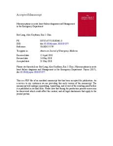
Figure 1. Arterial Vasodilatation and Renal Vasoconstriction in Patients with Sepsis. Endotoxemia stimulates the induction of nitric oxide synthase, which leads to nitric oxide–mediated arterial vasodilatation. The resultant arterial underfilling is sensed by the baroreceptors and results in an increase in sympathetic outflow and the release of arginine vasopressin from the central nervous system, with activation of the renin–angiotensin–aldosterone system (RAAS). These increases in renal sympathetic and angiotensin activities lead to vasoconstriction with sodium and water retention and a predisposition to acute renal failure.
162
n engl j med 351;2
The vasodilatory effect of constitutive endothelial nitric oxide synthase within the kidney might be expected to lessen the renal vasoconstriction induced by norepinephrine, angiotensin II, and endothelin during sepsis. However, the results of in vitro studies showed that the increase in the plasma nitric oxide concentration stimulated by inducible nitric oxide synthase during endotoxemia down-regulated endothelial nitric oxide synthase within the kidney.37 When cytokines activated inducible nitric oxide synthase, however, not only did the plasma nitric oxide concentration increase, but also the expression of inducible nitric oxide synthase increased in the renal cortex.38 In association with this increased expression of inducible nitric oxide synthase, a progressive increase in cGMP in the renal cortex oc-
www.nejm.org
july 8 , 2004
Downloaded from www.nejm.org at UNIV OF UTAH ECCLES on February 28, 2006 . Copyright © 2004 Massachusetts Medical Society. All rights reserved.
mechanisms of disease
curred during the initial 16 hours after exposure to endotoxin. At 24 hours, however, the plasma nitric oxide concentration remained high, though renal cGMP had decreased. Since cGMP is the secondary messenger for nitric oxide–mediated arterial vasodilatation, the down-regulation of this enzyme at 24 hours may also contribute to renal vasoconstriction during sepsis. Endothelial damage occurs during sepsis and may be associated with microthrombi and an increased concentration of von Willebrand factor in the circulation.39 Sepsis-related impairment of the endothelium may also attenuate or abolish the normal effect of endothelial nitric oxide synthase in the kidney to counteract the vasoconstrictor effects of norepinephrine, endothelin, and angiotensin II. The study of knockout mice, in which the expression of either endothelial nitric oxide synthase or inducible nitric oxide synthase has been ablated, has been helpful in elucidating the importance of endothelial damage during sepsis. Since there is no specific inhibitor of endothelial nitric oxide synthase, the effect of endotoxin (lipopolysaccharide) was tested in endothelial nitric oxide synthase–knockout mice, which have a significant increase in blood pressure and renal vascular resistance as compared with normal (control) mice. A small dose of endotoxin, which did not alter the glomerular filtration rate in the control mice, caused a profound decrease in the glomerular filtration rate in these knockout mice.40
though a soluble tumor necrosis factor receptor (TNFsRp55) afforded renal protection in murine endotoxemia,38 a prospective, randomized study of a monoclonal antibody against tumor necrosis factor a (the MONARCS [Monoclonal Anti-TNF: A Randomized Controlled Sepsis] trial) did not show any improvement in the survival of patients.41,42 Endotoxemia is known to be associated with the generation of oxygen radicals and thus may contribute to the early vasoconstrictor phase of acute renal failure. Endogenous scavengers of reactive oxygen species can attenuate renal tubular injury or renal vascular injury (or both) that is caused by re-
endotoxemia
Tumor Necrosis Factor a and Reactive Oxygen Species
Studies have also been undertaken in inducible nitric oxide synthase–knockout mice to determine the role of the high plasma nitric oxide concentration in the acute renal failure that is associated with endotoxemia. A dose of endotoxin (lipopolysaccharide) of 5 mg per kilogram causes a large and progressive rise in the plasma nitric oxide concentration in the normotensive mouse model by means of inducible nitric oxide synthase. However, in mice in which inducible nitric oxide synthase is ablated, this same dose of endotoxin fails to cause an increase in plasma nitric oxide.38 Nevertheless, these knockout mice still have a decrease in the glomerular filtration rate after receiving endotoxin, suggesting that cytokines such as tumor necrosis factor a can cause renal vasoconstriction even in the absence of inducible nitric oxide synthase. The role of tumor necrosis factor a in endotoxin-related acute renal failure has been tested in both animal and human studies. Aln engl j med 351;2
Figure 2. Effects of Systemic Arterial Vasodilatation in Patients with Sepsis and Acute Renal Failure. Sepsis and endotoxemia with acute renal failure can lead to early noncardiogenic pulmonary edema, hypoxia, and the need for mechanical ventilation. With prolonged ventilatory support, acute respiratory distress syndrome, multiple-organ dysfunction syndrome, and an extremely high mortality can occur. The goal is to intervene early to prevent excessive fluid administration and to lessen fluid overload by hemofiltration. This will prevent the need for long-term mechanical ventilation that could lead to damage to the pulmonary capillaries. It could also prevent tissue hypoxia and the acute respiratory distress syndrome and reduce the risk of death.
www.nejm.org
july 8, 2004
Downloaded from www.nejm.org at UNIV OF UTAH ECCLES on February 28, 2006 . Copyright © 2004 Massachusetts Medical Society. All rights reserved.
163
The
new england journal
active oxygen species during endotoxemia. However, the levels of the messenger RNA and protein of the endogenous scavenger extracellular superoxide dismutase, which is found predominantly in blood vessels and the kidney, have been noted to be decreased in mice during endotoxemia.43 In contrast, the mitochondrial scavenger manganese superoxide dismutase and the cytoplasmic scavenger copper–zinc superoxide dismutase were observed to be unaltered during endotoxemia. Exogenous oxygenradical scavengers were shown to protect against acute renal failure in this normotensive mouse model of endotoxemia. In a murine model of septic shock and acute renal failure, administration of a superoxide dismutase mimetic that had properties of oxygen-radical scavengers decreased deaths in the animals. Oxygen radicals also scavenge nitric oxide to produce peroxynitrite, an injurious reactive oxygen species. Furthermore, the decrease in endothelial nitric oxide synthase in the kidney when there is oxidant-related endothelial damage may contribute to the early vasoconstrictor phase of acute renal failure. Figure 3 depicts the potential good and bad effects of nitric oxide during sepsis. Nonspecific Inhibitors of Nitric Oxide Synthase
Studies in animals and humans have further examined the role of nitric oxide synthase in the decrease in the glomerular filtration rate during endotoxemia. No renal protection was found with the administration of a nonspecific inhibitor of nitric oxide synthase, Ng-nitro-l-arginine methyl ester, in an endotoxemic rat model of acute renal failure. In humans, the use of another nonspecific inhibitor of nitric oxide synthase, Ng-monomethyl-l-arginine, was found to increase mortality in patients with septic shock.44 Since this nonspecific inhibitor of nitric oxide synthase blocks both inducible and endothelial nitric oxide synthases, further studies with an inhibitor specific for inducible nitric oxide synthase, N6-(1-iminoethyl)-l-lysine, which would preserve any renal protective effect of endothelial nitric oxide synthase, were undertaken in the rat model.37 N6-(1-iminoethyl)-l-lysine appeared to be protective experimentally; however, these results require confirmation in clinical trials in humans. Cytokines, Chemokines, and Adhesion Molecules
The early vasoconstrictor phase of acute renal failure during endotoxemia may be followed by a proinflammatory phase, although there is probably an overlap in these processes. It is known that caspase
164
n engl j med 351;2
of
medicine
activates both interleukin-1b and interleukin-18 cytokines, and the resultant up-regulation of adhesion molecules contributes to neutrophil infiltration during endotoxemia. The importance of caspase in endotoxemia has been underscored by the observation that caspase-1–knockout mice are protected against renal failure that is induced by either ischemia45 or endotoxemia.46 Several chemokines are also expressed during endotoxemia in association with neutrophil and macrophage infiltration into the glomeruli and interstitium. The complex composed of a lipopolysaccharide and the lipopolysaccharide-binding protein activates the membrane-CD14 and toll-like receptors on cells, which up-regulate nuclear factor-kB (NF-kB), a nuclear transcription factor for the promoters of multiple cytokines, chemokines, and adhesion molecules.47Activation of NF-kB may therefore be a critical factor in the proinflammatory phase that involves a cytokine, chemokine, and adhesion molecule “storm,” which leads to acute renal failure and an increased rate of death. Blocking agents for NF-kB exist that could protect against endotoxemia better than targeting any individual cytokine, chemokine, or adhesion molecule.48 These substances need to be studied both in experimental models and in clinical studies in humans with concurrent sepsis and acute renal failure. Complement pathways are activated during sepsis by bacterial products such as lipopolysaccharide, C-reactive protein, and other stimuli. Complement C5a that is generated during sepsis seems to have procoagulant properties, and blocking C5a and C5a receptor in a rodent model of sepsis has been shown to improve survival.49-51 Although animal models of endotoxemia and sepsis have provided insights into sepsis and acute renal failure, the translation of these experimental results to patients with sepsis must be made with caution. Also, the mouse models in which sepsis was induced by the administration of lipopolysaccharide differed from the models achieved by cecal ligation and puncture.52,53
disseminated intravascular coagulation Sepsis affects the expression of complement, coagulation, and the fibrinolytic cascade. Sepsis can be viewed as a procoagulant state that can lead to disseminated intravascular coagulation with consumptive coagulopathy, thrombosis, and ultimately, hem-
www.nejm.org
july 8 , 2004
Downloaded from www.nejm.org at UNIV OF UTAH ECCLES on February 28, 2006 . Copyright © 2004 Massachusetts Medical Society. All rights reserved.
mechanisms of disease
orrhage. Disseminated intravascular coagulation has been associated with glomerular microthrombi and acute renal failure.54 Prospective, randomized trials have been undertaken to evaluate methods of intervening in the procoagulant process associated with sepsis. A major prospective, randomized study, the PROWESS (Recombinant Human Activated Protein C Worldwide Evaluation in Severe Sepsis) trial, showed that recombinant human activated protein C (drotrecogin alfa) significantly improved survival in patients with severe sepsis, as compared with those given placebo (75.3 percent vs. 68.3 percent, P=0.006).55 Results of renal-function tests were not reported in this trial.
early resuscitation Since the early vasoconstrictor phase of sepsis and acute renal failure is potentially reversible, it should be an optimal time for intervention. However, clinical studies performed in patients up to 72 hours after admission to the intensive care unit, in which attempts were made to optimize hemodynamics and monitor the patients with a pulmonary-artery catheter, not only were negative56-59 but showed increased mortality among patients with sepsis. In contrast, a randomized study of 263 patients with a mean serum creatinine concentration of 2.6 mg per deciliter (230 µmol per liter) on admission to the emergency department showed that early goaldirected therapy during the first six hours after admission was effective.60 The central venous oxygen saturation was continuously monitored as goaldirected therapy was instituted; in patients assigned to such interventions, the multiorgan dysfunction Figure 3. Good and Bad Effects of Nitric Oxide on the Kidney during Sepsis. score decreased significantly and in-hospital morThe induction of nitric oxide synthase and the generation of oxygen radicals during sepsis cause peroxynitrite-related tubular injury, systemic vasodilatatality decreased (30.5 percent, as compared with tion, and down-regulation of renal endothelial nitric oxide synthase. Endotox46.5 percent in the control patients, who received emia, however, may increase renal cortical inducible nitric oxide synthase, standard care; P=0.009). The goal-directed apwith a resultant increase in nitric oxide. The nitric oxide may afford protection proach included early volume expansion and adto the kidney by inhibiting platelet-aggregation–related glomerular microministration of vasopressors to maintain mean thrombi and causing cyclic guanosine monophosphate–mediated vasodilatation to counteract renal vasoconstriction with increased activity of the blood pressure at or above 65 mm Hg and transfusympathetic nervous system and angiotensin II during sepsis. Solid arrows sion of red cells to increase the hematocrit to 30 perindicate activation, and the dashed arrow and T bar inhibition. cent or more if central venous oxygen saturation was less than 70 percent. If these interventions failed to increase central venous oxygen saturation to greater than 70 percent, then therapy with dobutamine was tients compared the use of insulin to control blood instituted. glucose levels tightly (maintaining blood glucose levels between 80 and 110 mg per deciliter [4.4 and 6.1 mmol per liter]) with conventional treatment hyperglycemia and insulin (the use of insulin only if the blood glucose levels Hyperglycemia impairs the function of leukocytes exceeded 215 mg per deciliter [11.9 mmol per liter], and macrophages. A randomized study of 1548 pa- with the aim of maintaining glucose levels between n engl j med 351;2
www.nejm.org
july 8, 2004
Downloaded from www.nejm.org at UNIV OF UTAH ECCLES on February 28, 2006 . Copyright © 2004 Massachusetts Medical Society. All rights reserved.
165
The
new england journal
of
medicine
Multiple-organ failure with a proven focus of sepsis was also decreased (8 cases vs. 33 cases, P=0.02). Recent studies further support the importance of controlling blood glucose in critically ill patients but suggest a less stringent goal of maintaining blood glucose at a level of 145 mg per deciliter (8.0 mmol per liter) or less.62
glucocorticoids and mechanical ventilation Glucocorticoids have been known to enhance the pressor effects of catecholamines, but older studies in which septic shock was treated with large doses of glucocorticoid hormones for a short period of time did not show any benefit.63,64 However, a recent study65 in patients with septic shock showed that patients without a response to corticotropin (as defined by a rise in plasma free cortisol of less than 9 µg per deciliter at 30 or 60 minutes) who were treated for 7 days with intravenous boluses of 50 mg of hydrocortisone every 6 hours plus daily oral fludrocortisone (a 50-µg tablet) had a decrease in mortality at 28 days as compared with the placebo group (63 percent vs. 53 percent, P=0.02). In this randomized study, 229 of the 299 patients with septic shock who were enrolled were classified as not having a response. There was no difference in mortality among the 70 patients with a response to the short corticotropin study. Withdrawal of vasopressors was also significantly better at 28 days in those without a response (40 percent vs. 57 percent, Figure 4. Methods of Attenuating or Preventing Sepsis-Related Acute Renal Failure. P<0.001).65 Although this study did not report reArginine vasopressin (AVP) and hydrocortisone (50 mg every six hours for nal function results, it is known that septic shock is seven days) may be effective therapy for pressor-resistant hypotension and associated with acute renal failure in 38 percent of may decrease the likelihood of acute renal failure during septic shock. Early patients with negative cultures and 51 percent of padirected resuscitation of patients with sepsis may prevent the progression tients with positive cultures.2 from prerenal azotemia to acute tubular necrosis. Maintenance of blood gluOther studies show that the longer the duration cose levels below 145 mg per deciliter (8.0 mmol per liter) may decrease the incidence of acute renal failure, multiple-organ dysfunction syndrome, and of mechanical ventilation, the higher the mortality death. Finally, activated protein C can decrease disseminated intravascular in patients with sepsis and acute renal failure.31 One coagulation with glomerular and microvascular thrombi and thereby decrease study showed that daily interruption of a continuous mortality. T bars indicate inhibition. infusion of sedatives in critically ill patients who were undergoing mechanical ventilation shortened the time needed on the ventilator (7.3 vs. 4.9 days, 180 and 220 mg per deciliter [10.0 and 12.2 mmol P=0.004) and time in the intensive care unit (9.9 vs. per liter]).61 The group assigned to tight control of 6.4 days, P=0.02).66 blood glucose levels showed a decrease in mortality in the intensive care unit as compared with the group renal replacement receiving conventional treatment (4.6 percent vs. 8 percent, P<0.04), a 46 percent decrease in positive Patients with sepsis and acute renal failure are hyblood cultures, and a 41 percent decrease in acute percatabolic. Studies suggesting that increased dosrenal failure requiring dialysis or hemofiltration. es of dialysis improve survival in patients who are
166
n engl j med 351;2
www.nejm.org
july 8 , 2004
Downloaded from www.nejm.org at UNIV OF UTAH ECCLES on February 28, 2006 . Copyright © 2004 Massachusetts Medical Society. All rights reserved.
mechanisms of disease
hypercatabolic and have acute renal failure are persuasive. For example, survival was markedly improved with aggressive hemodialysis as compared with peritoneal dialysis in patients who had heatstroke, rhabdomyolysis, and acute renal failure.67 Hemofiltration has been shown to produce better survival rates than peritoneal dialysis in patients with acute renal failure associated with malaria and other infections.68 A recent study showed that daily hemodialysis as compared with alternate-day hemodialysis was associated with less systemic inflammatory response syndrome or sepsis (22 percent vs. 46 percent, P=0.005), lower mortality (28 percent vs. 46 percent, P<0.01) and a shorter duration of acute renal failure (mean [±SD], 9±2 vs. 16±6 days; P=0.001).69 Continuous renal-replacement therapy has increasingly been used to treat acute renal failure. A randomized study using continuous venovenous hemofiltration suggested that the ultrafiltration rate of 35 or 45 ml per kilogram per hour as compared with 20 ml per kilogram per hour improves survival in acute renal failure (P<0.001).70 Moreover, in patients with sepsis-related acute renal failure, better survival was observed with an ultrafiltration rate of 45 ml per kilogram per hour than with a rate of 35 mg per kilogram per hour. Meta-analysis of hemodialysis as compared with continuous renal-replace-
ment therapy in acute renal failure, however, has not yet shown an advantage for either mode of renalreplacement therapy.71 The benefit of the removal of cytokines by continuous renal-replacement therapy also remains to be proven as a method for improving survival in patients with sepsis and acute renal failure.
conclusions Acute renal failure is a common complication of sepsis and septic shock. Patients who have sepsisrelated acute renal failure have much higher mortality than patients with acute renal failure who do not have sepsis. Experimental models of endotoxemia and sepsis have provided insights into the pathogenesis of sepsis-related acute renal failure, but results from such models should be examined stringently before applying them to patients with sepsis. As shown in Figure 4, recent clinical studies indicate that interventions based on several proposed pathogenetic factors in sepsis-related acute renal failure may have a favorable effect on both the incidence of acute renal failure and the mortality of patients with acute renal failure. Supported by a grant (DK52599) from the National Institutes of Health. We are indebted to Jan Darling for editorial assistance.
references 1. Riedemann NC, Guo RF, Ward PA. The
enigma of sepsis. J Clin Invest 2003;112: 460-7. 2. Rangel-Frausto MS, Pittet D, Costigan M, Hwang T, Davis CS, Wenzel RP. The natural history of the systemic inflammatory response syndrome (SIRS): a prospective study. JAMA 1995;273:117-23. 3. Angus DC, Linde-Zwirble WT, Lidicker J, Clermont G, Carcillo J, Pinsky MR. Epidemiology of severe sepsis in the United States: analysis of incidence, outcome, and associated costs of care. Crit Care Med 2001; 29:1303-10. 4. Edelstein CL, Schrier RW. Pathophysiology of ischemic acute renal failure. In: Schrier RW, ed. Diseases of the kidney and urinary tract. 7th ed. Vol. 2. Philadelphia: Lippincott Williams & Wilkins, 2001:1041-69. 5. Landry DW, Oliver JA. The pathogenesis of vasodilatory shock. N Engl J Med 2001; 345:588-95. 6. Esson ML, Schrier RW. Diagnosis and treatment of acute tubular necrosis. Ann Intern Med 2002;137:744-52. 7. Brivet FG, Kleinknecht DJ, Loirat P, Landais PJ. Acute renal failure in intensive care
units — causes, outcome, and prognostic factors of hospital mortality: a prospective, multicenter study. Crit Care Med 1996;24: 192-8. 8. Schrier RW. Body fluid volume regulation in health and disease: a unifying hypothesis. Ann Intern Med 1990;113:155-9. 9. Schrier RW, Abraham WT. Hormones and hemodynamics in heart failure. N Engl J Med 1999;341:577-85. 10. Benedict CR, Rose JA. Arterial norepinephrine changes in patients with septic shock. Circ Shock 1992;38:165-72. 11. Cumming AD, Driedger AA, McDonald JW, Lindsay RM, Solez K, Linton AL. Vasoactive hormones in the renal response to systemic sepsis. Am J Kidney Dis 1988;11:2332. 12. Thiemermann C, Szabo C, Mitchell JA, Vane JR. Vascular hyporeactivity to vasoconstrictor agents and hemodynamic decompensation in hemorrhagic shock is mediated by nitric oxide. Proc Natl Acad Sci U S A 1993;90:267-71. 13. Titheradge MA. Nitric oxide in septic shock. Biochim Biophys Acta 1999;1411: 437-55.
n engl j med 351;2
www.nejm.org
14. Hollenberg SM, Broussard M, Osman J,
Parrillo JE. Increased microvascular reactivity and improved mortality in septic mice lacking inducible nitric oxide synthase. Circ Res 2000;86:774-8. 15. Hollenberg SM, Cunnion RE, Zimmerberg J. Nitric oxide synthase inhibition reverses arteriolar hyporesponsiveness to catecholamines in septic rats. Am J Physiol 1993;264:H660-H663. 16. Davies NW. Modulation of ATP-sensitive K+ channels in skeletal muscle by intracellular protons. Nature 1990;343:3757. 17. Keung EC, Li Q. Lactate activates ATPsensitive potassium channels in guinea pig ventricular myocytes. J Clin Invest 1991;88: 1772-7. 18. Morales D, Madigan J, Cullinane S, et al. Reversal by vasopressin of intractable hypotension in the late phase of hemorrhagic shock. Circulation 1999;100:226-9. 19. Landry DW, Levin HR, Gallant EM, et al. Vasopressin deficiency contributes to the vasodilation of septic shock. Circulation 1997;95:1122-5. 20. Wakatsuki T, Nakaya Y, Inoue I. Vaso-
july 8, 2004
Downloaded from www.nejm.org at UNIV OF UTAH ECCLES on February 28, 2006 . Copyright © 2004 Massachusetts Medical Society. All rights reserved.
167
The
new england journal
pressin modulates K(+)-channel activities of cultured smooth muscle cells from porcine coronary artery. Am J Physiol 1992;263: H491-H496. 21. Umino T, Kusano E, Muto S, et al. AVP inhibits LPS- and IL-1beta-stimulated NO and cGMP via V1 receptor in cultured rat mesangial cells. Am J Physiol 1999;276: F433-F441. 22. Zerbe RL, Henry DP, Robertson GL. Vasopressin response to orthostatic hypotension: etiologic and clinical implications. Am J Med 1983;74:265-71. 23. Kaufmann H, Oribe E, Oliver JA. Plasma endothelin during upright tilt: relevance for orthostatic hypotension? Lancet 1991;338: 1542-5. 24. Arnauld E, Czernichow P, Fumoux F, Vincent JD. The effects of hypotension and hypovolaemia on the liberation of vasopressin during haemorrhage in the unanaesthetized monkey (Macaca mulatta). Pflugers Arch 1977;371:193-200. 25. Bartelstone HJ, Nasmyth PA. Vasopressin potentiation of catecholamine actions in dog, rat, cat, and rat aortic strip. Am J Physiol 1965;208:754-62. 26. Cowley AW Jr, Liard JF. Vasopressin and arterial pressure regulation: special lecture. Hypertension 1988;11:I-25–I-32. 27. Kumar A, Haery C, Parrillo JE. Myocardial dysfunction in septic shock. Crit Care Clin 2000;16:251-87. 28. Fernandes Junior CJ, Iervolino M, Neves RA, Sampaio EL, Knobel E. Interstitial myocarditis in sepsis. Am J Cardiol 1994;74:958. 29. Schrier RW, Abraham E. Aggressive volume expansion and pseudo-ARDS. Hosp Pract (Off Ed) 1995;30(6):19, 23. 30. Sanz E, Lopez Novoa JM, Linares M, Digiuni E, Caramelo CA. Intravascular and interstitial fluid dynamics in rats treated with minoxidil. J Cardiovasc Pharmacol 1990;15:485-92. 31. Neveu H, Kleinknecht D, Brivet F, Loirat P, Landais P. Prognostic factors in acute renal failure due to sepsis: results of a prospective multicentre study. Nephrol Dial Transplant 1996;11:293-9. 32. Kikeri D, Pennell JP, Hwang KH, Jacob AI, Richman AV, Bourgoignie JJ. Endotoxemic acute renal failure in awake rats. Am J Physiol 1986;250:F1098-F1106. 33. Wang W, Falk SA, Jittikanont S, Gengaro PE, Edelstein CL, Schrier RW. Protective effect of renal denervation on normotensive endotoxemia-induced acute renal failure in mice. Am J Physiol Renal Physiol 2002;283: F583-F587. 34. Hohlfeld T, Klemm P, Thiemermann C, Warner TD, Schror K, Vane JR. The contribution of tumour necrosis factor-alpha and endothelin-1 to the increase of coronary resistance in hearts from rats treated with endotoxin. Br J Pharmacol 1995;116:3309-15. 35. Kon V, Badr KF. Biological actions and pathophysiologic significance of endothelin in the kidney. Kidney Int 1991;40:1-12.
168
of
medicine
36. Filep JG. Role for endogenous endothe-
lin in the regulation of plasma volume and albumin escape during endotoxin shock in conscious rats. Br J Pharmacol 2000;129: 975-83. 37. Schwartz D, Mendonca M, Schwartz I, et al. Inhibition of constitutive nitric oxide synthase (NOS) by nitric oxide generated by inducible NOS after lipopolysaccharide administration provokes renal dysfunction in rats. J Clin Invest 1997;100:439-48. 38. Knotek M, Rogachev B, Wang W, et al. Endotoxemic renal failure in mice: role of tumor necrosis factor independent of inducible nitric oxide synthase. Kidney Int 2001; 59:2243-9. 39. Reinhart K, Bayer O, Brunkhorst F, Meisner M. Markers of endothelial damage in organ dysfunction and sepsis. Crit Care Med 2002;30:Suppl:S302-S312. 40. Wang W, Mitra A, Poole BD, Falk SA, Schrier RW. Endothelial nitric oxide synthase (eNOS) deficient mice exhibit increased susceptibility to endotoxin-induced acute renal failure (ARF). J Am Soc Nephrol (in press). 41. Gallagher J, Fisher C, Sherman B, et al. A multicenter, open-label, prospective, randomized, dose-ranging pharmacokinetic study of the anti-TNF-alpha antibody afelimomab in patients with sepsis syndrome. Intensive Care Med 2001;27:1169-78. 42. Reinhart K, Karzai W. Anti-tumor necrosis factor therapy in sepsis: update on clinical trials and lessons learned. Crit Care Med 2001;29:Suppl:S121-S125. 43. Wang W, Jittikanont S, Falk SA, et al. Interaction among nitric oxide, reactive oxygen species, and antioxidants during endotoxemia-related acute renal failure. Am J Physiol Renal Physiol 2003;284:F532-F537. 44. Lopez A, Lorente JA, Steingrub J, et al. Multiple-center, randomized, placebo-controlled, double-blind study of the nitric oxide synthase inhibitor 546C88: effect on survival in patients with septic shock. Crit Care Med 2004;32:21-30. 45. Melnikov VY, Ecder T, Fantuzzi G, et al. Impaired IL-18 processing protects caspase-1-deficient mice from ischemic acute renal failure. J Clin Invest 2001;107: 1145-52. 46. Wang W, Reznikof L, Falk SA, et al. Caspase-1 knockout mice are resistant to endotoxemic acute renal failure (ARF). J Am Soc Nephrol 2003;14:350A. abstract. 47. Schor N. Acute renal failure and the sepsis syndrome. Kidney Int 2002;61:764-76. 48. Ulloa L, Ochani M, Yang H, et al. Ethyl pyruvate prevents lethality in mice with established lethal sepsis and systemic inflammation. Proc Natl Acad Sci U S A 2002;99: 12351-6. 49. Riedemann NC, Guo RF, Neff TA, et al. Increased C5a receptor expression in sepsis. J Clin Invest 2002;110:101-8. 50. Huber-Lang MS, Riedeman NC, Sarma JV, et al. Protection of innate immunity by
n engl j med 351;2
www.nejm.org
C5aR antagonist in septic mice. FASEB J 2002;16:1567-74. 51. Czermak BJ, Sarma V, Pierson CL, et al. Protective effects of C5a blockade in sepsis. Nat Med 1999;5:788-92. 52. Remick DG, Newcomb DE, Bolgos GL, Call DR. Comparison of the mortality and inflammatory response of two models of sepsis: lipopolysaccharide vs. cecal ligation and puncture. Shock 2000;13:110-6. 53. Fink MP, Heard SO. Laboratory models of sepsis and septic shock. J Surg Res 1990; 49:186-96. 54. Shimamura K, Oka K, Nakazawa M, Kojima M. Distribution patterns of microthrombi in disseminated intravascular coagulation. Arch Pathol Lab Med 1983;107: 543-7. 55. Bernard GR, Vincent J-L, Laterre P-F, et al. Efficacy and safety of recombinant human activated protein C for severe sepsis. N Engl J Med 2001;344:699-709. 56. Connors AF Jr, Speroff T, Dawson NV, et al. The effectiveness of right heart catheterization in the initial care of critically ill patients. JAMA 1996;276:889-97. 57. Opal SM, Cross AS. Clinical trials for severe sepsis: past failures, and future hopes. Infect Dis Clin North Am 1999;13:285-97. 58. Hayes MA, Timmins AC, Yau EHS, Palazzo M, Hinds CJ, Watson D. Elevation of systemic oxygen delivery in the treatment of critically ill patients. N Engl J Med 1994;330: 1717-22. 59. Gattinoni L, Brazzi L, Pelosi P, et al. A trial of goal-oriented hemodynamic therapy in critically ill patients. N Engl J Med 1995;333:1025-32. 60. Rivers E, Nguyen B, Havstad S, et al. Early goal-directed therapy in the treatment of severe sepsis and septic shock. N Engl J Med 2001;345:1368-77. 61. Van den Berghe G, Wouters P, Weekers F, et al. Intensive insulin therapy in critically ill patients. N Engl J Med 2001;345:1359-67. 62. Finney SJ, Zekveld C, Elia A, Evans TW. Glucose control and mortality in critically ill patients. JAMA 2003;290:2041-7. 63. Cronin L, Cook DJ, Carlet J, et al. Corticosteroid treatment for sepsis: a critical appraisal and meta-analysis of the literature. Crit Care Med 1995;23:1430-9. 64. Lefering R, Neugebauer EA. Steroid controversy in sepsis and septic shock: a meta-analysis. Crit Care Med 1995;23: 1294-303. 65. Annane D, Sebille V, Charpentier C, et al. Effect of treatment with low doses of hydrocortisone and fludrocortisone on mortality in patients with septic shock. JAMA 2002;288:862-71. 66. Kress JP, Pohlman AS, O'Connor MF, Hall JB. Daily interruption of sedative infusions in critically ill patients undergoing mechanical ventilation. N Engl J Med 2000; 342:1471-7. 67. Schrier RW, Henderson HS, Tisher CC, Tannen RL. Nephropathy associated with
july 8, 2004
Downloaded from www.nejm.org at UNIV OF UTAH ECCLES on February 28, 2006 . Copyright © 2004 Massachusetts Medical Society. All rights reserved.
mechanisms of disease
heat stress and exercise. Ann Intern Med 1967;67:356-76. 68. Phu NH, Hien TT, Mai NTH, et al. Hemofiltration and peritoneal dialysis in infection-associated acute renal failure in Vietnam. N Engl J Med 2002;347:895-902. 69. Schiffl H, Lang SM, Fischer R. Daily he-
modialysis and the outcome of acute renal failure. N Engl J Med 2002;346:305-10. 70. Ronco C, Bellomo R, Homel P, et al. Effects of different doses in continuous venovenous haemofiltration on outcomes of acute renal failure: a prospective randomised trial. Lancet 2000;356:26-30.
n engl j med 351;2
www.nejm.org
71. Tonelli M, Manns B, Feller-Kopman D.
Acute renal failure in the intensive care unit: a systematic review of the impact of dialytic modality on mortality and renal recovery. Am J Kidney Dis 2002;40:875-85. Copyright © 2004 Massachusetts Medical Society.
july 8, 2004
Downloaded from www.nejm.org at UNIV OF UTAH ECCLES on February 28, 2006 . Copyright © 2004 Massachusetts Medical Society. All rights reserved.
169