RESEARCH PAPER
Multiple Sclerosis 2008; 14: 330–342
Abnormal sensorimotor control, but intact force field adaptation, in multiple sclerosis subjects with no clinical disability Maura Casadio1,2, Vittorio Sanguineti1, Pietro Morasso1 and Claudio Solaro3 In MS subjects with no clinical disability, we assessed sensorimotor organization and their ability to adapt to an unfamiliar dynamical environment. Eleven MS subjects performed reaching movements while a robot generated a speed-dependent force field. Control and adaptation performance were compared with that of an equal number of control subjects. During a familiarization phase, when the robot generated no forces, the movements of MS subjects were more curved, displayed greater and more variable directional errors and a longer deceleration phase. During the force field phase, both MS and control subjects gradually learned to predict the robot-generated forces. The rates of adaptation were similar, but MS subjects showed a greater variability in responding to the force field. These results suggest that MS subjects have a preserved capability of learning to predict the effects of the forces, but make greater errors when actually using such predictions to generate movements. Inaccurate motor commands are then compensated later in the movement through an extra amount of sensory-based corrections. This indicates that early in the disease MS subjects have intact adaptive capabilities, but impaired movement execution. Multiple Sclerosis 2008; 14: 330–342. http://msj.sagepub.com Key words: motor adaptation; motor control; multiple sclerosis; robot therapy
Introduction In the early stages of MS, about 85% of the subjects show a relapsing–remitting course, that is, acute phases followed by clinical recovery. Relapses are often followed by partial or complete functional recovery. In about 40% of the patients, recovery is incomplete when evaluated after six months, with a residual deficit of 0.5 or more units of the expanded disability status scale (EDSS) score [1]. Brain plasticity is one potential contributor to the recovery of function after tissue damage, and
may limit the functional consequences of axonal loss. Clinical recovery is determined by many factors, such as increased expression of sodium channels, recruitment of silent pathways, remyelination. Cortical and subcortical reorganization may take place as well, thus playing a role in limiting the impact of tissue damage. Magnetic resonance imaging (MRI) and, even more, functional MRI (fMRI) studies have provided important insights on the mechanisms of tissue damage and functional recovery in MS subjects. Functional MRI studies in MS subjects with a relapsing–remitting course and minimal disability have shown that in simple motor
1 Department of Informatics, Systems and Telematics and Research Centre for Neuroscience and Neuroengineering, University of Genoa, via Opera Pia 13, Genoa 16145, Italy 2 Foundation ‘Don Gnocchi’, via Cisa Vecchia, 19038 Sarzana SP, Italy 3 Department of Neurology, Hospital PA Micone, Genoa, Italy Author for correspondence: Professor Vittorio Sanguineti, Department of Informatics, Systems and Telematics and Research Centre for Neuroscience and Neuroengineering, University of Genoa, via Opera Pia 13, Genoa 16145, Italy. E-mail:
[email protected] Received 18 April 2007; revised 30 July 2007; accepted 5 August 2007
© 2008 SAGE Publications Los Angeles, London, New Delhi and Singapore
Downloaded from http://msj.sagepub.com at Univ Degli Studi Di Genova on October 10, 2008
10.1177/1352458507085068
Abnormal sensorimotor control in MS subjects with no clinical disability tasks, these subjects recruit areas that (in healthy subjects) are typically associated to more complex tasks [2]. A recent study provided evidence that both inter- and intra-hemispheric motor reorganization occur in MS [3]. These findings point at a cortical and subcortical reorganization aimed at counteracting demyelination and/or neurodegeneration [4]. From the behavioral point of view, reorganization may lead to the development of compensatory strategies. Although compensatory strategies may leave performance relatively unaffected, they may be unveiled by experimental and analytical techniques that aim at investigating the mechanisms underlying sensorimotor control and adaptation. Robotic devices that were originally developed to study motor learning in normal subjects can be used to assess the motor functionality of neurological patients. Recently, an experimental protocol that had been widely used to investigate motor learning capabilities [5] was applied to subjects with degenerative cerebellar atrophy [6,7]. The results showed that these subjects completely lost their adaptation capability. Recently, it has been shown that MS subjects with little or no disability have subtle problems in gait and balance [8] and in controlling arm movements [9]. Multiple sclerosis subjects with mild disability also displayed a reduced ability to improve their performance with exercise in a visual tracking task [10]. However, it is unclear whether the ability of adapting to an unfamiliar environment is preserved in MS subjects with no disability and/or what is the origin of these problems. In this study, we asked whether sensorimotor adaptation in MS subjects might contribute to maintaining a normal level of motor performance. To this purpose, we investigated the ability of MS subjects with no disability to adapt to velocitydependent force fields. We also asked whether such adaptation differs from control subjects either in its outcome or in its dynamics.
Materials and Methods
Eleven subjects with clinically definite relapsing– remitting MS, according to Poser criteria [11], participated in this study (three males, eight females). Their mean age was 33.4 years (range 24–44) and the mean disease duration was five years (range 1–15). Patients were recruited at the Department of Neurology of the Hospital ‘P Antero Micone’ and at the Department of Neurosciences, Ophthalmology and Genetics, University of Genova. The inclusion criteria were: (i) EDSS [12] less than or equal to 1 http://msj.sagepub.com
and (ii) presence of neurological signs only, but no signs or symptoms at upper limbs and consequently, ‘normal’ score for the ‘arm’ portion of the Scripps’ neurological rating scale [13] for the sensory, motor and cerebellar systems. The exclusion criteria were: (i) relapses within the last three months; (ii) treatment with corticosteroids within the previous three months; (iii) mini mental state examination (MMSE) less than 24 and (iv) symptomatic oculomotor signs or visual acuity less than 8/10. The same neurologist examined all the patients. The performance of these subjects was compared with 11 controls – six males, five females, age 29.3 (range 22–41) – with no previous history of neurological dysfunction. Demographic and clinical details for each subject are listed in Table 1. The research conforms to the ethical standards laid down in the 1964 Declaration of Helsinki that protects research subjects. Each subject signed a consent form that conforms to these guidelines.
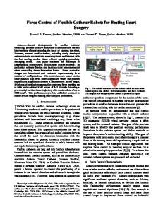
Experimental apparatus and task We used a planar robotic manipulandum (Braccio di Ferro, see Figure 1), specifically designed for robot therapy and for the evaluation of motor control and motor adaptation. The manipulandum – technical details are reported in Casadio et al. (2006) [14] – has a 80 ⫻ 40 cm elliptic workspace, and is actuated by two direct-drive brushless motors, mounted proximally to minimize inertia and frictional forces. The structure is extremely rigid, and robot geometry has been optimized for uniformity of the manipulability index and the force/torque ratio over the whole workspace. The force available at the handle in all directions is ⬎50 N (continuous) and ⬎200 N (peak). The rotations of the motors are measured by optical encoders, which allow to estimate the position of the handle with a spatial resolution ⬍0.1 mm over the whole workspace. The control architecture consists of an inner current loop Table 1
Subjects
331
Multiple sclerosis subjects
Subject
Age (y)
Sex
Hand
Disease duration (y)
EDSS
P1 P2 P3 P4 P5 P6 P7 P8 P9 P10 P11
34 32 44 38 45 27 32 37 22 32 24
F F F F F F M F F M M
R R R R R R R R R R R
4 4 3 15 5 5 3 6 7 2 1
0 1 0 1 0 1 0 0 1 0 0
Multiple Sclerosis 2008; 14: 330–342 Downloaded from http://msj.sagepub.com at Univ Degli Studi Di Genova on October 10, 2008
332
M Casadio et al.
Figure 1 Experimental set-up, seen from above: it shows the wrist holder, the arm weight support, the screen in front of the subject and the four-bar linkage of the robot.
(16 kHz), and an outer impedance control loop (1 kHz). We used a speed-dependent force field adaptation paradigm [5,7], which has been widely used to study sensorimotor co-ordination in unperturbed reaching movements and the adaptation induced by an unfamiliar artificial dynamic environment. Subjects were seated, with their torso and wrist restrained by means of suitable holders and grasped the handle of the manipulandum with their dominant hand. A low-friction sled on the horizontal surface of a table supported the forearm. The height of the seat was adjusted so that the arm could be kept horizontally at the level of the shoulder. Therefore, only shoulder and elbow could move and motion was restricted to the horizontal plane. The position of the seat was also adjusted in such a way that, with the hand pointing at the center of the workspace, the elbow joint was anteflexed about 90° and the shoulder was horizontally abducted about 45°. The task consisted of reaching movements in eight different directions, starting from the same center position. The targets were presented on a 19⬙ LCD computer screen, placed in front of the subjects, about 1 m away, at eye level. Targets were displayed as green circles (diameter 2 cm) against a black background. The current position of the hand in the workspace was also continuously displayed,
as a yellow circle (diameter 0.4 cm). The nominal amplitude of the movements (distance of the targets from the center position) was 10 cm. The sequence of target presentations alternated the central target and one of four peripheral targets, (corresponding to the directions: 0°, 45°, 90° and 135°) generated in random order. As in Diedrichsen et al. (2005) [15], the endpoint of each movement was used as the starting point for the subsequent movement. This corresponded to four additional directions (180°, 225°, 270° and 315°) for the return movements. In order to decrease movement variability, the subjects were encouraged to keep an approximately constant timing. We set the desired duration to 500 ⫾ 50 ms. If the estimated duration was inside this range, a positive feedback/reward to the subject (a pleasant sound) was provided. If the measure was below or above that range, no sound was generated and the color of the target was changed to either white or red, respectively. We also informed the subjects that reaction time was not important – they could wait as long as they wanted after target appearance before starting each movement – but when ready, they had to perform a single, rapid movement toward the target. The experiment was organized into target sets, each consisting of a sequence of target presentations in which each peripheral target occurred 12 times, for a total 12 ⫻ 4 ⫽ 48 center-out movements, plus the corresponding 48 return movements. The experimental protocol included three phases: (i) null field, in which the robot generates no force (five target sets); (ii) force field, in which the force field was turned on (five target sets) and (iii) aftereffect (two target sets). Each set lasted approximately 5 min, and the subjects were allowed to rest between one set and another. The null-field phase had the purpose of establishing a background level of performance. During the force field phase, the manipulandum generated a velocity-dependent curl field [7,16], in which perturbing forces were perpendicular to the instantaneous movement direction and had a magnitude proportional to the hand speed. More specifically, the force applied to the hand was generated as: (1)
where b is a viscous coefficient. We chose a value, b ⫽ 13 N/m/s, which is large enough to induce a detectable adaptation, but small enough to prevent muscle fatigue. This value corresponds to peak forces of 4–6 N at the hand. The hand velocity vector, x, ˙ was calculated online from the rotations of the joints, measured by the encoders.
Multiple Sclerosis 2008; 14: 330–342 Downloaded from http://msj.sagepub.com at Univ Degli Studi Di Genova on October 10, 2008
http://msj.sagepub.com
Abnormal sensorimotor control in MS subjects with no clinical disability During the field sets, we inserted ‘catch trials’, that is, trials in which the force field was unexpectedly turned off. The probability of catch trials was set to 1/6, corresponding to two catch trials per direction per target set.
Movement analysis Hand trajectories were sampled at 100 Hz and smoothed by using a sixth order Savitzky–Golay filter with a 170 ms window (cut-off frequency: ~11 Hz), which was also used to estimate the subsequent time derivatives of the trajectory. Movement onset was defined as the first time when speed exceeded 10% of the maximum speed. Likewise, end of movement was defined as the first time when speed fell below 10% of the maximum speed. This criterion allows to identify primary movements. Usually, late adjustments to the endpoint (secondary submovements) were not observed and are not analysed here. For each movement, we estimated the time elapsed between, respectively, movement onset and the time when speed reaches a peak (acceleration duration) and between this same time and that of movement termination (deceleration duration). We also estimated the aiming error as the difference between the target direction and the actual movement direction, estimated from the trajectory in the early phase of the movement. The aiming error was calculated within two different time intervals after movement onset: 100 ms and 300 ms. During the first 100 ms, movements can be assumed to be under open-loop control; therefore, we took the 100-ms aiming error as an indicator of the performance of the feed-forward component of control. As regards the 300-ms aiming error, we took it as a measure of lateral deviation from the straight line (i.e., an indicator of path curvature) [7]. The 100-ms aiming error has a ‘systematic’ component, related to target direction, which reflects the inability to account for the anisotropy of arm (and robot) inertia and a random component, which reflects the variability of the performance from trial to trial. To distinguish among these effects, for each movement direction we took both the average aiming error (the ‘systematic’ component) and its SD (the ‘random’ component). To test for changes in motor performance during the null-field phase, we carried out a three-way repeated-measures ANOVA, by looking at the effects of disease (control versus MS), movement direction (0°, 45°, 90°, 135°, 180°, 225°, 270°, 315°) and time (early versus late target set of the null phase) for all the above quantities, namely 100-ms aiming error (systematic and random part), acceleration and deceleration duration and 300-ms aiming http://msj.sagepub.com
333
error. To test whether the differences (if any) among control and MS subjects were still present at the end of familiarization, we looked (contrast analysis) at the effect of disease at the end of familiarization (late target set). Finally, to assess the adaptation capability of MS subjects in comparison to controls, the same analysis was repeated for the force field phase.
Analysis of force field adaptation To test for adaptation, during each phase of the experiment we looked at the time course of each indicator. In particular, for each phase we looked at the changes of each indicator between the first and the last target set. Subjects may react to the perturbations introduced by the curl field in different ways: (1) they may just ignore them, and accept the distortion introduced by the field, a viable alternative as this kind of perturbation does not specifically affect the achievement of the target; (2) they may resist the perturbation by increasing their hand stiffness (e.g., by co-contracting the muscles), without changing the underlying motor commands and (3) they may compensate the perturbation by predicting it, through the formation of a suitable internal model. The latter option implies that subjects modify their motor commands. The first alternative can be ruled out if we can demonstrate that the movements made under the curl field do change over repetitions, a finding that is well established for healthy subjects [5], but needs to be demonstrated for MS patients. The second alternative can be ruled out by observing the catch trials. If the observed adaptation to the force field were due to a mere increase in hand stiffness, catch trials should display no deviation. In contrast, if error reduction is a consequence of an adapted feed-forward control, the unexpected disappearance of the field in the catch trials should result in errors in the opposite direction. In this study, we used the following definition of a learning index (LI), originally proposed by CriscimagnaHemminger et al. (2003) [16] and used by Smith et al. (2005) [7]:
(2) where yf and yc are the 300-ms aiming errors in the field trials and in the catch trials, respectively. Learning index is zero (no learning) if the error in catch trials is zero; it is one if the error in field trials is zero. Aiming error measures were adjusted for any bias that may have been present during the last Multiple Sclerosis 2008; 14: 330–342
Downloaded from http://msj.sagepub.com at Univ Degli Studi Di Genova on October 10, 2008
334
M Casadio et al.
null-field set. Therefore, errors in a field set always refer to variations with respect to the errors in the late null set. To test for differences in the LI of MS subjects and controls, we focused on the late value of the LI, and ran a one-way ANOVA with disease (control versus MS) as factor.
State–space model of adaptation Recently, it has been suggested [17] that sensorimotor adaptation is better characterized in terms of its temporal evolution rather than just in terms of its outcome. A recent theory [18–20] describes how, in force field adaptation, the effect of errors experienced in a movement in one specific direction can be related to the previous history of experienced movements. Let us denote the error on trial n as y(n). This may be any measure of movement inaccuracy, for instance the 300-ms aiming error. We may express this quantity as a function of the force perturbation, f(n), and of an internally stored ‘motor command’ that – for that direction – would compensate the perturbation, zd(n), where d is the movement direction in that trial. The observed temporal evolution of y(n) can be modeled by a simple linear state–space model:
(3)
The parameter Dd indicates how much angular error results from a perturbation in direction d in a naive participant (i.e., with zd(n) ⫽ 0). This parameter may be related to the hand compliance (the inverse of stiffness), and corresponds to the contribution of co-contraction to the response to force field perturbation. The quantity r(n) is an output or ‘execution’ noise term, which accounts for the portion of the observed movement error that is not predicted by the model. The state equation (Equation 3, top) states that at each trial the experienced error, y(n), modifies the internal state z(n) – that is, the internally stored ‘motor command’ for each movement direction – by an amount that is determined by the rate of adaptation, B. The vector quantity q(n) is a state–space or ‘adaptation’ noise term, which accounts for the portion of state update that does not depend on the previous error. Note that the state equation (Equation 3, top) is an eight-dimensional vector equation (because there are eight movement directions) whereas the output equation (Equation 3, bottom) is scalar.
On every trial, the state vector is updated for all possible movement directions – in other words, movements in one particular direction contribute to the adaptation of the internal model in all directions – but only the state component that corresponds to that particular movement direction contributes to the error at that trial. Therefore, B is an 8 ⫻ 8 matrix, where the element bij denotes the contribution of experiencing an error in direction j on the internally stored motor command for direction i. Each column of B represents the error generalization function, that is, how an error in one direction contributes to adaptation of the internal model in all directions. Further details about the model are reported in Appendix A. To test for changes in MS subjects and control, for each model parameter we ran a one-way ANOVA with disease (control/MS) as factor.
Results Figure 2 shows typical hand trajectories recorded during the adaptation process (early and late phases, respectively), with respect to the different experimental conditions (null field, force field and after-effect). The figure suggests that the overall motor and adaptation patterns are similar in control and MS subjects, but there might be detectable performance differences. Multiple sclerosis subjects display subtle but detectable abnormalities To assess whether and how the motor performance of MS subjects differs from that of controls, we initially focused on the null-field trials (N trials), when subjects had to familiarize with the robotic device, but the latter generated no forces. The early portion of the movements can be characterized in terms of the spatial distribution of the peak hand acceleration, for the different target directions. Previous studies with normal subjects [21] reported a systematic directional variation of peak acceleration, possibly due to an inaccurate account of the anisotropy of arm inertia. In our experiment, we found qualitatively similar variations in peak acceleration in MS subjects and controls. In addition, MS subjects displayed a much greater trial-by-trial variability; see Figure 3A. To quantify these differences, we initially focused on the 100-ms aiming error (both systematic and random components), which is largely determined by peak acceleration. As regards the effect of disease, we found that in MS subjects both the systematic and, even more, the random component are significantly greater than in controls;
Multiple Sclerosis 2008; 14: 330–342 Downloaded from http://msj.sagepub.com at Univ Degli Studi Di Genova on October 10, 2008
http://msj.sagepub.com
Abnormal sensorimotor control in MS subjects with no clinical disability
Figure 2 Typical trajectories toward the eight targets (center-target distance ⫽ 10 cm) in the different phases of the experiment. From Top to Bottom: null field (early and late trials); force field (early and late trials) and early after effects. Gray traces denote presence of forces; black traces indicate no forces. Black traces in the force field phase denote the catch trials. Control subject (left column); MS subjects (right column). For clarity of display, trajectories have been shifted to originate from the same central position.
respectively, F(1,20) ⫽ 19.441, P ⫽ 0.00027 and F(1,20) ⫽ 29.099, P ⫽ 0.00003. In other words, in MS subjects the directional errors are greater and more variable than in controls. We also found that in MS subjects the acceleration and, even more, the deceleration portions of the movements last significantly longer than in controls [F(1,20) ⫽ 8.1135, P ⫽ 0.00993 and F(1,20) ⫽ 16.090, P ⫽ 0.00069, respectively]. Path curvature (i.e., the 300-ms aiming error) too is significantly greater in MS subjects [F(1, 20) ⫽ 13.429, P ⫽ 0.00154]. In summary, the movements of MS subjects that tend to start in the wrong direction are slower, more curved and display more variability. http://msj.sagepub.com
335
Figure 3 Subjects display directional errors in the early part of the movement. (A) Spatial distribution of peak acceleration in XY space, for two typical control (top) and MS subjects (bottom). Scale bar: 2 m/s2; (B) Directional dependence of the systematic and random component of the 100ms aiming error, for Control (gray) and MS (black) subjects; Thin and thick lines indicate, respectively, early and late familiarization (null field) phase. Scales: ⫾ 25 deg and (C) Time change of the systematic (left) and random (right) components of the 100-ms aiming error, during the familiarization phase. Error bars denote SE.
We also analysed the effect of time to assess whether subjects modified their motor performance while getting familiar with the task and the experimental conditions – for example, the inertial and friction forces of the manipulandum. We found that, over time, movements significantly decrease the 100-ms aiming error – both systematic [F(1,20) ⫽ 13.818, P ⫽ 0.0014) and random [F(1,20) ⫽ 6.6194, P ⫽ 0.01816], components; the durations of the acceleration [F(1,20) ⫽ 35.436, P ⬍ 0.00001) and of the deceleration phases [F(1,20) ⫽ 46.280, P ⬍ 0.00001], and the lateral deviation [F(1,20) ⫽ 54.663, P ⬍ 0.0001]. We then looked at the disease ⫻ time interaction. A significant interaction would be the evidence that the rate of familiarization differs in MS subjects and controls. We found no significant interaction for the 100-ms aiming error (systematic part) and for acceleration duration. In contrast, MS subjects displayed a greater decrease in aiming error variability [F(1,20) ⫽ 5.8762, P ⫽ 0.02495] and in the duration of the deceleration phase [F(1, 20) ⫽ 10.528, P ⫽ 0.00406); see Figure 3C. Multiple Sclerosis 2008; 14: 330–342
Downloaded from http://msj.sagepub.com at Univ Degli Studi Di Genova on October 10, 2008
336
M Casadio et al.
We also looked at the differences between control and MS subjects at the end of familarization (late target set of the null phase). We found that MS subjects still have a greater 100-ms aiming error (both systematic – P ⫽ 0.011 – and random components – P ⫽ 0.0023) than controls. Moreover, their movements are still more curved (P ⫽ 0.039) and have a prolonged deceleration phase (P ⫽ 0.004) whereas the durations of the acceleration phase are no longer different. This means that, while all subjects gradually improve their performance, MS subjects display a greater improvement in trajectory repeatability and a greater decrease in the duration of the deceleration portion of the movement. As regards the effect of direction, all subjects display a significant direction dependence in the 100-ms aiming error – both systematic [F(7,140) ⫽ 25.169, P ⬍ 0.00001] and random parts [F(7,140) ⫽ 43.481, P ⬍ 0.00001], which is consistent with that of the peak acceleration; see Figures 3A and 3B. A similar dependence was found for acceleration duration [F(7,140) ⫽ 58.304, P ⬍ 0.00001) and, to a lesser extent, deceleration duration [F(7,140) ⫽ 12.354, P ⬍ 0.00001). For no indicator direction dependence was not found to be significantly affected by disease. In addition, in their movements MS subjects display a greater variability. Moreover, even though MS subjects display a greater improvement, at the end of the familiarization phase there is still a discrepancy in the performance of MS subjects and controls (see Figure 3C).
Multiple sclerosis subjects have an intact ability to adapt to force fields The qualitative observation of the trajectories in the different phases of the experiment (see Figure 2) suggests that MS subjects are still capable of adapting to this unfamiliar dynamic environment. A look at the time course of the performance indicators suggests that early exposition to the force field induces a sudden increase in deceleration duration and lateral deviation. However, these changes are gradually compensated during the force field phase. When the force field is turned off, these same indicators undergo a second abrupt change in the opposite direction, which gradually vanishes during the last (wash-out) phase. They eventually recover the magnitudes observed at the end of the baseline phase; see Figure 4. A very different behavior is observed in the 100ms aiming error and in acceleration duration, which both decrease monotonically along all the phases of the experiment. In the initial portion of the movement, hand speed is small, and therefore
the speed-dependent force field has little effect on the 100-ms aiming error. However, its monotonic decrease over the whole experiment suggests that subjects keep adapting to the unfamiliar dynamics of the robot arm over the whole experiment, irrespective of the presence/absence of the force field. Statistical analysis of the adaptation capability of MS subjects in comparison to controls resulted in significant effects of disease for the 100-ms aiming error [F(1,20) ⫽ 8.0521, P ⫽ 0.010173], the 300-ms aiming error [F(1,20) ⫽ 7.9221, P ⫽ 0.01071], the deceleration duration [F(1,20) ⫽ 16.211, P ⫽ 0.000661], but not in the acceleration duration. The effect of disease in the field trials confirms the findings related to the null-field phase: MS subjects tend to make greater directional errors, and their movements have a prolonged deceleration phase. Moreover, we found a significant effect of time for deceleration duration [F(1,20) ⫽ 67.839, P ⬍ 0.0001) and the 300-ms aiming error [F(1,20) ⫽ 11.128, P ⫽ 0.00329], but not in the 100-ms aiming error and in the acceleration duration. In no indicators we found significant interactions between disease and time. This means that both MS and control subjects effectively adapt to the force field. The effect of the force field does not affect the early phase of the movement much, when speed is lower. This is why there is no adaptation in the 100-ms aiming error and in the acceleration duration. Moreover, the lack of significant interaction between disease and time suggests that there are no differences in MS subjects and controls in the amount of their adaptation. In other words, in spite of the differences in motor performance, MS subjects and controls do not differ significantly in their ability to adapt and in their rate of adaptation. This is also evident from Figure 4. This result is further confirmed when looking at the rate of adaptation for each individual subject measured by the LI defined in Equation 2. Statistical analysis shows that there is no significant difference in comparing the two populations of subjects. Figure 5 confirms in graphical terms that the temporal evolution of the 300-ms aiming error in the field trials and catch trials, as well as that of the LI, is almost the same in MS subjects and controls.
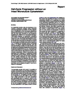
Trial-by-trial adaptation The state–space model provides additional information on the way adaptation evolves in time. For each subject, we estimated the model parameters (compliance, learning rate, variance of the adaptation and execution noise). A comparison of the
Multiple Sclerosis 2008; 14: 330–342 Downloaded from http://msj.sagepub.com at Univ Degli Studi Di Genova on October 10, 2008
http://msj.sagepub.com
Abnormal sensorimotor control in MS subjects with no clinical disability
337
Figure 4 Time course of movement performance indicators (mean ⫾ SE) during the different phases of the experiment (N: null field, F: force field), for control (gray) and MS subjects (black). (A) 100-ms aiming error; (B) 300-ms aiming error. (C); Acceleration duration and (D): Deceleration duration.
Figure 5 Learning indicators. (A) Temporal evolution of the LI (mean ⫾ SE, see text) during the force field phase, for control (gray) and MS subjects (black) and (B) LI, per subject.
parameter estimates in control and MS subjects suggested no significant differences in arm compliance among MS subjects and controls. In contrast, we found small differences in the learning rate http://msj.sagepub.com
(P ⫽ 0.027, P ⫽ 0017 and P ⫽ 0.038 for, respectively, 0°, 45° and 180°). In particular, in MS subjects the learning rate is smaller at 0° and 45° and greater at 180°; see Figure 6. Multiple Sclerosis 2008; 14: 330–342
Downloaded from http://msj.sagepub.com at Univ Degli Studi Di Genova on October 10, 2008
338
M Casadio et al.
Figure 6 State–space model of adaptation. (A) Observed (gray) and predicted (black) trial-by-trial patterns of lateral deviation (300-ms aiming error), for a typical control subject. Light gray circles indicate catch trials. Scale bar: 20 deg; (B) Same for a typical MS subject; (C) Average compliance ellipse for control (black) and MS subjects (gray); (D) Direction dependence (mean ⫾ SE) of the learning rate, for control (black) and MS subjects (gray); (E and F) Variances of the adaptation noise (E) and the execution noise (F); average (gray) and per subject (squares).
Multiple Sclerosis 2008; 14: 330–342 Downloaded from http://msj.sagepub.com at Univ Degli Studi Di Genova on October 10, 2008
http://msj.sagepub.com
Abnormal sensorimotor control in MS subjects with no clinical disability With regards to noise variances, we found no significant differences in the adaptation noise. This means that the state–space model explains the dynamics of adaptation equally well in MS subjects and controls. In contrast, we found that MS subjects display a significantly greater (P ⫽ 0.0006) execution noise. This means that sometimes the adaptive response to force fields may be inappropriate, and this occurs more often in MS subjects than in control subjects.
Discussion We used a robot manipulandum to assess sensorimotor performance and adaptation capability in MS subjects with no disability – for these subjects, functional brain imaging studies suggest evidence of cortical and subcortical reorganization. When the robot generated no forces, the movements of MS subjects were more curved, displayed greater and more variable directional errors and a longer deceleration phase. During the force field phase, both MS and control subjects gradually learned to predict the robot-generated forces. The rates of adaptation were similar, but MS subjects showed a greater variability in responding to the force field.
Descending commands are abnormal in MS subjects Analysis of the motor performance during the nullfield trials shows that MS subjects with no disability make greater errors than controls. More specifically, their movements start in the wrong direction. Also, they have a prolonged deceleration phase and are more curved. This is consistent with previous findings [9]. In addition, here we report that MS subjects display a greater variability in the direction of start of their movements. The peculiar dependence on direction displayed by the 100-ms aiming error and the acceleration duration may reflect the fact that both MS and control subjects inaccurately account for the anisotropy of the inertia of the arm and the robot system. This finding is well established in normal subjects [21]; MS subjects display a similar pattern, but with a greater error magnitude. In addition, these subjects generate motor commands that are not only systematically inappropriate, but also more variable from trial to trial. Nevertheless, such errors do not prevent subjects from reaching the targets. This is likely because these errors are compensated, at least in part, by making on-line corrections, based on the available visual and/or proprioceptive information. This may explain the greater duration of the deceleration phase. http://msj.sagepub.com
339
Can the above abnormalities be due to degradation of performance caused by, for instance, muscle fatigue? If this were the case, these effects would have been even greater at the end of the force field adaptation phase because under this condition subjects are required to generate much greater muscle forces than those needed in the null-field condition. However, this was not the case; rather, the observed abnormalities point to defective control. We also found that in both MS subjects and controls motor performance improves across repetitions. Multiple sclerosis subjects even display a greater improvement in aiming error variability and in deceleration duration. This suggests that MS subjects and controls are equally good at familiarizing with the dynamics of the manipulandum, and that the performance of MS subjects may benefit from exercise. However, the latter never achieve the performance of controls. These results are also consistent with similar findings of a deteriorated balance and gait function in MS subjects with no disability [8].
Multiple sclerosis subjects preserve their ability to adapt to unfamiliar dynamic environments In spite of these subtle abnormalities in motor coordination, we found that the ability to adapt to an unfamiliar dynamic environment is substantially preserved in MS subjects. In particular, MS subjects and controls display similar levels of arm compliance, and very little differences in the rates of adaptation. Therefore, MS subjects effectively learn to predict the force field generated by the robot rather than just trying to resist to perturbations by stiffening their arm. The most significant difference we found between MS subjects and controls is in the extent to which the observed trial-by-trial motor errors during force field adaptation are predicted by the state–space model. Variability in the process of force field adaptation may come from at least two different sources [15,17]. It may be due to an inability to learn how to predict the current perturbation (adaptation noise), to an inability to effectively use such prediction (execution noise), or both. We found that the variance of adaptation noise does not differ significantly from controls, whereas the variance of execution noise is significantly greater. This latter finding suggests that MS subjects are capable of acquiring an accurate internal representation of the unfamiliar dynamics (curl field), but they make more errors when actually using such internal representation during movement execution. As a consequence, in MS subjects motor commands are inappropriate more often than in controls, and therefore require an extra amount of Multiple Sclerosis 2008; 14: 330–342
Downloaded from http://msj.sagepub.com at Univ Degli Studi Di Genova on October 10, 2008
340
M Casadio et al.
corrections, which are performed by appropriately exploiting the available sensory information. Such a difficulty in movement execution is confirmed by the finding of a greater variability observed in the 100-ms aiming error. Movement execution problems may also be related to the lack of attention observed in MS subjects [22]. The experiment presented here allows to discriminate between evaluation of motor performance and assessment of adaptation capability (i.e., the ability to predict the consequences of our actions). Our results suggest that MS subjects with no disability have problems with movement execution but not with motor adaptation. Nevertheless, adaptation capability may be affected in subjects with a more severe impairment. Recently, Leocani et al. (2007) [10] reported that MS subjects with a more severe impairment (EDSS ⬇ 5) display a greater difficulty than controls in tracking a moving target, and also display a smaller improvement with time of their tracking performance.
Preserved adaptation capability and brain reorganization We demonstrated that in MS subjects with no disability, in some cases after a long disease duration, the ability to learn new movements is similar to that of healthy controls. This suggests a preserved potential for the brain to reorganize. There is an increasing evidence that cortical reorganization contributes to maintain normal levels of function after a relapse. Such reorganization would have important implications for the development of future treatments, aimed at promoting neuronal plasticity and at improving the clinical outcome of the disease. MRI techniques allow to assess the anatomical neuronal damage and its consequences on cortical adaptation, but provide little information on the corresponding motor performance and on the residual ability to adapt. Functional MRI techniques have been extensively used to study cortical reorganization in MS subjects. In an fMRI study, Rocca et al. (2005) [2] showed that a repetitive finger flexion-extension task resulted in the recruitment of different brain areas in MS subjects at various stages of the disease. With respect to healthy controls, nondisabled subjects with relapsing– remitting MS (a class of subjects that corresponds to those tested in the present study) displayed increased activations in various cortical areas. Moreover, a comparison of performance and fMRI activations in simple and complex motor tasks [23] suggested that in early-stage MS subjects, finger flexion-extension leads to the recruitment of brain
areas that in healthy subjects are associated with more complex tasks (e.g., object manipulation). Other studies have addressed the contribution of cortical adaptation to visual recovery after optic neuritis, showing a clear time course: a low fMRI response during the acute phase is followed by an increase in the volume of the activated regions, which suggests a transient adaptive change. Two studies [24,25] have demonstrated that visual function at baseline was inversely related to optic nerve damage and directly related to the extent of response on fMRI, thus suggesting a dynamic spatiotemporal cortical change. These results support the notion that if a neural pathway is affected by MS lesions, the corresponding cortical areas are more largely activated in order to maintain the finalized activity. Taken together, these results suggest that in early MS a substantial cortical reorganization takes place, possibly triggered by widespread tissue damage. Such reorganization might contribute to maintaining normal levels of function. Moreover, such a reorganization involves areas that are related to the processing of sensorimotor information [23,26]. This is consistent with our findings that MS subjects achieve a close-to-normal motor function by performing a greater proportion of microadjustments (as witnessed by the greater curvature and duration of the deceleration phase), to compensate for the partly incorrect descending commands (as witnessed by the greater aiming errors). It should be noted that the brain areas which display extra activation in MS subjects with no disability [2] are consistent with those that were found to correlate with the processing of motor errors in normal subjects [15], during a task similar to that described here. This indirectly supports the notion that extra activation may reflect the need for extra corrections, in the sense that corrections would correlate with extra activation in the brain areas related to error and/or sensory processing, an/or in greater activation in the areas related to motor commands.
Fatigue Fatigue is a major concern with MS subjects. In the design of the experimental protocol, we carefully took this problem into account. Subjects were allowed to rest between consecutive blocks of trials. However, only one MS subject did and this supports the fact that the task was not fatiguing and in fact was well tolerated. Moreover, Figure 4 and the corresponding statistical analysis shows that there was no degradation of performance at the end of the adaptation phase as compared to the final portion of the null phase. This, again, suggests that
Multiple Sclerosis 2008; 14: 330–342 Downloaded from http://msj.sagepub.com at Univ Degli Studi Di Genova on October 10, 2008
http://msj.sagepub.com
Abnormal sensorimotor control in MS subjects with no clinical disability subjects displayed no problems in performing the task in the late phase of the experiment. Indeed, a small improvement was often observed.
Implications for robot-assisted rehabilitation Overall, these results suggest that sensorimotor adaptation protocols may represent a useful tool to measure the motor performance after the adaptation, which follows brain lesions. Robotic devices are also a promising tool for neuromotor rehabilitation. They may be used to guide or assist the movements of a patient and in close interaction with him, just like a human physical therapist [27]. A prerequisite for the success of both human and robot therapy is that patients preserve their ability to adapt to novel dynamic environments, an ability related to the feed-forward component of control. Robot therapy would be an ideal tool for implementing this strategy, also taking into account that it is possible to integrate in the robot training sessions motor evaluation primitives that are essential for monitoring the disease and finely tune the interaction parameters that characterize the training sessions.
341
Because the force field was kept constant in all trials in which it was applied, but the actual force experienced varied depending on the movement velocity, following [20] the force input to the model, f(n), was represented as a discrete scalar (force field magnitude), and was modeled with values of 0 or 1, corresponding to catch or field trials, respectively. Thus, the compliance computed from this model has units of angular error and can be interpreted as the aiming error directly induced by the force field. However, this quantity is intrinsically related to the hand compliance matrix, and the relationship between the two is a simple multiplicative constant – T/b, where T ⫽ 300 ms is the time after movement onset, and b ⫽ 13 Nm/s is the field viscosity, see [7] for more details. We used this conversion to report the compliance ellipses in Figure 6. The noise terms were assumed to be Gaussian and with zero mean, that is, r(n) ~ N(0,R) and q(n) ~ N(0,Q), and we assumed R ⫽ R2 and Q ⫽ I Q2, which adds another two parameters to the model (i.e., R2 and Q2). We used a nonlinear optimization procedure to find B, D, R and Q that maximize, for every subject, the log-likelihood of the observed aiming error sequence [17,29].
References Acknowledgements This work is partly supported by the Italian Multiple Sclerosis Foundation (FISM) and by the Italian Ministry of Education, University and Research (MIUR).
Appendix A. State–space computational model The type of state–space computational model of the trial-to-trial changes in motor output described in the Methods section was previously used to characterize the adaptation to curl fields in healthy subjects [18–20], and in subjects with cerebellar atrophy or Huntington’s disease [7]. Our model is similar to the model used in [7], with the only addition of the output and state–space noise terms. To reduce the number of parameters, we assumed that arm compliance in the horizontal plane is described by a symmetric 2 ⫻ 2 matrix [28]. Since the eight-dimensional compliance vector D is completely determined by arm compliance, we constrained D to three free parameters. Furthermore, we assumed that the error generalization functions (columns of B) are essentially symmetric (i.e., the amount of generalization to ⫺45° is essentially the same as generalization to ⫹45°, see [7,18]. Therefore we enforced symmetry on B, which constrained it to five free parameters. http://msj.sagepub.com
1. Lublin FD, Baier M, Cutter G. Effect of relapses on development of residual deficit in multiple sclerosis. Neurology 2003; 61: 1528–32. 2. Rocca MA, Colombo B, Falini A, Ghezzi A, Martinelli V, Scotti G et al. Cortical adaptation in patients with MS: a cross-sectional functional MRI study of disease phenotypes. Lancet Neurol 2005; 4: 618–26. 3. Wang J, Hier DB. Motor reorganization in multiple sclerosis. Neurol Res 2007; 29: 3–8. 4. Filippi M, Rocca MA. MRI evidence for multiple sclerosis as a diffuse disease of the central nervous system. J Neurol 2005; 252(Suppl 5): v16–24. 5. Shadmehr R, Mussa-Ivaldi FA. Adaptive representation of dynamics during learning of a motor task. J Neurosci 1994; 14: 3208–24. 6. Maschke M, Gomez CM, Ebner TJ, Konczak J. Hereditary cerebellar ataxia progressively impairs force adaptation during goal-directed arm movements. J Neurophysiol 2004; 91: 230–8. 7. Smith MA, Shadmehr R. Intact ability to learn internal models of arm dynamics in Huntington’s disease but not cerebellar degeneration. J Neurophysiol 2005; 93: 2809–21. 8. Martin CL, Phillips BA, Kilpatrick TJ, Butzkueven H, Tubridy N, McDonald E, Galea MP. Gait and balance impairment in early multiple sclerosis in the absence of clinical disability. Mult Scler 2006; 12: 620–8. 9. Solaro C, Brichetto G, Casadio M, Roccatagliata L, Ruggiu P, Mancardi GL et al. Subtle upper limb impairment in asymptomatic multiple sclerosis subjects. Mult Scler 2007; 13: 428–32. 10. Leocani L, Comi E, Annovazzi P, Rovaris M, Rossi P, Cursi M et al. Impaired short-term motor learning in multiple sclerosis: Evidence from virtual reality. Neurorehabil Neural Repair 2007; 21: 273–8. 11. Poser CM, Paty DW, Scheinberg L, McDonald WI, Davis FA, Ebers GC et al. New diagnostic criteria for
Multiple Sclerosis 2008; 14: 330–342 Downloaded from http://msj.sagepub.com at Univ Degli Studi Di Genova on October 10, 2008
342
12. 13. 14. 15. 16.
17. 18.
19. 20. 21.
M Casadio et al.
multiple sclerosis: guidelines for research protocols. Ann Neurol 1983; 13: 227–31. Kurtzke JF. Rating neurologic impairment in multiple sclerosis: an expanded disability status scale (EDSS). Neurology 1983; 33: 1444–52. Sipe JC et al. A neurologic rating scale (NRS) for use in multiple sclerosis. Neurology 1984; 34: 1368–72. Casadio M, Morasso PG, Sanguineti V, Arrichiello V. Braccio di Ferro: a new haptic workstation for neuromotor rehabilitation. Technol Health Care 2006; 13: 1–20. Diedrichsen J, Hashambhoy Y, Rane T, Shadmehr R. Neural correlates of reach errors. J Neurosci 2005; 25: 9919–31. Criscimagna-Hemminger SE, Donchin O, Gazzaniga MS, Shadmehr R. Learned dynamics of reaching movements generalize from dominant to nondominant arm. J Neurophysiol 2003; 89: 168–76. Cheng S, Sabes PN. Modeling sensorimotor learning with linear dynamical systems. Neural Comput 2006; 18: 760–93. Donchin O, Francis JT, Shadmehr R. Quantifying generalization from trial-by-trial behavior of adaptive systems that learn with basis functions: theory and experiments in human motor control. J Neurosci 2003; 23: 9032–45. Scheidt RA, Dingwell JB, Mussa-Ivaldi FA. Learning to move amid uncertainty. J Neurophysiol 2001; 86: 971–85. Thoroughman KA, Shadmehr R. Learning of action through adaptive combination of motor primitives. Nature 2000; 407: 742–7. Gordon J, Ghilardi MF, Cooper SE, Ghez C. Accuracy of planar reaching movements. II. Systematic extent errors resulting from inertial anisotropy. Exp Brain Res 1994; 99: 112–30.
22. Mainero C, Pantano P, Caramia F, Pozzilli C. Brain reorganization during attention and memory tasks in multiple sclerosis: insights from functional MRI studies. J Neurol Sci 2006; 245: 93–8. 23. Filippi M, Rocca MA, Mezzapesa DM, Ghezzi A, Falini A, Martinelli V et al. Simple and complex movement-associated functional MRI changes in patients at presentation with clinically isolated syndromes suggestive of multiple sclerosis. Hum Brain Mapp 2004; 21: 108–17. 24. Langkilde AR, Frederiksen JL, Rostrup E, Larsson HB. Functional MRI of the visual cortex and visual testing in patients with previous optic neuritis. Eur J Neurol 2002; 9: 277–86. 25. Toosy AT, Werring DJ, Bullmore ET, Plant GT, Barker GJ, Miller DH et al. Functional magnetic resonance imaging of the cortical response to photic stimulation in humans following optic neuritis recovery. Neurosci Lett 2002; 330: 255–9. 26. Filippi M, Rocca MA, Mezzapesa DM, Falini A, Colombo B, Scotti G et al. A functional MRI study of cortical activations associated with object manipulation in patients with MS. Neuroimage 2004; 21: 1147–54. 27. Prange GB, Jannink MJ, Groothuis-Oudshoorn CG, Hermens HJ, Ijzerman MJ. Systematic review of the effect of robot-aided therapy on recovery of the hemiparetic arm after stroke. J Rehabil Res Dev 2006; 43: 171–84. 28. Mussa-Ivaldi FA, Hogan N, Bizzi E. Neural, mechanical and geometric factors subserving arm posture in humans. J Neurosci 1985; 5: 2732–43. 29. Ghahramani Z, Hinton GE. Parameter estimation for linear dynamical systems. University of Toronto, Toronto, 1996.
Multiple Sclerosis 2008; 14: 330–342 Downloaded from http://msj.sagepub.com at Univ Degli Studi Di Genova on October 10, 2008
http://msj.sagepub.com